1st Trimester Obstetric Ultrasound
1st Trimester Obstetric Ultrasound
Goals
Views
Technique and normal findings
Normal anatomy
Normal, non-pregnant anatomy
Normal, pregnant anatomy
Measuring gestational age
Abnormal findings
Ectopic pregnancy
Spontaneous abortion
Pearls and pitfalls
Goals
- Evaluate for intrauterine pregnancy
- Estimate the gestational age of the fetus and predict likely due date
- Evaluate for potential ectopic pregnancy
Probe: Curved transducer [Providers may use endocavitary probe for very early pregnancy as needed, provided adequate probe sanitation measures are in place]
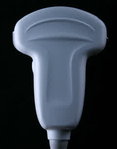
Patient positioning: supine or left lateral decubitus
Views
- Long axis view of uterus (index marker towards mother’s head)

- Sagittal view of the uterus
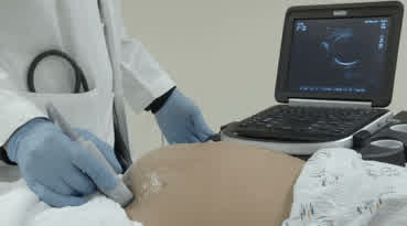
Technique and Normal Findings
Normal anatomy
Normal, non-pregnant anatomy
- In a longitudinal view, urine in the bladder looks like a black, or hypoechoic, triangle on the right side of the screen next to the uterus.
- The uterus is shaped like a bean and appears grey, or moderately echogenic (arrow).
- The normal, non-pregnant uterus has a thin hyperechoic white line in the center that is the endometrium. The * denotes the cervix.
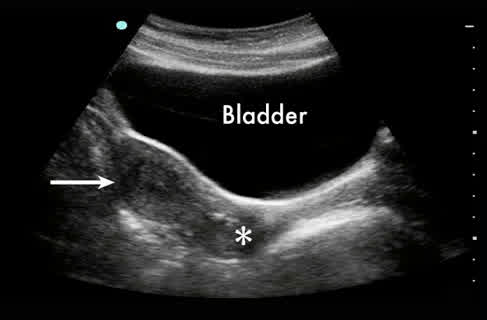
Normal anatomy in sagittal view. Note the bladder, uterus (arrow) and cervix (*)
Normal, pregnant anatomy
- Early in the pregnancy, a normal fetus lives in a sack of fluid, the gestational sac, inside the uterus.
- The yolk sac, a perfect circle that looks like a bright ring (arrow), is the first sign that the gestational sac contains a fetus. It is visible via trans-abdominal imaging around 5 weeks following the last menstrual period.
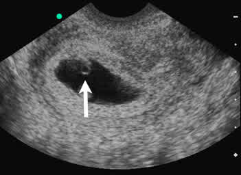
Uterus with a yolk sac (arrow)
- As the fetus gets older, more grey tissue can be seen and is called a fetal pole (arrow) connected to the yolk sac (arrowhead). On average, it is visible via trans-abdominal imaging around 7 weeks following the last menstrual period.
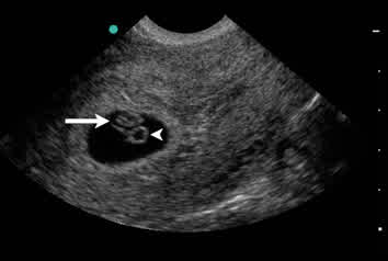
Uterus with a yolk sac (arrowhead) and fetal pole (arrow)
- It is possible to see a fetal heart beat at about 8 weeks from the mother’s last period.
Measuring gestational age
The Crown Rump Length (CRL) is the most accurate way to measure the age of the fetus in the first trimester
- Point the index marker to the mother’s head. The other end of the transducer should rest against the pubic bone.
- Find a good image of the fetus on ultrasound, then push the Freeze button. The fetus should be measured when it looks the longest and both the head and the fetus’s bottom can be seen in the same picture. Do not include the yolk sac or limbs in the measurement.
- For all gestations <14 weeks, select CRL on the available calculations list (this will differ by machine).
- The age of the fetus appears at the bottom of the ultrasound screen if you are in “OB” scanning mode. Pushing Freeze again will return to active scanning.
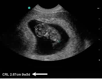
CRL measured with calipers. Gestational age calculation appears at the bottom of the screen (arrow).
Abnormal Findings
Ectopic pregnancy
- In an ectopic pregnancy, the uterus will be empty.
- If the ectopic pregnancy has ruptured, there may be blood extending from the pelvis into the abdomen. Free fluid may accumulate in the right upper quadrant between the liver and kidney, the splenorenal recess in the left upper quadrant, and/or the pelvis.
- You may also see a mass, echogenic abnormality, or actual pregnancy outside the uterus, but even without these findings, an empty uterus plus free fluid in the pelvis should raise suspicion for an ectopic pregnancy.
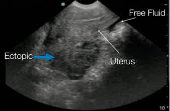
Ruptured ectopic pregnancy. Note the uterus with endometrial stripe at the top of the screen.
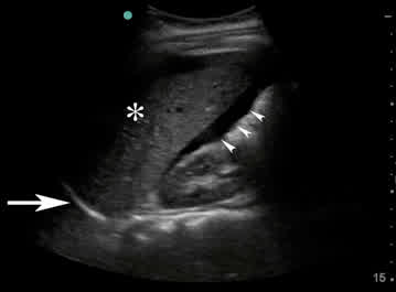
Blood (arrowheads) between the liver (*) and kidney. The diaphragm is marked with an arrow.
Spontaneous abortion
- If there is a fetus in the uterus but the mother complains of bleeding or pain, there is a risk of spontaneous abortion. She must be examined to make sure there is no infection, and that she has not sustained trauma.
- If the mother has already miscarried and tissue has been passed from the vagina, she may have some retained products of conception or tissue inside the uterus that can be seen on ultrasound and can cause infection.
- If retained products of conception are present, consider using colour Doppler to assess for retained placental parts that may be actively bleeding.
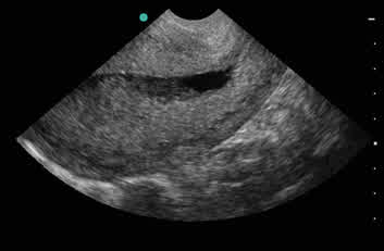
Retained products of conception within the uterus
Pearls and pitfalls:
- A trans-abdominal ultrasound is best performed when the patient has a full bladder, which provides a good window for viewing the uterus.