2nd and 3rd Trimester Obstetric Ultrasound
Partners In Health Ultrasound Series: 2nd and 3rd Trimester Obstetric Ultrasound
Goals
Views
Technique and normal findings
Evaluation of fetal presentation
Evaluation of the placenta
Gestational age
Evaluation for multiple fetuses
Evaluation of amniotic fluid
Abnormal findings
Placenta previa
Abnormal amniotic fluid volume
Evaluation of uterine fundus large for expected gestational age
Pearls and pitfalls
Goals
- Determine the age of the pregnancy
- Evaluate for placenta previa
- Assess for causes of enlarged fundal size
- Evaluate fetal heart rate
Probe & Mode: Curved/abdominal transducer, OB mode
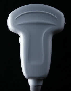
Patient positioning: supine or left lateral decubitus
Views
- Long axis view of uterus (index marker towards mother’s head)
- Sagittal view of uterus
Technique and Normal Findings
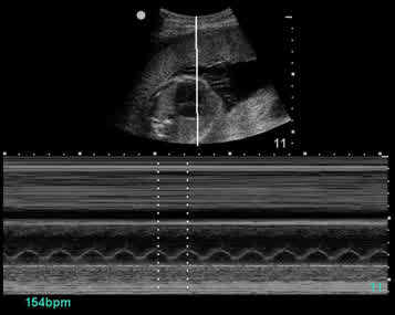
Fetal Heart Rate (FHR): Begin each 2nd/3rd trimester scan with evaluation of FHR for early identification of any distress before proceeding with the rest of the scan
- A normal FHR is between 120 and 180 beats per minute.
- To calculate the FHR, either watch the beating fetal heart and count manually, or use the ultrasound machine in M-mode to do the calculation.
Fetal heart rate measurement using M-mode
- For this technique, use “OB” mode under scan type in the Patient section.
- Find the fetal heart and maximise its size on the screen by changing the depth and possibly using the Zoom button.
- Press the M mode button and move the reference line over the beating heart. Then press M Mode again.
- When the fetal heart tracing has continued for several heartbeats, press the Freeze button
- Press the Calc button and select FHR.
- Move the dotted line over one peak of one heart beat and press Select. A second dotted line will appear and should be moved to the next peak.
- The calculated FHR will appear in the lower right corner of the screen
Evaluation of fetal presentation
- Ultrasound can be used to evaluate which part of the fetus is projecting towards the internal cervical os, or fetal presentation.
- The cephalic (also known as vertex) presentation, in which the fetal head presents towards the internal cervical os, is considered normal and occurs in about 97% of births.
- Abnormal presentations include breech presentation (in which the fetal rump presents towards the internal cervical os), shoulder presentation, and cord presentation.
To evaluate fetal presentation,
- Place the probe low over the mother’s pelvis, just superior to the pubic bone
- Assess the presenting fetal parts to see if the hyperechoic oval-shaped skull rim is seen, or if some other presenting part is visualised.
- If the head is not visualised next to the os, scan in the longitudinal and sagittal planes until the head is located.
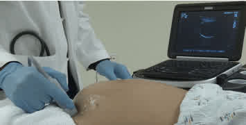
Probe low over mother’s pelvis. Oval-shaped skull bone on ultrasound screen confirms vertex/cephalic presentation.
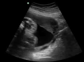
Non-cephalic presentation. Presenting part of fetus is not head.
Evaluation of the placenta
- The growing placenta is often seen on ultrasound by about 10-12 weeks gestational age. It appears as a moderately hyperechoic (grey) structure between the amniotic sac and uterine wall, to which it is adherent.
- The placental location is of prime importance. Scan the entire uterus in an organised fashion to visualise the whole placenta, with special attention to whether the placenta is located anteriorly or posteriorly and to where the inferior edge lies.
- If the inferior edge of the placenta is still visualised and the probe is very low over the mother’s pubic bone, near her bladder, strongly consider low-lying placenta or placenta previa. A trans-vaginal ultrasound should be arranged to confirm location.
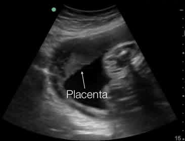
Anterior implantation of placenta
Gestational age
- The most accurate time to determine the gestational age of the pregnancy using ultrasound is in the 1st trimester (<14 weeks) using the Crown-Rump Length measurement.
- Ultrasound measurements of gestational age in the 2nd or 3rd trimester are thought to be accurate to within 2 weeks in the second trimester and to within 3 weeks in the third trimester.
To measure gestational age in the 2nd or 3rd trimester, make sure you are in OB mode:
- Scan the entire uterus to determine the fetal head location and general position of the fetus.
- Once the fetal head is seen, begin with Biparietal diameter (BPD). Scan and freeze the image of the fetal head in the axial plane (transverse) to identify these landmarks: the third ventricle, the thalamus (*), and the skull bone. Caliper measurements should be made perpendicular to the falx cerebri, from the outer edge of one parietal bone to the inner edge of the opposite parietal bone. Save the image and measurement.
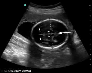
Biparietal diameter measurement. Note the cavum septi pellucidi (arrowheads), falx cerebri (arrow) and thalami (*)
- Head circumference: Using the same frozen image, place the calipers around the outer edge of the skull bone in the best fit position. Save the image and measurement.
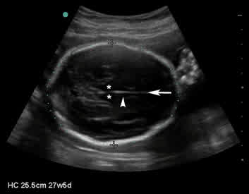
Head circumference measurement with thalamus (*), third ventricle (arrowhead), falx cerebri (arrow) and calipers measuring outer edge of the skull bone
- Abdominal circumference (AC): This is the least accurate measure as a single measurement, but used with other measures can improve overall accuracy. Ensure that the measurement of the fetal abdomen is in an axial plane creating a circular cross section of the abdomen, and that the following landmarks can be identified: transverse spine, the umbilical vein within the liver (arrowhead), and the stomach (*). The calipers are placed around the outer skin edge of the abdomen. Save the image and measurement.
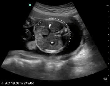
Abdominal circumference measurement. Note the landmarks of the stomach (*) and umbilical vein within the liver (arrowhead)
- Femur length (FL): Identify the femur and measure its length, including only the bony portions of the diaphysis and metaphysis. Save the image and measurement. Be sure not to measure the humerus.
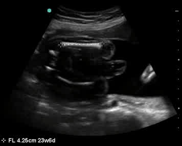
Femur length measurement
- Press the Report button to obtain an average of all measurements taken (BPD, HC, AC, and FL) to give a single estimation of gestational age and a single estimated due date
Evaluation for multiple fetuses
Any mother whose uterus is larger than expected for gestational age should be evaluated for multiple fetuses
- For every ultrasound exam performed, always scan through the entire uterus to ensure there is only one fetus.
- If there is suspicion for more than one fetus, look for easily identifiable structures such as the heart or head to confirm the number of fetuses.
- If two fetuses are identified, note whether the fetuses are diamniotic and have separate sacs, or are sharing the same sac. This may require advanced knowledge of obstetric imaging and referral. Note the presence and number and position of any placental tissues.
Evaluation of amniotic fluid
Any mother whose uterus is larger or smaller than expected for gestational age should have her amniotic fluid volume assessed
- Determining whether there is an appropriate amount of amniotic fluid around a fetus is important in the second and third trimester.
- Amniotic fluid volume may be estimated visually to see if it looks normal, excessive, or low based on the sonographer’s opinion. Amniotic fluid volume may also be measured using ultrasound by using the AFI (Amniotic Fluid Index) or the DVP (Deepest Vertical Pocket).
Amniotic fluid index (AFI): One technique for measuring the amniotic fluid is the AFI, which adds fluid measures from all four quadrants of the uterus to create a numeric score.
- For this measurement, visually divide the gravid uterus into four quadrants using the mother’s umbilicus and linea nigra as landmarks for the centre line. In the OB mode on the machine, select AFI.
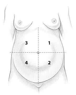
The gravid uterus is divided into four quadrants. The mother’s upper left quadrant is number 1, lower left is number 2, right upper quadrant is number 3 and right lower quadrant is number 4.
- Amniotic fluid pockets in each quadrant are measured from the superficial edge of the pocket to the deepest fetal structure. The measurements in centimetres are added together to produce the AFI.
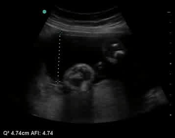
Amniotic fluid pockets in each quadrant are measured from the superficial edge of the pocket to the deepest fetal structure.
- In general, an AFI of 8-20 cm is considered normal. Oligohydramnios can be defined as an AFI less than 5 cm, while values greater than 24 cm suggest polyhydramnios.
- Measure only where there is a clear pocket of fluid free of fetal parts or cord. Keep the probe perpendicular to the skin surface of the mother and be careful to avoid applying pressure, which will displace fluid and affect measurements.
Deepest vertical pocket (DVP): Another method for assessing amniotic fluid volume on ultrasound is to measure one single deepest vertical pocket after scanning the whole uterus to find the best place to measure.
- On the machine, select the calipers function to measure between two points.
- Scan through the patient’s uterus in the longitudinal and sagittal planes to locate the pocket of maximal depth of amniotic fluid that is free of umbilical cord and fetal parts.
- Using the calipers, measure the depth of this pocket.
- In general, a DVP of 2-8 cm is considered normal. A DVP less than 2 cm indicates oligohydramnios. A DVP greater than 8 cm indicates polyhydramnios.
Abnormal Findings
Placenta previa
- Patients with unexplained vaginal bleeding must be evaluated for placenta previa.
- Examine the position of the placenta; in placenta previa, the inferior portion approaches or crosses over the internal os of the cervix.
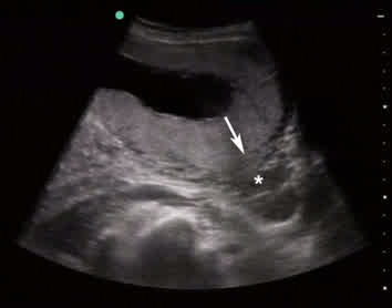
In placenta previa, the placenta crosses over the internal os (arrow) of the cervix (*)
Abnormal amniotic fluid volume
- Fetuses with less fluid than normal, or oligohydramnios [AFI<5, DVP<2cm], may be at risk for preterm birth, renal agenesis (absence of the kidneys), intrauterine fetal demise, and infection. Typically, low fluid is caused when the membranes rupture and leak slowly.
- Excess amniotic fluid, or polyhydramnios [AFI>24, DVP>8cm], is also a problem that should be addressed. There are many causes of polyhydramnios, including maternal diabetes, fetal anemia, and certain birth defects.
Evaluation of uterine fundus large for expected gestational age
- In assessing a patient whose uterine fundus measures large for their expected gestational age by physical exam, it is important to evaluate for excess fluid (see “Evaluation of amniotic fluid”) and multiple fetuses (see “Evaluation for multiple fetuses”) that may increase the size of the uterus.
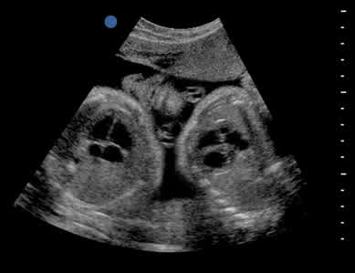
Twin pregnancy showing 2 hearts
Pearls and Pitfalls
- Never use Doppler mode on the fetal heart. M mode is safe and applies less energy to the growing heart, but Doppler concentrates high energy and heat on the delicate growing heart of the fetus and should not be used.
- An example documentation of an obstetric ultrasound would be: “Live, singleton intrauterine pregnancy with fetal heart rate 150 bpm by M mode. Estimated gestational age using BPD, and FL is 32 weeks 4 days. Placenta anterior ̶ no previa. AFI 12.”