Acute Asthma in Adults
Acute asthma in adults
Introduction
Identification
Severity assessment
Management based on severity
Relevant history, examination
Investigations
Admission/referral criteria
Discharge criteria/checklist
Follow up: acute and long-term
Preventative measures
Advice for caregivers
Treatment Algorithm
Introduction/ Description
Asthma is a chronic inflammatory disease that is characterized by reversible airflow limitation secondary to airway inflammation and bronchial hyper-responsiveness resulting in reduced airway diameter. Inflammation is induced by a variety of stimulants (allergens) and may be acute, sub-acute or chronic. Airway obstruction leads to increased resistance to airflow, decreased expiratory flow rates and hyperinflation, with subsequent ventilation-perfusion mismatch.
Identification/ Recognition/ Diagnosis of Asthma
Cardinal asthma symptoms are cough, wheeziness, chest tightness and dyspnoea. The perception of the severity of asthma symptoms differs between patients and influences health-seeking behaviour. An acute asthma attack / exacerbation is characterized by:
- Progressive and abruptly worsening asthma symptoms
- Progressive decline in response to B2 agonists despite increased use of rescue bronchodilator therapy
- Progressive deterioration in lung function (decrease in PEF measurement).
- Status asthmaticus is a severe and continuous asthma exacerbation requiring emergent treatment. These patients present with life-threatening or near-fatal asthma
Differential diagnosis
- Foreign body obstruction
- Vocal cord dysfunction
- Heart failure
- Anaphylaxis
- COAD / emphysema
- Pulmonary embolism
- Pneumothorax
- Allergic bronchopulmonary aspergillosis
- Bronchiectasis
- Inhalation injury
- Severity assessment (triage)
Severity assessment (triage)
| Mild/Moderate | Severe Asthma | Life-Threatening Asthma |
| Clinical: Talks in phrases May prefer sitting to lying down May be agitated No accessory muscle use | Clinical: Talks in words Sits hunched Usually agitated Using accessory muscles | Clinical: Drowsy; confused Absence of wheeze/silent chest Paradoxical thoraco-abdominal movement Exhaustion |
| Vitals: Increased RR Pulse 100 - 120 bpm Pulsus paradoxus <20 mmHg O2sats in RA 90 - 95% PEF >60% predicted or best | Vitals: RR > 30/min Pulse > 120 bpm Pulsus paradoxus 20 - 40 mmHg O2 sats in RA < 90% PEF 33 - 60% predicted or best | Vitals: Bradycardia or arrythmia Hypotension Absence of pulsus paradoxes PEF < 200L/min or < 33% predicted or best Unable to perform PEF |
| ABG: (not usually necessary) PaO2 > 8 kPa; PaCO2 < 4.6 kPa | ABG: PaO2 > 8 kPa; PaCO2 < 4.6 kPa | ABG: PaO2 < 8 kPa; PaCO2 4.6 - 6 kPa |
Management based on severity
Immediate action:
Non-drug treatment
Assess exacerbation severity, protect airway, supply oxygen, ensure adequate ventilation and IV access. Call for help if features of a severe / life-threatening asthma attack are present. Perform a focused initial assessment. Check for anaphylaxis.
Drug treatment (ranked from best to least evidence for efficacy)
- First line: Oxygen, inhaled B2 agonists and systemic corticosteroids
- Second line: add inhaled Ipratropium bromide to if features of acute severe or life-threatening asthma attack, or if poor initial response to first line.
- Third line: add intravenous MgSO4if features of acute severe or life-threatening asthma attack, or if poor response to bronchodilators
- Fourth line: add intramuscular adrenaline and/ or intravenous Aminophylline (low evidence) and Salbutamol (low evidence) if poor response to above and features of acute severe or life-threatening asthma attack remain
- Adjunctive treatment
- Antibiotics: Routine administration of antibiotics not recommended in acute asthma attack in adults, unless evidence of bacterial infection present (fever, purulent sputum, CXR changes)
- Intravenous fluids: Provide for maintenance and replacement requirements, and need for caloric intake in patients unable to take oral fluids
- Ketamine: Produces additional bronchodilation and relaxation of smooth muscle beyond other adjunctive treatment, possibly avoiding need for RSI. Indicated for life-threatening / near fatal asthma; for failed standard treatment; if imminent intubation considered (visibly tiring, worsening respiratory muscle fatigue, inability to speak, altered mental status, PaCO2 ≥ 5.6 mmHg)
Drug tables/ dose charts
Oxygen
- Give supplementary oxygen by nasal cannula, facemask or NRB if hypoxic, cyanosed, or severe acute asthma attack and no oximetry availableC
- If oximetry available maintain > 92%C
- In- and out-of-hospital nebulised B2 agonists should preferably be oxygen drivenA
Inhaled B2 agonists
- Nebulised 5 mg salbutamol or 1 mg fenoterol every 20 minutes until a satisfactory response is achieved
- No nebulizer available: 10 – 20 puffs (100 mcg/puff) salbutamol MDI via spacer every 20 minutes PRN, taking several deep breaths from spacer after every 2 puffs
Systemic corticosteroids
- Oral prednisone 40mg (0.5 – 1 mg/kg, max 60mg) PO stat, then daily
- Or if severe breathlessness/ unable to swallow: hydrocortisone 100 – 200 mg IVI stat, then 6 hourly
Inhaled ipratropium bromide
- Nebulised ipratropium bromide 0.5 mg 4 hourly, in addition to salbutamol nebs, until a satisfactory response
- Or if no nebulizer available: up to 20 puffs (20 mcg/puff) ipratropium bromide MDI via LVS every 20 minutes PRN, taking several deep breaths from spacer after every 2 puffs
MgSO4
- IV MgSO4 1 – 2 g (40mg/kg) in 200 mL 0.9% saline over 20 minutesB
- One additional dose may be given > 12 hours after 1st dose if necessary
- Nebulised MgSO4is not recommendedA
Adrenaline
- Up to 3 doses of 1:1000 adrenaline 0.3 – 0.5 mg IM every 20 minutes (lower evidence if non-anaphylactic cause)
- Caution in elderly and patients with cardiac disease; risk of ischaemia and arrhythmias
Methylxanthines (low evidence)
- Loading dose: aminophylline 5 mg/kg IVI over 30 minutes (2.5 mg/kg if patient already taking theophylline)
- Maintenance: aminophylline infusion at 0.5 mg/kg/hr
Intravenous B2 agonists (low evidence)
- Loading dose: salbutamol 15 mcg/kg (max 0.25 mg) in 200 mL 0.9% saline IVI over 10 minutes
- Maintenance: salbutamol infusion at 3 – 20 mcg/min
Ketamine
- Breathing spontaneously: ketamine 0.25 – 0.5 mg/kg IVI slow bolus every 20 min PRN
- Intubated: continuous ketamine infusion 0.5 – 2 mg/kg/hr
- Side effects: laryngospasm (incidence 0.02%; attempt to relieve spasm with pressure behind angle of mandible, below ear; intubate if procedure fails); hallucinations (administer midazolam 0.05 mg/kg IV, max 2 mg); hypertension (decrease dose / dosing interval); salivation (suction, decrease dose / dosing interval, consider atropine).
Monitoring
- Perform regular monitoring of heart rate, blood pressure and respiratory rate
- Use oximetry to record oxygen saturation and maintain sats > 92%.
- Measure and record PEF at 15 – 30 minute intervals until a clear response is to treatment is achieved. Change in PEF response to initial therapy is one of the best ways to assess treatment response for acute asthma.3 Early response to treatment (PEF at 30 minutes) is the most important predictor of outcome and need for hospitalization.3
Relevant history, Examination
History
- History of current illness e.g.: URTI, treatment non-adherence, chest pain, haemoptysis, autonomic symptoms, fever, change in cough characteristics
- Pattern of use of B2 agonists ± controller therapy
- Past medical history: elicit any high risk features of severe asthma
- Smoking history: if stopped, when?
- Family history of asthma/ atopy
- Known trigger factors e.g.: pets, URTIs, seasonal change
- Medications, including recent systemic steroid use
High risk features in patients with an acute asthma attack
- Previous intubation or ICU admission for an asthma attack
- Regular use of 2 or more MDIs, or excessive use of B2 agonists / rescue therapy (> 1canister/month)
- Currently using or recently stopped systemic steroid use
- Not currently using inhaled steroids, or poor adherence with to inhaled steroids
- >1 hospital admission in the previous year for asthma-related illness
- >2 EC visits in the previous year for asthma-related illness, or a recent ECvisit in the previous month for asthma-related illness
- Brittle asthma (sudden severe attack with few warning features, despite regular Rx)
- History of non-adherence ± psychosocial issues (social isolation, psychological problems, cognitive impairment)
- Comorbid illness, including pregnancy
Examination
- Vitals: HR, BP, RR, sats, level of consciousness, presence of pulsus paradoxus
- General: Signs of respiratory distress, able to talk, cyanosis, clubbing, oedema
- Chest: Signs of hyperinflation, tracheal deviation, subcutaneous emphysema, presence of focal or bilateral wheeze, bibasal crepitations
- CVS: Pedal oedema, jugular venous distension, apex beat, character of heart sounds, presence of murmurs ± bruits
- Abdomen: Presence of abdominal breathing, liver span, ascites
- After initial B2 agonist administration, record the best of 3 PEF attempts. If breathless and unable to perform PEF, classify as severe asthma attack.
Investigations
- Routine CXR, arterial blood gases and empiric antibiotics are not recommended
- Perform CXR if the patient has an atypical presentation, if the response to initial treatment is poor and their clinical condition is deteriorating, to exclude complications (lobar collapse, infection, pneumothorax, pneumomediastinum), to exclude alternative diagnoses (heart failure, COAD) or if intubation is planned.
- Perform ABG measurements if any life-threatening features are present. Repeat ABG measurements within one hour of starting treatment if:
- Initial PaO2 is < 8 kPa unless arterial oxygen saturation is > 92%; or
- Initial PaCO2 is normal or raised; or
- Deterioration or not improved within 4 – 6 hours
- Monitor serum potassium and blood glucose concentrations.
- Check serum theophylline concentration if aminophylline is continued for more than 24 hours (aim for concentrations of 10 – 20 mg/L or 55 – 110 mol/L)
Admission/ referral criteria
- Criteria for hospital admission include:
- Patients with any features of a life-threatening or near-fatal event
- Patients with any features of a severe episode > 2 hours after initial Rx
- Patients with a mild/ moderate event with high-risk features.
- Arrange immediate transfer to an acute care facility if the patient has signs of an acute severe attack, has an altered mental state, is tiring or has a silent chest (monitor in a high care/resus setting while awaiting transfer).
Discharge criteria/ checklist
- Assessments for adequate response to treatment should be performed frequently. Subjective and objective evidence of improvement must be sought. Improvement in symptoms, vital signs and PEF should be documented in the patient’s file prior to discharge from the EC. Clinical criteria for EC discharge:
- Subjective symptoms markedly improved
- Not requiring further B2 agonist treatment
- Pulse < 120; RR 20; sats > 92% in room air
- PEF > 60% of predicted or best
- No prolonged symptoms prior to current EC visit
- No recurrence after recent exacerbation
- Good social factors (access, medications, transport)
- Adherence to medication likely
Follow-up: acute and long-term
- Prior to discharge, arrange ongoing treatment. Regular controller therapy must be prescribed or the current controller dose increased for 2 – 4 weeks to prevent recurrence of attacks. Prescribe a short course of oral corticosteroids (Prednisone
- 20 – 40 mg PO daily x 7 – 14 days). Ensure availability of rescue B2 agonist therapy. Check inhaler technique and adherence. Early follow-up must be arranged for all patients, preferably within 2 – 7 days. Low risk patients may follow-up with their family doctor or local clinic. All hospitalized patients, those with high-risk features ± recurrent EC presentations, should be referred to a respiratory specialist as well.
Preventative measures
- Formulate an asthma action plan with the patient. Examples of Asthma Action Plans can be found at these websites:
- National Heart, Lung and Blood Institute: http://www.nhlbi.nih.gov/files/docs/public/lung/asthma_actplan.pdf
- Asthma Australia: http://www.asthmaaustralia.org.au/asthma_action_plan.aspx
- American Lung Association: http://www.lung.org/lung-disease/asthma/taking-control-of-asthma/AsthmaActionPlan-JUL2008-high-res.pdf
- Asthma UK: http://www.asthma.org.uk/advice-personal-action-plan
Advice for caregivers/ patient information leaflet
- www.asthma.org.uk: Database of patient information and asthma tools
- www.nationalasthma.org.au: Database of patient information and asthma tools, links to other international asthma organizations and resources
- www.pulmonolgy.co.za: Basic asthma information, links to previous South African asthma guidelines
- www.allergysa.org: Basic patient information
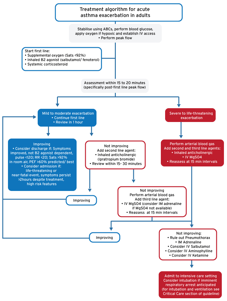
Treatment algorithm for acute asthma exacerbation in adults
Drug table | |
| Nebulized 5mg or MDI 10-20 puffs (via spacer), continuous or q20min |
| Nebulized 1 mg continuous or q20min |
| 40mg (0.5-1mg/kg, max 60mg) stat |
| 100-200mg stat and then q6h |
| Nebulized 0.5mg or MDI 10-20 puffs (via spacer) q4h |
| 1-2g (40mg/kg) over 20min |
| 1:1000, 0.5mg IM q20min (up to 3 doses) |
| Load 5mg/kg over 30min (2.5mg/kg if on theophylline) Maintenance at 0.5mg/kg/hour |
| Load 0.25mg (slow) Maintenance at 3-20µg/kg/hour |
References
Scottish Intercollegiate Guidelines Network & British Thoracic Society. SIGN 141: British Guideline on the Management of Asthma. (2014).
South African Medicines Formulary. 530–533 (Health and Medical Publishing Group of the South African Medical Association, 2010).
Lalloo, U. G. et al. Guideline for the management of acute asthma in adults: 2013 update. South African Med. J. 103, 189–198 (2013).
Goyal, S. & Agrawal, A. Ketamine in status asthmaticus: A review. Indian J. Crit. Care Med. 17, 154–61 (2013).
Global Initiative for Asthma. Pocket Guide for Asthma Management & Prevention. (2014).
Martin, R. J. Alternative and experimental agents for the treatment of asthma [Internet]. UpToDate. 2014 [cited 2014 Nov 12]. Available from: http://www.uptodate.com.ez.sun.ac.za/contents/alternative-and-experimental-agents-for-the-treatment-of-asthma?source=see_link&anchor=H4#H4
Morris, M. J. Asthma [Internet]. MedScape. 2014 [cited 2014 Nov 14]. Available from: http://emedicine.medscape.com/article/296301-overview
Weingart, S. D. Preoxygenation, reoxygenation, and delayed sequence intubation in the emergency department. J Emerg Med [Internet]. Elsevier Inc.; 2011 Jul [cited 2014 Aug 31];40(6):661–7. Available from: http://www.ncbi.nlm.nih.gov/pubmed/20378297
Weingart, S. D. et al. Delayed Sequence Intubation: A Prospective Observational Study. Ann Emerg Med [Internet]. Elsevier; 2014 Oct [cited 2014 Oct 25]; Available from: http://www.annemergmed.com/article/S0196064414013651/fulltext