Acute Coronary Syndromes
Acute coronary syndromes
Introduction
Identification
Severity assessment
Anatomic Distribution of ECG Changes
Management based on severity
Relevant history/examination
Investigations
Referral criteria
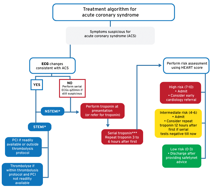
*** Serial troponin measurement triggers (taken 3 to 6 hours apart)
Isolated elevations of troponin can be due to numerous other conditions that affects the heart
- Initial value elevated: positive if a repeat test shows that an increase or decrease ≥20% have occurred
- Initial value normal followed by a positive value is also positive
Introduction/Description
Acute coronary syndrome (ACS) implies a continuum of conditions compatible with myocardial ischemia as a result of abrupt reduction in coronary blood flow. Myocardial infarction (MI) is defined as a rise or fall of troponin above the normal reference in the setting of a strong clinical suspicion of ACS. An MI is either represented as a ST elevation (STEMI) or a non-ST elevation (NSTEMI) which is an important difference as treatment also differs accordingly. A third ACS diagnosis, Unstable Angina (UA) exists where a troponin rise or fall is not seen, but clinical & ECG evidence strongly suggests ACS. Unstable angina and NSTEMI are collectively referred to as NSTEACS.
Identification/ recognition/ diagnosis of problem
There are several findings that suggest either for or against ACS. None of these are failsafe & should be seen as a guide rather than an absolute certainty. History is key, but cannot effectively rule out ACS in isolation. If you are unsure, the best evidence suggests going with your gut instinct, serial ECGs & cardiac enzymes.
Clinical factors that INCREASE likelihood of ACS include:
- CP radiating bilaterally > right > left
- Diaphoresis associated with CP
- Nausea & vomiting associated with CP
- Pain with exertion
Clinical factors that DECREASE likelihood of ACS include:
- Pleuritic pain
- Positional pain
- Sharp, stabbing pain
- Pain reproducible with palpation
The ACS diagnosis triangle: The main components of an ACS diagnosis rest upon the history & presentation, the ECG findings & the cardiac enzyme results. ACS is considered if any two of these suggest cardiac ischaemia. It is strongly considered if cardiac enzymes are positive.
Differential Diagnosis
- Life threatening: pulmonary embolus, dissecting aortic aneurysm.
- Other: pneumothorax, pericarditis, pleurisy, pneumonia, pleural effusion, abdominal – oesophagitis, cholecystitis, oesophageal rupture, PUD, musculoskeletal & chest wall
- ECG STEMI mimics: pericarditis, myocarditis, benign early re-polarisation, LBBB, LVH, ventricular aneurysm, Brugada syndrome, ventricular paced rhythm, hyperkalaemia, raised ICP
Severity assessment (triage)
All patients presenting with chest pain should have an ECG performed early & then presented to a clinician able to make a treatment decision. The ECG should be interpreted in light of the acute history & clinical findings.
ECG findings in suspected STEMI-ACS
- New ST elevation measured at the J point in two contiguous leads with cut-points:
- ≥0.1 mV (1 mm) in all leads other than leads V2–V3 where the following cut points apply:
- ≥0.2 mV (2 mm) in men ≥40 years
- ≥0.25 mV (2.5 mm) in men <40 years
- ≥0.15mV (1.5 mm) in women
- LBBB is no longer considered an absolute indication for acute reperfusion unless the following is seen:
- LBBB plus haemodynamic instability
- LBBB plus Sgarbossa criteria (≥3 points = 90% specificity of STEMI; sensitivity of 36%)
- 5 points: STE ≥ 1 mm in a lead with a positive QRS complex (concordance)
- 3 points: ST depression ≥1 mm in lead V1, V2, or V3
- 2 points: STE ≥ 5 mm in a lead with a negative (discordant) QRS complex
- ST depression in ≥2 precordial leads (V1–V4) may indicate transmural posterior injury. Multilead ST depression with STE in lead aVR may indicate left main or proximal LAD occlusion
- ECG findings suggesting high risk for deterioration
- Antero-septal infarct (V1-4): LV dysfuntion; CCF; Infranodal 2nd or 3rd degree block
- Inferior and Right Ventricular infarct: Hypotension (esp with Nitro & Morphine); 1st degree heart block; AF/Flutter; Premature atrial contactions; Papillary muscle rupture (new murmur)
ECG findings in suspected NSTEACS
- New horizontal or down-sloping ST depression ≥0.05 mV (0.5 mm) at the J point in two contiguous leads
- T inversion ≥0.1 mV (1 mm) in two contiguous leads with prominent R wave or R/S ratio >1.
- A normal ECG does not rule out NSTEACS. Consider serial measurements particularly if symptoms persist
High risk clinical findings
- HR > 100 BPM & SBP < 90 mmHg
- Pulmonary oedema; S3 gallop or signs of shock (cool, clammy, confused)
- AV block on initial ECG
- Tachyarrhythmias present (non-sustained VT)
Anatomic Distribution of ECG Findings
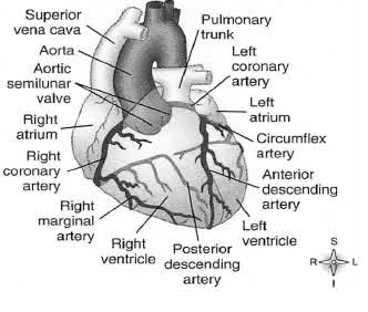
| ANATOMIC DISTRIBUTION | ECG FEATURES | VESSEL | COMPLICATIONS |
| Septal MI | Lead V1 and V2 | Left Anterior Descending | Conduction abnormalities, BBB |
| Anterior MI | V3,V4 | Left Coronary Artery (LCA) | Bad prognosis, High risk of sudden death, CCF, complete heart block |
| Inferior MI | II, III, AVF | Right Coronary Artery (RCA) | Distended neck veins with clear lungs, RV infarct |
| Lateral MI | V5, V6, I, AVL Reciprocal changes V1-V3 | LCA, circumflex | LV dysfunction, AV nodal block |
| RV MI | Lead V4R | RCA | Hypotension – caution with morphine, nitrates Bradycardia |
| Posterior MI | ST depression V1 – V4 Tall R in V1, V2 Lead V8, V9 | LCA, circumflex or RCA, post descending | LV dysfunction |
Management based on severity
Immediate action
Non-drug treatment
- Stabilise ABC's, O2 titrated to normoxia (sats 94-98; note that hyperoxia can be harmful in ACS), IV access and attach monitors
Drug treatment
- Give Aspirin 300mg after the diagnosis has been made
- Give P2Y12 receptor blocker to all patients once the reperfusion strategy (percutaneous coronary intervention, fibrinolysis, or no reperfusion) has been chosen. For patients receiving fibrinolysis, use clopidogrel 300mg. An uncommon exception to this approach is patients for whom early coronary artery bypass graft surgery is anticipated; in these patients, give asprin without P2Y12 receptor blocker.
- Adjunctive treatment: Treat pain with small, regular doses IV morphine. Nitrates sublingual or IV may relieve pain and restore flow where ACS is complicated by vasospasm. Maintain blood glucose below 11mmol/L.
- NSTEACS: do not start treatment until NSTEACS diagnosed (characteristic ECG changes, with or without troponin rise or fall above reference). Full anti-coagulation can cause harm through bleeding from other sites. After assessing contra-indications give Enoxaparin as follows:
- For patients <75 years of age, use a loading dose of Enoxaparin 30mg IV bolus followed by 1mg/kg subcutaneously every 12 hours (maximum of 100mg). The first subcutaneous dose should be administered with the IV bolus.
- For patients >75 years of age, do not use a loading dose but give 0.75mg/kg subcutaneously every 12 hours (maximum of 75mg).
- STEMI: Reperfusion should be offered if patients present within 12 hours of symptom onset. Reperfusion therapy is reasonable for symptom onset within 12 to 24 hours who have ongoing clinical symptoms, and/or ECG evidence of ongoing ischemia (PCI is preferred in this group).
- Percutaneous coronary intervention (PCI): Should ideally be offered if available within 120 min from first health care presentation, contra-indications to thrombolysis present, STE resolution not seen on 90 min post thrombolysis ECG. Other indications include: extensive anterior infarction, infero-posterior infarct with associated RV infarct and cardiogenic shock. A 90 min door to balloon time is ideal. Early cardiology discussion in selected patients is therefore essential
- Thrombolysis: Offer if no contra-indications to thrombolysis present and PCI not readily available (predicted breach of 120 min from first health care presentation). A 30 minute door to thrombolysis time is ideal. Contra-indications are listed below. It is recommended to take informed consent prior to starting thrombolysis although this is not always feasible in the emergency setting. Resolution post-thrombolysis is suggested by:
- Improved chest pain
- Rapid normalisation of ST segments (halved in 90 min)
- Reperfusion arrhythmias
- Early but short lasting rise in serum markers (8-12hrs)
Contraindications to thrombolysis
Absolute
- Known structural CNS lesion (AV malformation; Tumour)
- Any prior Intracerebral Haemorrhage
- Ischaemic stroke within the last 3 months (excluding the last 3 hours)
- Significant head or facial injury within the last 3 months
- Suspicion of aortic dissection
- Active bleeding (excluding menses) or bleeding disorders
Relative
- History of chronic, severe, poorly controlled HTN (SBP > 180 or DBP > 100) on admission
- History of ischaemic stroke previous to 3 months
- Dementia or other known intracranial pathology not noted above
- Traumatic or prolonged (> 10 minutes) CPR within the last 3 weeks
- Non-compressible vascular procedures within the last 3 weeks
- Major surgery within the last 3 weeks
- Internal bleeding within the last 3-4 weeks
- Pregnancy
- Active PUD
- Current use of anticoagulants (higher INR = Greater risk of bleeding)
- Prior exposure > 5 days to Streptokinase or allergic reaction to Streptokinase (use tPA)
Added tables/ dose charts
Nitrates: GTN infusion
- GTN 50mg in 50 mL to run at 0 – 12 mL/h (0 – 200 µg/min)
- Start at 5 mL/h and titrate up or down in 0.5 – 1 mL/h increments
- Avoid lowering the systolic blood pressure below 90 – 100 mmHg
- GTN 50mg in 200 mL to run at 0 – 48 mL/h (0 – 200 µg/min)
- Start at 20 mL/h and titrate up or down in 2 – 4 mL/h increments
- Avoid lowering the systolic blood pressure below 90 – 100 mmHg
Thromolysis: Streptokinase
- Resus or monitored area, IV access: 1 for infusion and 1 for fluid boluses
- Obtain baseline BP, then repeat every 3-5 minutes
- 1.5 million IU in 200 ml Saline 0.9% over 30 minutes
- Be prepared for hypotension: If occurs, place patient head down and give fluid boluses
- Do not anti-coagulate post-thrombolysis
Thrombolysis: Alteplase (tPA)
- 15 mg bolus
- Followed by 0.75 g/kg over next 30 min (max 50 mg)
- Followed by 0.5 mg/kg over 60 min (max 35 mg), for total dose 100 mg
- Anti-coagulate post-thrombolysis using enoxaparin as for NSTEACS
Relevant history/ examination
History
- Signs and Symptoms:
- Site: Where exactly is the pain?
- Onset: When did it start, was it constant/intermittent, gradual/ sudden?
- Character: What is the pain like e.g. sharp, burning, tight?
- Radiation: Does it radiate/move anywhere?
- Associations: Is there anything else associated with the pain e.g. sweating, vomiting, dizziness, syncope
- Time course: Does it follow any time pattern, how long did it last?
- Exacerbating/relieving factors: Does anything make it better or worse?
- Severity: How severe is the pain on a scale of 1-10?
- Allergies
- Medications: Ask about adherence. Ask about VIAGRA use.
- Past Medical History: IHD; Diabetes; HTN
- Illicit drug use – COCAINE and Amphetamines.
- Contraindications to thrombolysis and aspirin
Examination
- ABC approach.
- Examine for precipitants (e.g. infection; arrhythmias; anaemia) and consequences of ischaemia (e.g. cardiac failure).
- Consider differential diagnosis
- Measure and record vital signs
- Point of care Haemoglobin and glucose
Investigations
- ECG at presentation, repeated after 20 min.
- Troponin should be performed at presentation and repeated after 3 to 6 hours. Isolated elevations of troponin can be due to numerous other conditions
- Initial value elevated: positive if a repeat test shows that an increase or decrease ≥20% have occurred
- Initial value normal followed by a positive value is also positive
- Two negative values rules out MI, however unstable angina should be considered if clinical presentation and ECG changes remain suspicious. A 12 hour troponin is advisable in these instances
- FBC, renal function
- A chest x-ray should be considered
Referral criteria
STEMI: Early referral should be considered if:
- 24 hour primary PCI is available within 120 min from first health care presentation,
- Contra-indications to thrombolysis are present,
- STE resolution not seen on 90 min post thrombolysis ECG.
- Other indications include: extensive anterior infarction, infero-posterior infarct with associated RV infarct and cardiogenic shock.
NSTEACS: The HEART score should be used to risk stratify patients
The HEART score
| History: | |
| Highly suspicious | 2 |
| Moderately suspicious | 1 |
| Slightly suspicious | 0 |
| ECG | |
| Significant ST-depression | 2 |
| Non specific re polarisation disturbance | 1 |
| Normal | 0 |
| Age | |
| ≥ 65 years | 2 |
| 45 – 65 years | 1 |
| ≤ 45 years | 0 |
| Risk Factors | |
| ≥ 3 risk factors or history of atherosclerotic disease | 2 |
| 1 or 2 risk factors | 1 |
| No risk factors known | 0 |
| Troponin | |
| ≥ 3x normal limit | 2 |
| 1 – 3x normal limit | 1 |
| ≤ normal limit | 0 |
HEART Score | Risk of MACE | Suggested action |
0 - 3 | 0.9 % | Alternative diagnosis most likely and can be safely discharged if the alternative does not require inpatient treatment Write a comprehensive referral letter to the primary care provider to consider outpatient follow up for provocative testing and/ or specialist review |
4 - 6 | 12 % | Start ACS treatment if alternative diagnosis less likely than ACS Admit to medicine |
7 - 10 | 65 % | Alternative diagnosis UNLIKELY Start ACS treatment Admit to medicine or consider discussion for early invasive strategies |
MACE:major Adverse Cardiac Events (AMI; PCI; CABG; Death). TheHEART score assesses risk of MACE 6 weeks after EC presentation
Contact numbers
- Metro West: GSH Cardiology C26: 021 404 2020
- Metro East: TBH Cardiology 021 938 4911
This guideline was approved in December 2017 and will be reviewed in December 2018 or sooner if important evidence becomes available.
References
Edwards M, Chang AM, Matsuura AC, et al. Relationship between pain severity and outcomes in patients presenting with potential acute coronary syndromes. Ann Emerg Med. 2011;58(6):501-7
Body R, Carley S, Wibberley C, et al. The value of symptoms and signs in the emergent diagnosis of acute coronary syndromes. Resuscitation. 2010;81(3):281-6
Swap CJ, Nagurney JT. Value and limitations of chest pain history in the evaluation of patients with suspected acute coronary syndromes. JAMA. 2005;294(20):2623-9
Goodacre S, Locker T, Morris F, et al. How useful are clinical features in the diagnosis of acute, undifferentiated chest pain? Acad Emerg Med. 2002;9(3):203-8
Panju AA, Hemmelgarn BR, Guyatt GH, et al. The rational clinical examination. Is this patient having a myocardial infarction? JAMA. 1998;280(14):1256-63
Thygesen K, Alpert JS, Jaffe AS, et al. Third universal definition of myocardial infarction. Eur Heart J. 2012;33(20):2551-67
Life in the fast Lane. Glyceryl Trinitrate. http://lifeinthefastlane.com/book/critical-care-drugs/gtn-ntg/ (accessed 25 April 2014)
O'Gara PT, Kushner FG, Ascheim DD, et al. 2013 ACCF/AHA guideline for the management of ST-elevation myocardial infarction: a report of the American College of Cardiology Foundation/American Heart Association Task Force on Practice Guidelines. Circulation. 2013;127(4):e362-425
Sgarbossa EB, Pinski SL, Barbagelata A, et al. Electrocardiographic diagnosis of evolving acute myocardial infarction in the presence of left bundle-branch block. N Engl J Med. 1996;334(8):481-7
Life in the fast Lane. Myocardial Ischaemia. http://lifeinthefastlane.com/ecg-library/myocardial-ischaemia/ (accessed 8 April 2014)
Gatien M, Perry JJ, Stiell IG, et al. A clinical decision rule to identify which chest pain patients can safely be removed from cardiac monitoring in the emergency department. Ann Emerg Med. 2007;50(2):136-43
Lin A, Kerr D, Kelly AM. Is cardiac monitoring during transport of low-risk chest pain patients from the emergency department necessary? Emerg Med Australas. 2007;19(3):229-33
Shah AS, Newby DE, Mills NL. High sensitivity cardiac troponin in patients with chest pain. BMJ. 2013;347:f4222
Thygesen K, Mair J, Giannitsis E. How to use high-sensitivity cardiac troponins in acute cardiac care. Eur Heart J. 2012;33(18):2252-7
Sathyanarayan S, Awan S, Lynch P. Troponin negative chest pain patients readmitted within 6 months with Troponin positive chest pain. Emergency Services Directorate, Plymouth Hospitals NHS Trust. Audit number CA_2013/14/1992, 2013
Backus BE, Six AJ, Kelder JC, et al. A prospective validation of the HEART score for chest pain patients at the emergency department. Int J Cardiol. 2013 Oct 3;168(3):2153-8
Amsterdam EA, Wenger NK, Brindis RG, et al. 2014 AHA/ACC Guideline for the Management of Patients With Non-ST-Elevation Acute Coronary Syndromes: A Report of the American College of Cardiology/American Heart Association Task Force on Practice Guidelines. J Am Coll Cardiol. 2014. pii: S0735-1097(14)06279-2
For further information on the drugs listed in the above guidelines, click on the drug name and you will be directed to the EMGuidance drug monograph.