Acute Decompensated Heart Failure
Acute decompensated heart failure
Introduction
Identification
Management
Relevant history, Examination
Investigations
Referral criteria
Discharge criteria/checklist
Follow-up
Treatment Algorithm
Introduction/ Description
Heart failure is a complex clinical syndrome that results from any structural or functional impairment of ventricular filling or ejection of blood.
Identification/recognition/diagnosis of problem
The acute decompensated heart failure patient appears extremely ill, restless, poorly perfused and sweaty (peripheries often cold and clammy), tachypnoeic, tachycardic, hypoxic with increased work of breathing (using respiratory accessory muscles) and frothy sputum (may be pink/ blood tinged).
Causes of heart failure:the FAILURE mnemonic
- Forgot medication
- Arrhythmia/ Anaemia
- Ischaemia/ Infarction/ Infection
- Lifestyle: taken too much salt
- Upregulating of cardiac output: pregnancy, hyperthyroidism
- Renal failure/ Retention of fluid: Steroids, NSAIDs
- Endocardium: valve abnormalities
Differential Diagnosis
- Acute Renal Failure
- Acute Respiratory Distress Syndrome
- COPD, emphysema
- Myocardial Infarction
- Pneumonia
- Pneumothorax
- Neurogenic pulmonary oedema
- Pulmonary Embolism
- Pulmonary Fibrosis
Management
Immediate action
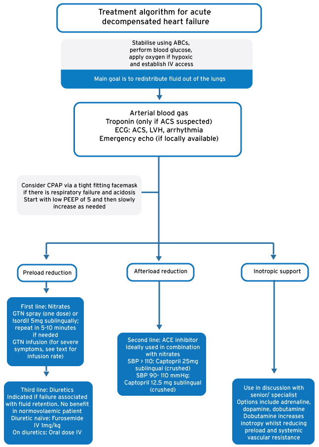
- Stabilise using ABCs, perform blood glucose, apply oxygen if hypoxic and establish IV access.
- Main goal is to redistribute fluid out of the lungs
- Decrease preload (right-sided over filling) using nitrates (± furosemide)
- Decrease afterload (“unload” the left side of the heart) using ACE inhibitors
- Consider non-invasive Positive Pressure Ventilation (CPAP) in respiratory failure
- Increase contractility (left ventricular systolic function) using inotropes
- Treat the cause aggressively
Dose charts
| Preload reduction: Give nitrates | |
|
|
| Consider furosemide | |
|
|
| Consider morphine | |
|
|
| Afterload Reduction: Give ACE inhibitors | |
|
|
| Inotropic support | |
| |
| Non-invasive Positive Pressure Ventilation (NPPV) | |
| |
Relevant history, Examination
History
- Assess clinical severity by history and physical examination
- Assess cardiac structure and function
- Determine the aetiology, with particular attention to reversible causes
- Evaluate for coronary disease and myocardial ischemia
- Evaluate the risk of life-threatening arrhythmia
- Identify any exacerbating factors
- Identify comorbidities which influence therapy
- Identify barriers to treatment adherence
- Presence of paroxysmal nocturnal dyspnoea or orthopnoea
- Presence of dyspnoea on exertion
New York Heart Association Functional Classification should be noted
- Class I No limitation of physical activity. Ordinary physical activity does not cause undue fatigue, palpitation, or dyspnoea.
- Class II Slight limitation of physical activity. Comfortable at rest, but ordinary physical activity results in fatigue, palpitations, or dyspnoea.
- Class III
- IIIA: Marked limitation of physical activity. Comfortable at rest, but less than ordinary activity causes fatigue, palpitation, or dyspnoea.
- IIIB: Marked limitation of physical activity. Comfortable at rest, but minimal exertion causes fatigue, palpitation, or dyspnoea.
- Class IV Unable to carry on any physical activity without discomfort. Symptoms of cardiac insufficiency present at rest. If any physical activity is undertaken, discomfort is increased.
Examination
- weight and vital signs with assessment for orthostatic changes
- Presence and degree of crackles, S3 gallop, jugular venous pressure elevation, hepatic enlargement and tenderness, positive hepatojugular reflux, oedema and ascites
Investigations
- Arterial blood gas: early indication of respiratory status (e.g. acidosis, failure)
- Renal function, FBC
- Glucose and urine analysis (point of care)
- Cardiac Markers (troponin) only if clinically indicated
- ECG: look for
- Evidence of myocardial infarction or ischaemia
- Arrhythmia
- Left ventricular hypertrophy or other chamber enlargement (etiology)
- Chest X-Ray
- Echo: if trained in use, focussed emergency cardiac ultrasound can give valuable information about left ventricular function
Referral criteria
- Although nitrates and ACE inhibitor (and furosemide if used) can be initiated within primary care, referral to secondary or tertiary care will be required to continue treatment. Observation is often required for an extended period of time within a high care setting. Transfer/ referral may be required to accommodate this if not locally available. Escalation of care to intubation will require intensive care referral.
Discharge criteria/checklist
- No longer hypoxic on room air
- Vital signs have returned to within normal parameters
- Return to baseline effort tolerance
- Cause of failure identified and appropriately addressed
- Patient understanding of dietary sodium restriction checked
- Patient understanding of medical regimen checked
- Medication adjusted as required
- Follow up arrangements made as required
Follow-up: elements to focus on in follow-up
- Functional capacity and activity level
- Changes in body weight
- Patient understanding of and compliance with dietary sodium restriction
- Patient understanding of and compliance with medical regimen
- History of arrhythmia, syncope, presyncope, palpitation
- Adherence and response to therapeutic interventions
- The presence or absence of exacerbating factors for HF, including worsening ischemic heart disease, hypertension, and new or worsening valvular disease.
Treatment algorithm for acute decompensated heart failure
References
Rossiter D, Blockman M (eds.). South African Medicines Formulary. 11th Ed. Cape Town: Health and Medical Publishing Group; 2014
Cameron P, Jelinek G, Kelly A, et al. Textbook of Adult Emergency Medicine. 4th Ed. Elsevier; 2015