Acute Glomerulonephritis (AGN) in Children
Guideline to the approach to acute glomerulonephritis in children
Lead Author: Dr Christel du Buisson
Acknowledgements: Dr PJ Sinclair, Dr P Nourse, Dr P Gajjar & Prof M McCulloch
These guidelines were created in May 2015.
Guidance to managing acute glomerulonephritis (AGN) in children
Approach algorithm
Acute Glomerular Nephritis in Children: Emergency Algorithm
Introduction
Clinical presentation
Examination and assessment
Differential diagnosis
Investigations
Management
Emergency Management
Referral criteria
Appendix A
Guidance to managing Acute Glomerulonephritis (AGN) in children
General information on AGN:
A childhood renal disease that includes the following presenting features:
- Painless macroscopic haematuria (cola/tea coloured urine) or microscopic haematuria
- Oedema: mild to severe
- Hypertension: can be so severe as to cause hypertensive encephalopathy
- Oliguria or Anuria
Approach Algorithm
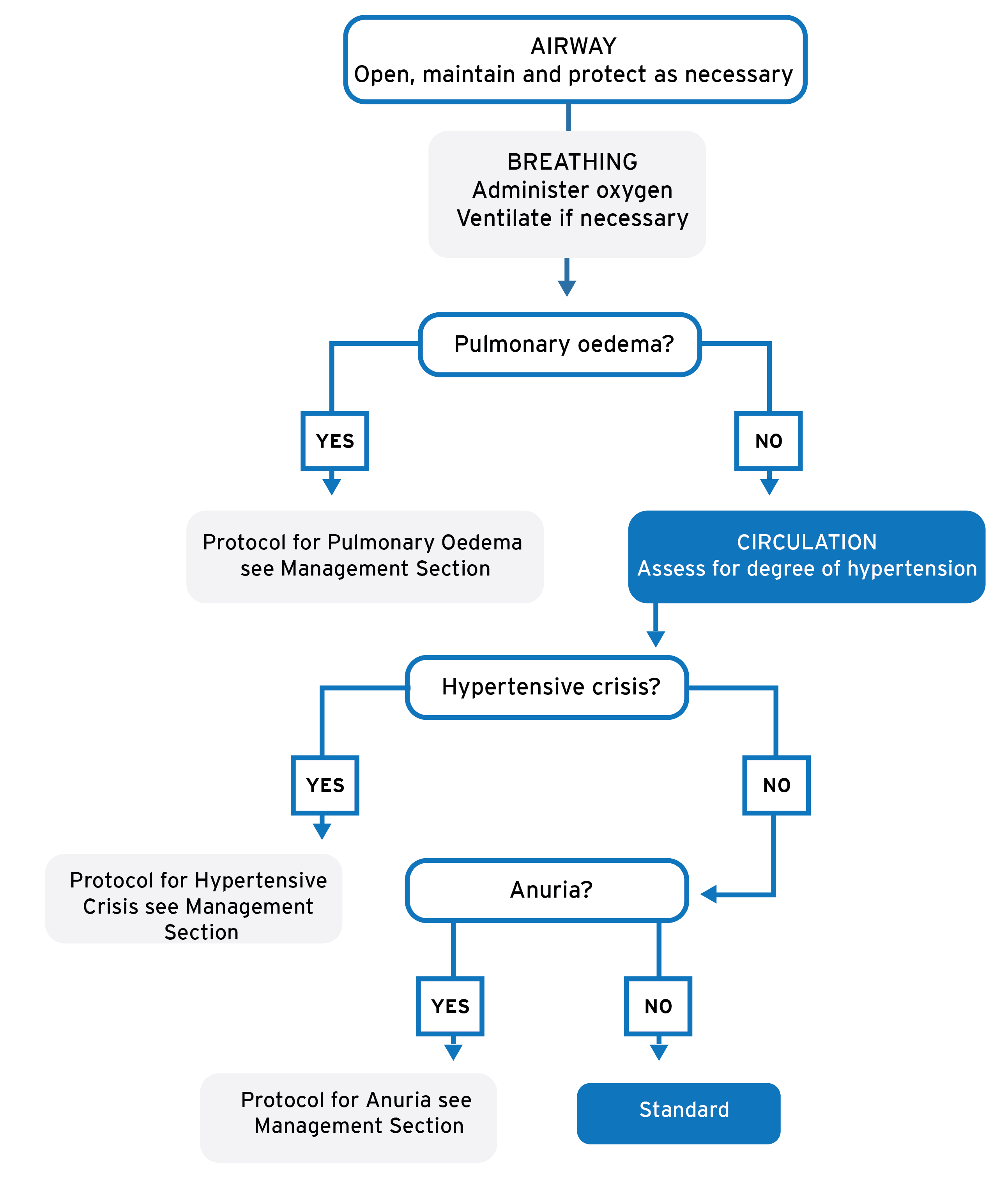
Click on image to enlarge
Acute Glomerular Nephritis in Children: Emergency Algorithm
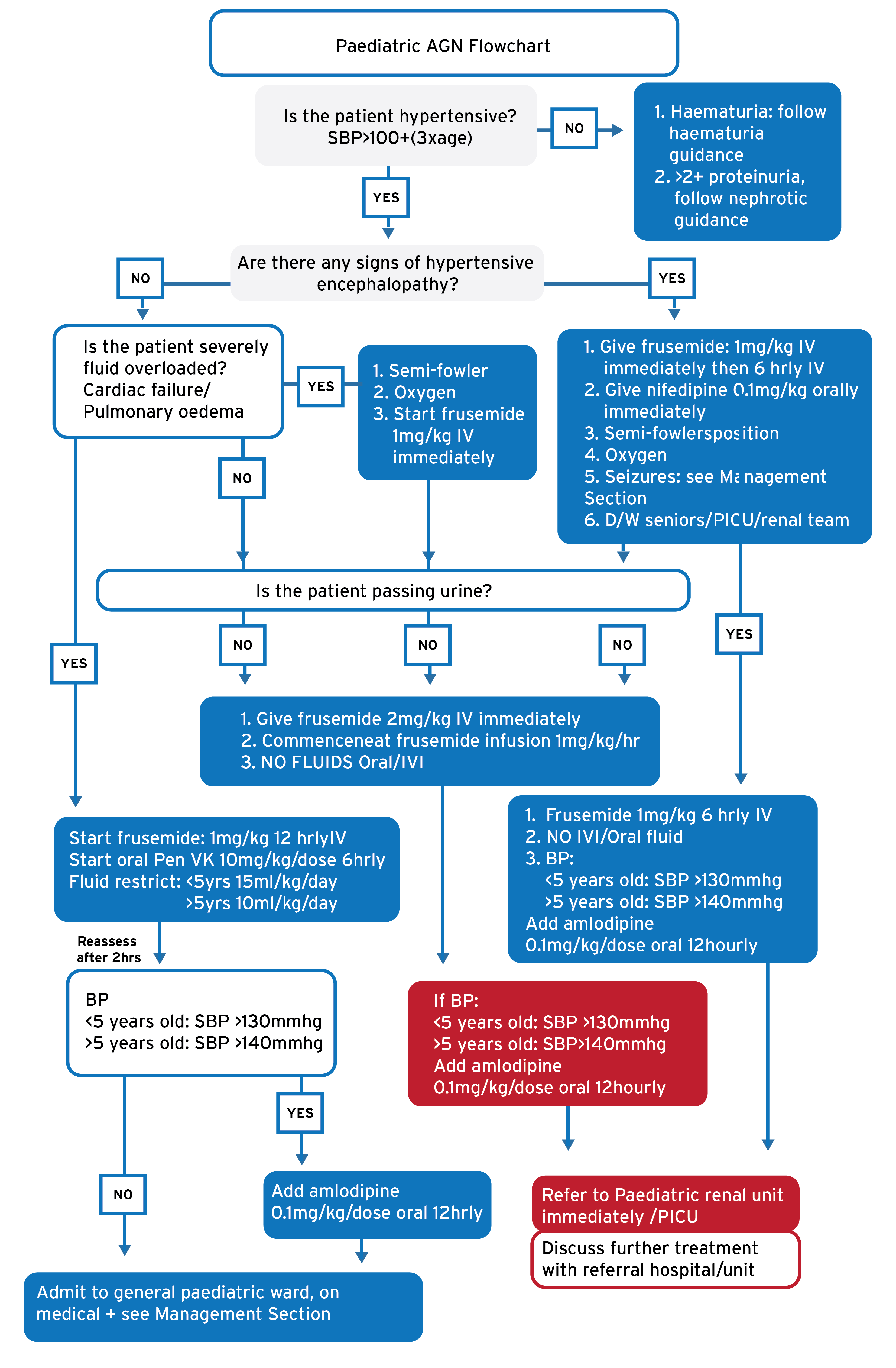
Click on image to enlarge
Introduction
The nephritic syndrome (high blood pressure, haematuria, oliguria) is usually due to acute glomerulonephritis (AGN). However all three features may not be present in all cases of AGN.
- AGN has varied presentations so it must be thought of in many differential diagnoses in children.
- Most cases are due to prior streptococcal infection (acute post-streptococcal APSGN). Impetigo is present in >50% of cases, or a history of upper respiratory tract infection/pharyngitis 7-21 days prior to presentation
- Majority of patients with APSGN are asymptomatic and will be referred for haematuria (see haematuria guidance)
- Less common but more serious glomerulonephritides (e.g. SLE, Hep B nephropathy) may present in a similar way and the differential diagnosis must be kept in mind
- AGN is unusual before the age of 2 years as is recurrence of this condition
- A nephritic-nephrotic syndrome (both features of nephritic & nephrotic syndrome) can mimic APSGN clinically, especially if the child also has impetigo, which is not uncommon.
- A rare complication is Rapidly Progressive Glomerulonephritis (RPGN), which should be presumed if a child with APSGN presents with features of nephritis & is in renal failure. Children with suspected RPGN should be referred to a paediatric nephrologist immediately.
Clinical presentation
Suspect AGN in children with recent onset of:
- Painless haematuria colour change noted in urine - cola /tea/red colour
- Oedema: sudden onset; facial or pedal or generalised
- Shortness of breath: sign of cardiac failure/pulmonary oedema
- Oliguria: may have noted a decreased urinary frequency
- Encephalopathy: severe headache, blurred vision, seizure(s), coma. focal neurological signs (especially cranial nerves, combined with features of raised intracranial pressure)
- Prior throat or skin infection - but note evidence of recent streptococcal infection (by far the most common cause of AGN) is not always present on history or examination.
All children with these presentations must have their blood pressure done.
Red Flags- Look for evidence of the three main acute complications of AGN:
- Pulmonary oedema
- Hypertensive encephalopathy (as above)
- Acute severe renal failure (anuria).
If any are present, initiate acute management & investigation (see below under Management)
BEWARE :
Hypertensive encephalopathy can present without any oedema
Pulmonary oedema can present without any peripheral oedema and it can mimic severe pneumonia & acute asthma
Examination and Assessment
Chief features of examination & assessment
- Fluid overload. This could present with any of the following signs: oedema, cardiac failure, raised JVP &/ or pulmonary oedema
- Pallor: Usual seen in severely fluid overloaded patients, due to haemodilution
- Hypertension: Due to fluid overload.
- For a chart of normal values of systolic & diastolic BP see Appendix A
- A quick bedside calculation of the 99th centile of systolic blood pressure (SBP) for a child can be done using the formula: SBP=100+(3 x age), thus anything above this value will definitely be abnormal.
- Cardiac: Features of fluid overload, with hyperdynamic circulation, displaced apex beat and if severe, even cardiac failure. No pericardial effusions.
- Respiratory: If severely fluid overloaded, there may be respiratory distress due to pulmonary oedema. This can exist without cardiac failure. No pleural effusions.
- Abdomen: if in cardiac failure, could have a tender hepatomegaly. No ascites.
- Encephalopathy: severe headache, blurred vision, seizure(s), coma. focal neurological signs
- Renal:
- Macroscopic haematuria: Cola colour/Tea colour, but could be frank (red)
- Dipstix: Microscopic haematuria
- Dipstix: 1-2+ Proteinuria in the presence of 4+haematuria or macroscopic haematuria
- Look for evidence of more complicated or chronic renal disease: growth failure, pale nails, anasarca, cardiomegaly, hypertensive features on fundoscopy, auto-immune rashes
Differential Diagnosis
- If all the features of AGN are present, vasculitides or chronic renal disorders are the main differentials to consider
- If only certain features are present, then think about cardiac diseases (fluid overload), vascular and renal diseases (hypertension), protein depletion states (oedema), haematuria (infections, other renal disease)
Investigations
Bedside – Urine Sample
- Macroscopic: Colour - Cola colour/tea/red
- Urine dipstix: Confirmation of haematuria. When macroscopic haematuria/4+ is present, 1-2+ proteinuria is likely
- Culture: Only if leucocytes and nitrates where positive on initial dipstix. If a bag specimen was obtained, a separate clean catch or in/out catheter specimen must be obtained for culture, otherwise a contaminated sample would be send. Midstream sample is perfect for urine culture.
Laboratory
FBC, Na, K, Urea, Creatinine, ASOT/antiDNase B
C3 (Other tests only if differential diagnosis significantly likely)
Imaging
Chest X-ray - only if symptoms and signs of cardiac failure with/without pulmonary oedema
Management
General Management
Note:
All children with AGN will need admissions to hospital.
Deal with severe complications as below, if present. If no severe complications present, give standard care as follows
- Always remember Airway, Breathing, Circulation as for any other primary survey
- Blood pressure monitoring:
- If the patient has hypertensive encephalopathy/cardiac failure/pulmonary oedema: check BP every 30 min
- If hypertensive but no other signs of end organ injury or severe fluid overload: check BP 2 hourly
- ALL PATIENTS will need to be fluid restricted as indicated on flow diagram
- Strict NO SALT diet. If patients are able eat, they can, they are only fluid restricted.
- Bed Rest
- Daily weight
- STRICT URINE OUTPUT MONITORING:
- If anuric or not potty trained-Catheterise
- If potty trained & pass urine-monitor all urine passed
- Review blood results
- Any patient with decreased eGFR (estimated GFR) calculated using Schwartz Formula:
- eGFR= 40 x height(cm)/Cr
- normal range =80-120ml/min/1.73
- if not already discussed for referral, should be discussed for immediate referral to a paediatric ward
Emergency Management
Pulmonary oedema emergency management
- Put patient in semi-Fowler’s position
- Give oxygen
- Insert an IV line
- Give IV furosemide 1mg/kg IV stat
- If poor response to the above, prepare to intubate & transfer to ICU
- Request Chest X-ray
- Monitor in high-care bed
- Monitor pulse, respiration, BP & urine output
Hypertensive crisis/Encephalopathy emergency management
Place in ‘recovery position’ until the child is waking
- Assess and manage ABC
- Give oxygen
- Check dextrostix
- Control seizures according to protocol
- Give furosemide 1mg/kg IV
- Give another 2mg/kg over an hour if no urine is passed within 30 mins
- Give nifedipine sublingually or orally 0.1mg/kg stat.
- Labetalol infusion: if SBP has not decreased by at least 25% of the difference between the measured SBP and the 99th centile upper limit of SBP for age [calculated as: 100+(3xage)] within 30mins of nifedipine & frusemide start a labetalol infusion: 0.25mg/kg/hour (this can be increased up to 3mg/kg/hour if required). Discuss urgently with consultant.
Example: 4 yr old child with SBP is 160mmHg. The 99th upper limit of normal BP for this child is: (100+(3x4) = 112mmHg. Subtract this from the measured SBP: 160-112 = 48mmHg. Within 30 minutes of giving the nifedipine & frusemide the SBP should drop by at least 25% of 48 i.e. 12mmHg. If not start labetalol infusion.
- Move the child to a high care bed
- Monitor BP every 10 minutes
- If SBP remains high after 2 hours give amlodipine 0.1mg/kg
Anuria emergency management
- Catheterise
- No fluids orally or intravenously
- Give frusemide 2mg/kg IV stat
- Discuss urgently with senior staff or paediatrics
Arrange transfer or admission:
If there is delay in transferring the child to a ward, keep the child on continuous cardiovascular & oxygen saturation monitoring. Review regularly for occurrence or re-occurrence of signs of severe disease.
Treat any further rise in BP to >20mmHg above the 95th centile for age with a further dose of nifedipine 0.1mg/kg sublingual oral.
Referral criteria (L1,2,3)
Referral criteria to paediatric nephrologist:
- Query regarding diagnosis AGN or atypical presentation
- Any atypical picture, nephritic/nephrotic (significant proteinuria/low albumin).
- Hypertensive emergency – any signs of end organ injury:
- cardiac failure
- pulmonary oedema
- hypertensive encephalopathy
- Still hypertensive/requiring diuretics after 48hours
- Renal failure: Decrease in estimated GFR (see eGFR Schwartz formula above)
Children who have any complications or meet criteria above need to be referred to level 2 or 3
Other children can be admitted to the paediatric ward of a district hospital
Appendix A
| Age of child | 95th Percentile of Systolic and Diastolic Blood Pressure | |
| First 12 hours | First week | |
| newborn prem | 65/45 mmHg | 80/50 mmHg |
| newborn full term | 80/50 mmHg | 100/70 mmHg |
| Systolic mmHg | Diastolic mmHg | |
| 6 weeks - 6 years | 115 | 80 |
| 8 years | 120 | 82 |
| 9 years | 125 | 84 |
| 10 years | 130 | 86 |
| 12 years | 135 | 88 |
| 14 years | 140 | 90 |
References:
- Up To Date Post-streptococcal glomerulonephritis –Patrick Niaudet, MD. Updated July 2014. Available from: http://www.uptodate.com
- D van Brussen. Streptococcal pharyngitis in children: to treat or not to treat. Eur J Pediat(2014) 173:1275-1283
- T Matthew Eison. Post-streptococcal acute Glomerulonephritis in children-Clinical features and pathogenesis. Paediatric Nephrology(2011)26:165-180
- Z Bircan. Post-streptococcal Glomerulonephritis with pulmonary oedema and microscopic haematuria. Paediatric Nephrology(2005)20:1204