Acute Kidney Injury
Acute kidney injury
Introduction
Identification
Severity assessment
Management based on severity
Relevant history
Investigations
Referral criteria
Discharge criteria/checklist
Treatment Algorithm
Introduction/ Description
Abrupt loss of kidney function that results in the retention of nitrogenous waste products and the dysregulation of extracellular volume, electrolytes and acid-base homeostasis.
Identification/recognition/diagnosis of problem
Patients often present with non-specific symptoms. Decrease urinary output can be described as is a decreased effort tolerance and dyspnoea. Acute kidney injury is usually first clinically suspected and later confirmed by renal function tests. The initial blood gas is often the first indication of a potential renal problem. Common causes that should invoke a high suspicion is described in the box below:
Pre-renal/ decreased renal blood flow (75%) | Intrinsic Renal (20%) | Post-renal (5%) |
|
|
|
Severity assessment (Triage)
The RIFLE classification provides a useful assessment of severity
| RIFLE classification of acute kidney injury | ||
| Stage | Serum creatinine | Urine output |
| Risk | Increase of 1.5- 2 times baseline | < 0.5 mL/kg/h for 6 hours |
| Injury | Increase of 2- 3 times baseline | < 0.3 mL/kg/h |
| Failure |
| < 0.3 mL/kg/h |
| Loss |
| -- |
| End-stage | Complete loss of function > 3 months | -- |
Management based on severity
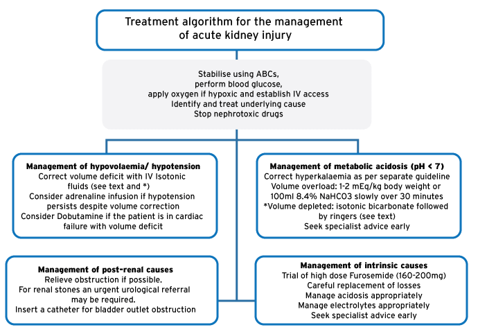
- Stabilise using ABCs; blood glucose, apply oxygen if hypoxic & establish IV access.
- Find and treat the underlying cause.
- Stop any nephrotoxic drugs.
Management: Hypovolemia/ hypotension
Aggressively treat the cause of volume loss or hypotension as the resulting reduced perfusion lead to recurrent ischaemic injury.
- Correct volume deficit with IV Isotonic fluids (see below)
- Consider adrenaline infusion if hypotension persists despite volume correction
- Consider Dobutamine if the patient is in cardiac failure with volume deficit. Starting Dobutamine suggests escalation of care to a high care environment. It should ideally be started in consultation with the specialist team.
Management: Metabolic Acidosis
- Correct hyperkalaemia as per separate guideline
- Use of Sodium bicarbonate is only indicated if the pH < 7.0. Treatment with sodium bicarbonate should prompt an urgent discussion with the specialist team.
Fluid choice in renal failure
- Patient fluid overloaded/ fluid retention: Start with a dose of 1-2 mEq/kg body weight or 100ml 8,4% slowly over 30 minutes.
- Patient under-filled (dehydrated) and non-anion gap acidosis (i.e. no keto- or lactic acidosis): use Isotonic 1.26% bicarbonate (1000 mL 5% dextrose water with 150 mL 8.4% Sodium bicarbonate added): preferred starting resuscitation fluid for patients with hyperkalaemia and acidosis (pH < 7) in renal failure. Calculate the bicarbonate deficit (0.4 x weight in kg x [24 – patient’s HCO3- level]) and divide by 150 mEq/L which will give you the volume of isotonic bicarbonate required in litres. Round this DOWN to the nearest full litre (example: a result of 1.7 L would mean you’ll use 1 L, likewise a calculation requirement of 0.6 L would mean that 1.26% bicarbonate is not used). Continuing to use isotonic bicarbonate after giving this volume would cause a metabolic alkalosis. Isotonic bicarbonate should be avoided in an anion gap acidosis (e.g. lactic or keto-acidosis)
- Ringers lactate (or similar): preferred follow on fluid for patients with hyperkalaemia and acidosis, to restore further volume deficit as clinically indicated. Ringers lactate does not cause lactic acidosis or increase potassium
- Saline 0.9%: Should be avoided in hyperkalaemic, acidotic renal failure. Saline 0.9% may increase potassium.
Management: Post-renal causes
- Relieve obstruction if possible. For renal stones an urgent urological referral may be required. Insert a catheter for bladder outlet obstruction. It is important to be consider post-obstructive diuresis
Management: Intrinsic causes
- Careful replacement of losses
- Manage acidosis appropriately
- Manage electrolytes appropriately
- Seek advice of referral physician
Relevant history, Examination
History
Concentrate in the history on factors known to lower renal perfusion and nephrotoxic drugs (e.g. aminoglycosides, NSAIDs, Tenofovir). Specifically target causes listed earlier.
Examination
- General: Blood pressure, Patient weight, Urine output
- Look for signs of dehydration & hypovolaemia, including dry mucous membranes, orthostatic dizziness, tachycardia & hypotension
- Look for signs of fluid overload, including a raised JVP, Bibasal crackles, S3 gallop & peripheral oedema)
- Skin: Rashes, infections, petechiae, purpura, ecchymosis
- Look for signs of metabolic acidosis, including Kussmaul breathing and tachypnoea
Investigations
- Essential: urine dipstick, renal function, haemoglobin, white cell count, blood gas.
Acute kidney injury is likely if a blood gas shows a metabolic acidosis in the absence of raised ketones or lactate, and no history to suggest toxicity from drugs that commonly cause etabolic acidosis (e.g. tricyclic antidepressants, salicylates, etc.)
- Consider also calcium, magnesium, phosphate
- ECG if suspected Hyperkalaemia
- Kidney ultrasound
- Urine microscopy (may provide clues to diagnosis)
- Other tests to be guided by onward specialist team
Referral criteria
Admission criteria
Admit under specialist team if any of the following present:
- Electrolyte abnormalities
- Correction of fluid losses in hypovolemic pre-renal failure
- Fluid overload and acute kidney injury
- Poor follow-up
- Correction of underlying cause required
- Work-up for cause of acute kidney injury
- Renal replacement therapy considered
Local admission
Local admission is appropriate for uncomplicated acute kidney injury due to Hypovolemia. Stabilising and initial correction of acid and electrolyte imbalances should be commenced whilst awaiting transfer to specialist team
Referral for renal replacement therapy should be considered in:
- Diuretic resistant pulmonary oedema
- Hyperkalaemia refractory to optimal medical therapy
- Metabolic acidosis refractory to optimal medical therapy
- Uremic complications (pericarditis, encephalopathy, bleeding)
- Toxins requiring dialysis (lithium, salicylates, theophylline)
Discharge criteria/checklist
- No electrolyte abnormalities present
- Improvement in renal function
- Improvement in symptoms and normal vital signs
- Appropriate outpatient follow up arranged
Treatment algorithm for the management of acute kidney injury
References
Hoste AJ, Clermont G, Kersten A et al. RIFLE criteria for acute kidney injury are associated with hospital mortality in critically ill patients: a cohort analysis. Critical Care 2006,10:R73 (doi:10.1186/cc4915)
Van Biesen W, Vanholder R, Lameire N. Defining acute renal failure: RIFLE and beyond. Clin J Am Soc Nephrol 1: 1314–1319, 2006
Ramrakha P, Moore K. Oxford handbook of acute medicine. Second Edition 2005. Acute renal failure p 378-387
Khalil P, Murty P, Palevski P. The patient with acute kidney injury. Prim Care Clin Office Pract: 35 (2008) 239–264
Rossiter D, Blockman M (eds.). South African Medicines Formulary. 11th Ed. Cape Town: Health and Medical Publishing Group; 2014
Cameron P, Jelinek G, Kelly A, et al. Textbook of Adult Emergency Medicine. 4th Ed. Elsevier; 2015