Acute Otitis Externa
Acute Otitis Externa
Clinical presentation
Necrotising (malignant) otitis externa
Predisposing factors
Investigations
Distinguish from other causes of Otorrhea
Management
Regular aural toilet
Topical agents
Topical preparations
Analgesia
Moderate to severe OE
Beware of allergy
Acute (and chronic) otitis externa is relatively common.
Acute otitis externa is inflammation of the skin of the external ear canal, which may also involve the pinna or tympanic membrane. Onset is usually rapid.
Clinical presentation
The classic features of otitis are ear pain, visible inflammation of the ear canal and/or pinna and a discharge from the ear. A typical sign is pain elicited by manipulation of the pinna.
The inflammation may be localised to a small area of the ear canal or meatus; inflammation may also be more generalised and involve the pinna.
On internal inspection the canal may be red, inflamed and swollen. You may see a pussy discharge or debris in the canal. Look out for areas of necrosis, abscess formation or underlying perforation. All of these should prompt urgent referral to an ENT specialist.
Other signs to look for are: oedema, itching, vesication, cranial nerve deficits and decreased hearing.
One form of otitis externa deserves special mention: necrotising (malignant) otitis externa.
Necrotising (malignant) otitis externa
This is a particularly severe form of otitis externa that usually only affects immunocompromised individuals – classically diabetics. The otitis becomes so severe that it spreads through the skin of the ear canal and eventually causes osteitis of the skull surrounding the external meatus. Soft tissue and cartilage necrosis is also common. Pseudomonas are commonly isolated.
These patients are very ill and systemic sepsis and local destruction are common complications. Untreated, more than half of patients will die.
It is critical to identify these patients and refer them to an ENT surgeon urgently. Consult the ENT surgeon regarding the most appropriate antibiotic treatment.
Predisposing factors
The two most common predisposing factors are minor trauma from over-zealous cleaning of the ears and excessive water exposure, such as with swimmers and surfers.
| PREDISPOSING FACTORS |
| Minor ear trauma (cleaning) |
| Prolonged water exposure |
| Excessive heat and moisture |
| Immunocompromise (i.e. Diabetes mellitus) |
| Structural abnormalities of the ear |
| Abnormal skin (psoriasis, eczema etc.) |
In a predisposed individual, the inflammation may then be caused by either infection or non-infective inflammatory conditions.
Common bacterial causes include Staph. aureus, Pseudomonas (esp. diabetic patients) and Streptococci.
Keep in mind that viruses (i.e. herpes zoster) and fungi (i.e. aspergillus) may also cause otitis.
Investigations
An uncomplicated case of otitis externa in an otherwise healthy patient can be managed without expensive special investigations.
Consider microscopy and culture of the ear if the patient is immunocompromised or the disease is severe, recurrent or not responding to treatment.
Distinguish from other causes of Otorrhea
Two important conditions should be distinguished from otitis externa:
- Acute Otitis Media
- Chronic suppurative otitis media with or without cholesteatoma
A few important questions and physical signs should help you to make the correct diagnosis:
1.) Is there an associated itch or discomfort?
Yes- usually suggested of otitis externa (OE)
2.) Is there any pain?
Yes- usually otitis externa.
In Chronic Suppurative Otitis Media (CSOM) without cholesteatoma the perforation does not allow pus to build up; thus is painless.
CSOM with cholesteatoma is usually also painless, unless there is the complication of mastoiditis or dural involvement.
3.) How long has the ear been wet?
A longer history suggest CSOM rather that OE
4.) Does the ear dry up intermittently?
Yes- CSOM without cholesteatoma or OE.
If cholesteatoma is present. It is unlikely that any treatment will stop the infective process.
5.) Symptoms of complications involving the middle ear? i.e. Hearing loss, Facial weakness with vertigo
Suggests CSOM with cholesteatoma. These require emergency action and surgery. Refer to ENT surgeon.
6.) Is the patient a cotton-bud abuser?
Yes- Suggests OE
Management
The critical components of care are avoidance of any predisposing factors, good aural hygiene, topical treatments and analgesia.
Regular aural toilet
The ear canal should be gently cleaned under direct vision at least once a week. Clear as much of the discharge and debris out as possible. Do not insert a probe or curette deeply into the canal if you cannot see exactly where it is going: perforation of the TM or damage to the middle ear are dangerous complications.
If the canal cannot be cleaned or you are concerned about damage to deeper structures, refer the patient to an ENT surgeon.
Topical Agents
Topical agents are the treatment of choice for almost all cases of otitis externa.
The most commonly used preparation is the combination of hydrocortisone, neomycin sulphate and polymyxin B.
This combination results in first attempt cure rates of 87-97%, but has some problems with hypersensitivity and concerns regarding ototoxicity with neomycin if the TM is perforated.
An alternative is acetic acid with or without topical steroids. Adding a steroid drop to acetic acid improves cure rates.
Newer topical treatments: fluoroquinolones: ofloxacin and ciprofloxacin. These agents cover Gram negative and Gram positive pathogens. Cure rates are similar as to the above two strategies but hypersensitivity and ototoxicity is much less likely.
| CONSIDER SYSTEMIC ANTIBIOTICS IF |
| Immunocompromised patient |
| Severe infection with local spread |
| Systemically ill patients |
Use Amoxicillin or co-trimoxazole, consider Co-amoxyclav if the patient is ill.
Topical Preparations:
- Soothe: Glycerine and ichthamol
- Antiseptic: Acetic acid
- Steroid: Hydrocortisone
- NOT if cause = Fungus
- Antimicrobial: Fluroquinolones (oflaxacin, ciprofloxacin)
- BEWARE Allergy
Consider an antistaphylococcal agent if furunculosis is present in external auditory meatus (e.g. cloxacillin)
Analgesia
The pain of otitis externa can be severe. Make sure you prescribe adequate analgesia.
REMEMBER: NO EAR BUDS! EVER!
Moderate to severe OE
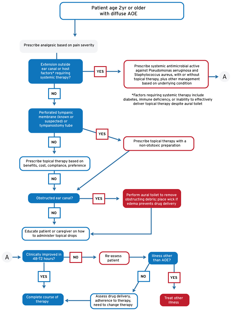
If the external canal is severely swollen and topical medications cannot reach the deep section of the canal, treatment should include micro-suction and the insertion of an ear wick.
These patients should be discussed with an ENT surgeon.
Beware of allergy
Patients not responding (within 48-72hrs) to appropriate therapy may be allergic to the constituents of the drops or consider alternative diagnosis.
Refer ENT surgeon
A flow diagram suggesting management of otitis externa.
References:
- Roland PS et al. Clinical practice guideline: Cerumen impaction. Otolaryngology – Head and Neck Surgery 2008: 139, S1-S21.
- Corbridge R et al. Oxford Handbook of ENT and Head and Neck Surgery. Oxford university Press;2006
- Marx JA et al. Rosen’s Emergency Medicine: Concepts and clinical practice, 7th ed. St. Loius, MO: Mosby; 2009