An Approach to a Breast Mass and Other Common Breast Complaints
Doctor, I have a lump in my breast! An approach to a breast mass and other common breast complaints
Introduction
Clinical Findings on Breast Examination
Differential Diagnosis of a Breast Lump
Differential Diagnosis of Breast Thickening
Differential Diagnosis of Skin Dimpling
Differential Diagnosis of Nipple Discharge
Differential Diagnosis of Breast Pain
Breast Diagnostics
Imaging Techniques
Biopsy Techniques
Guidelines to Special Investigation
Management of Fibroadenomas
Gynecomastia
Breast Cancer
Context: Outpatients
Introduction
The central clinical question is whether the lump is breast cancer or not. Since breast cancer is the commonest cancer to affect women in most countries, this is a frequent scenario.
History taking is aimed at identifying symptoms suggestive of a benign breast lump and evaluating the patient's risks for breast cancer. Take a gynaecological history as well.
The most powerful risk factors for the development of breast cancer are age (postmenopausal), gender (women! obvious!), previous breast carcinoma or predisposing breast pathology (DCIS/LCIS/atypical ductal hyperplasia), a family history of breast or ovarian carcinoma, and previous radiotherapy to the chest wall.
Undress the patients, and examine systematically both breasts, the axillae and the supraclavicular fossa. Use the palm of your hand (not your fingertips) and evaluate for lumps, tenderness, adenopathy; look for signs of skin dimpling, ulcerations, skin oedema, or fixity.
More than half of all patients who present with a breast lump have a normal examination (i.e. they don't have a lump!)- the breasts are markedly variable in consistency and nodularity, and some patients and less experienced clinicians can over-interpret normal breast consistency.
The patient's age and clinical context is critically important- the older the patient, the more likely the diagnosis of breast cancer. A number of benign conditions occurring elsewhere (such as lipomas, sebaceous cysts) can also cause breast lumps.
Clinical Findings on Breast Examination
| Clinical Findings on Breast Examination | |
Normal | Clinical findings vary enormously depending on patients’ age, pregnancy status, adiposity. The upper outer quadrant and the infra-mammary fold are areas of increased breast density, which is normal. Young women have much more dense, “nodular” breast tissue. |
| Thickening | Area of increased breast density, which is different from the surrounding tissue and is asymmetrical, but is not as clearly defined as a breast lump. If you were to excise it, you would struggle to define its exact extent and may remove a large area. |
| Lump | A localised swelling, protuberance, bulge or bump in the breast that feels different from the breast tissue around it or the breast tissue in the same area of the other breast. If you are to biopsy it, you would be able to exactly define where the lump ends and the normal parenchyma starts. |
| Skin Dimpling | Best seen in a sitting position with the arms elevated- notice a small dimple in the skin overlying a breast lump- it is strongly associated with breast carcinoma- very few benign breast conditions cause dimpling. |
| Nipple Discharge | Spontaneous, persistent, blood-stained single duct discharge merits investigation. Bilateral, clear, non-spontaneous discharge is common and clinically irrelevant. |
Differential Diagnosis of a Breast Lump
Common | Clues |
| Breast carcinoma | Presents as a painless, firm lump. More overt signs (adenopathy, skin oedema, ulcerations)- occur in advanced cases. |
| Breast cyst | Perimenopausal patients, smooth lump, tender to pressure |
| Fibroadenoma | Adolescent and young patients, well defined, smooth very mobile mass |
| Breast abscess | Commonest in late pregnancy or breastfeeding/ usually obvious clinical diagnosis based on findings of erythema, tenderness, fluctuation, purulent discharge |
| Periductal mastitis | 95% are smokers, causing chronic retro-areolar inflammatory masses and recurrent abscess and fistulae |
| Fat necrosis | Follows a traumatic event (direct injury i.e. seatbelt injury after MVA or assault) Can be very similar clinically to a breast carcinoma- hard, craggy, irregular |
| Uncommon and Weird | Clues |
| TB | Commonest in HIV+ patients, either as a chronic abscess / or with axillary adenopathy/sinus |
| Galactocele | In breastfeeding |
| Phyllodes tumours | Feels like a large fibroadenoma in an older patient, but can become enormous |
| Metastases to the breast | Melanoma or lymphoma can present with suspicious breast masses |
| Don’t want to miss | Clues |
| Breast carcinoma | Be absolutely sure that a “benign lump” is properly evaluated, and histology reports are representative |
Differential Diagnosis of a Breast Lump in Different Clinical Contexts
Context | Diagnosis | Comments |
Adolescent | Fibroadenoma | Breast cancer is exceedingly rare in this context. Young women have dense, nodular breasts which can be difficult to evaluate, this can lead to over-referral to breast clinics |
Pregnancy | Lactational abscess Galactocele Breast carcinoma | Pregnant women have dense, nodular breasts which can be difficult to evaluate. Breast cancer can be easily missed in this context. Fibroadenomas can occur in this context, and typically hypertrophy during pregnancy |
Middle age | Breast carcinoma Cyst Fat necrosis Periductal mastitis | In patients older than 40, breast cancer starts to be a probable diagnosis Fibroadenoma is very uncommon Look at benign diagnoses with “suspicion” & evaluate fully |
Elderly | Breast carcinoma | Almost all breast lumps in this age group are malignant |
Male | Gynaecomastia Breast carcinoma | 1% of breast cancer occurs in men Gynaecomastia can be caused by drugs (spironolactone/ steroids/ ARV’s) or liver failure |
HIV + | Breast carcinoma TB Lymphoma | Consider TB and lymphoma is patients with marked adenopathy The most likely cause remains breast carcinoma |
Differential Diagnosis of Breast Thickening
Common | Clues |
Normal | Bilateral symmetrical UOQ thickening |
Fibrocystic breast change | Areas of fibro-cystic change can feel different to surrounding tissue. This is common in perimenopausal patients |
Carcinoma | Carcinoma may be less easy to feel in breast parenchyma that is thick, and nodular Lobular carcinoma typically presents with ill-defined thickening, rather than a well-demarcated mass |
Periductal mastitis | 95% are smokers, causing chronic retro-areolar inflammatory masses and recurrent abscess and fistulae |
Don’t want to miss | Clues |
Breast carcinoma | - |
Differential Diagnosis of Skin Dimpling
Common | Clues |
Carcinoma | Most skin dimpling is caused by carcinomas, even small impalpable lesions |
Surgical scars | Prior surgery can leave “gaps” of breast parenchyma that distort skin appearance |
Don’t want to miss | Clues |
Breast carcinoma | Look for occult primary lesions if nil palpable |
Differential Diagnosis of Nipple Discharge
Common | Clues |
Intraductal papilloma | Single duct, spontaneous blood-stained or clear serous discharge |
DCIS / carcinoma | Single duct, spontaneous blood-stained or clear serous discharge Carcinoma may have an associated irregular mass or thickening |
Periductal mastitis | Smoker, recurrent infections, usually a yellow purulent discharge from the nipple or peri-areolar fistula |
Ductal ectasia | >1 duct, fluid can be dark green/ brown |
Don’t want to miss | Clues |
Breast carcinoma | Look for occult primary lesions if nil palpable. |
Differential Diagnosis of Breast Pain
| Common | Clues |
| Physiological mastalgia | >95% of all cases of breast pain are physiological and do not correlate to any underlying breast pathology. This is commonest in adolescent and around menopause but may occur at any age. |
| Less common | Focal, localized, persistent breast tenderness may reflect a local breast condition |
| Breast abscess | Obvious clinical diagnosis in almost all cases |
| Fat necrosis | On occasion, this can be tender |
| Breast cyst | A tender breast lump, in perimenopausal woman |
| Fibroadenoma | A small proportion can be tender to pressure, but most are asymptomatic |
| Referred Pain | Costo-chondral tenderness typically just lateral to the sternum, on focal pressure = Tietze’s syndrome. Herpes zoster giving referred pain from dermatomal innervation. Thrombosed vein within the breast parenchyma. On palpation, it feels like a tender “chord” of tissue- Mondor’s disease |
| Breast carcinoma | A locally advanced, ulcerated carcinoma can cause local pain. An impalpable or early breast carcinoma is typically NOT painful. If a patient has mastalgia AND breast cancer it is more commonly a coincidence, rather than cause and effect |
| Don't want to miss | Clues |
| Breast carcinoma | Examine patients thoroughly, and reassure about the benign nature of mastalgia Any clinical signs must be evaluated on their merits |
Breast Diagnostics
Triple assessment is the gold standard for the evaluation of a breast mass: ie (1) clinical evaluation must be supported by appropriate (2) imaging and (3) tissue biopsy.
Always consider if all 3 aspects of the assessment are concordant (in agreement) or discordant (don't make sense!)- The bottom line is: if the investigations are CONCORDANT (ie if clinical, biopsy and imaging features are malignant- the final diagnosis will be malignant, and conversely if clinically and all the investigations are benign then the diagnosis will be benign!) or DISCORDANT (i.e. the biopsy reports a malignant lesion, but the imaging looks benign, or vice versa) - discordant reports mandate either repeat investigations or surgery to clarify the diagnosis.
Imaging Techniques
| Mammogram | US | MRI |
| Strengths | ||
|
|
|
| Weaknesses | ||
|
|
|
| False +ve | ||
| <5% | <5% | 10% |
| False -ve | ||
| 5-10% | 5-10% | <5% |
Be aware that a normal mammogram does not exclude cancer with 100% confidence - approximately 10% of malignancies are missed on imaging (10% false negative rate) - never disregard a suspicious clinical sign, even if radiology is reassuring - a biopsy is a critical part of the "triple assessment".
Characteristic Features of Breast Cancer on Imaging
| Mammography | Ultrasound |
|
|
BI-RADS Classification: all reports should classify the findings according to the BI-RADS system
| Assessment | Management | Cancer Probability |
| 1 - negative | Follow up only if clinically indicated | 0% |
| 2 - benign | Follow up only if clinically indicated | 0% |
| 3 - probably benign | Review in 6/12 or routine screening (if indicated) | <2% |
4 - suspicious a- low suspicion b- intermediate suspicion c- high suspicion | Tissue diagnosis | 2-10% 10-50% 50-95% |
| 5 - highly suggestive of malignancy | Tissue diagnosis | >95% |
| 6 - known biopsy - proven malignancy | - | n/a |
Biopsy Techniques
FNA cytology | |||
Strengths | Weakness | False +ve | False -ve |
Cheapest Widely available Easy to learn Can be done in consultation rooms | Indadequate sampling Cytology report only Cannot offer definitive treatment on the result Cannot distinguish infiltrating cancer from DCIS | <1% | 5-10% (mostly related to sampling errors) |
Needle core biopsy | |||
Strengths | Weakness | False +ve | False -ve |
Gold standard Not expensive Can be done in a consultation room Obtain a sample for histology examination | More complex to learn Needles 10x >expensive than FNA Lab costs greater | <0.05% | <5% if done correctly |
Excision biopsy | |||
Strengths | Weakness | False +ve | False -ve |
Large volume of tissue obtained = representative | Expensve Requires procedure/theatre room Hospital costs Leaves a scar | <0.01% | <0.1% |
Punch biopsy | |||
Strengths | Weakness | False +ve | False -ve |
Suitable for skin lesions only Cheap Histology examination | Limited clinical use to skin abnormalities only (i.e. Paget's disease of the nipple | - | - |
Biopsy results should be reported as follows (IACC standards):
Cytology | |
C1 | Insufficient material |
C2 | Benign |
C3 | Atypical probably benign |
C4 | Suspicious, probably in situ or invasive carcinoma |
C5 | Malignant |
Histology | |
B1 | Insufficient material |
B2 | Benign |
B3 | Atypical probably benign |
B4 | Suspicious, probably in situ or invasive carcinoma |
B5 | Malignant |
The most definitive test to confirm or exclude cancer is always HISTOLOGY - if the lesion has been correctly biopsied (either by core needle or excision biopsy) this gives unequivocal information about the diagnosis; if the patient has a malignancy, it gives the treating team details about histological and genetic profiling subtype, hormone receptors, tumour grade, vascular invasion - all of which help plan treatment. None of these details is reliably obtained from cytology aspiration (or from radiological investigations!!).
Guidelines to Special Investigation
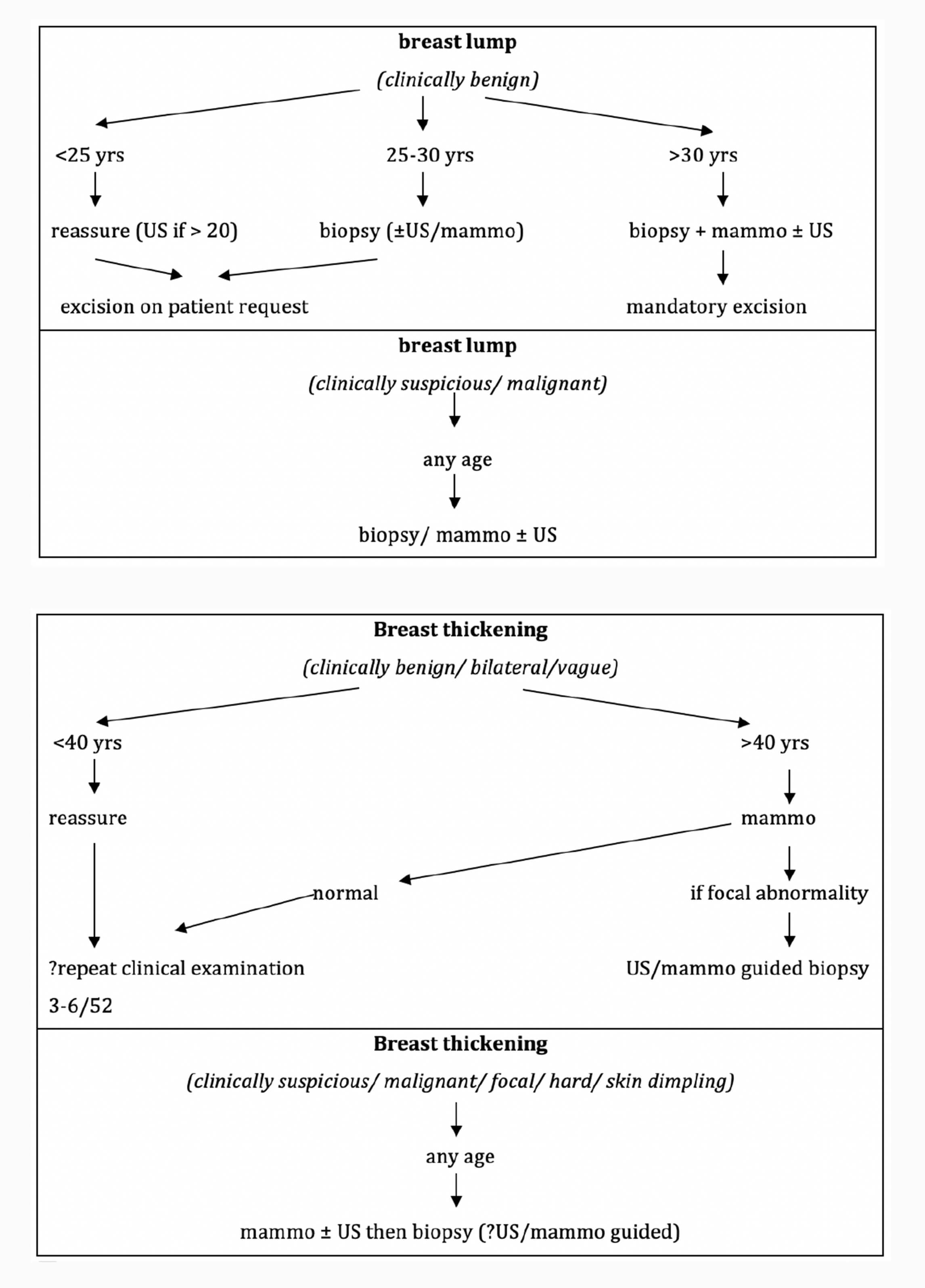
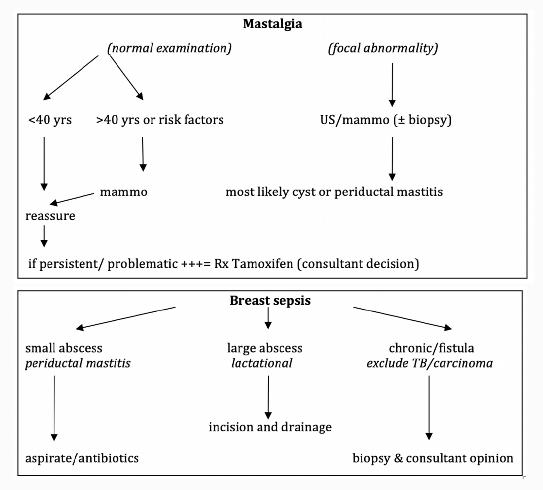
Click on the above images to enlarge
Management of Fibroadenomas
Clinical features of a typical FA
- <3cm
- well defined, smooth
- mobile
- firm
- may be mildly tender or multiple
Management of a typical fibroadenoma under 3cm
< 20 yr old | |||
Investigations | Management | Follow up if not excised | Indications for surgery |
Nil | Reassure | None | Not recommended |
20-25 yr old | |||
Investigations | Management | Follow up if not excised | Indications for surgery |
Ultrasound | Reassure | None if the US and clinical exam are consistent with FA | Patient request |
25-30 yr old | |||
Investigations | Management | Follow up if not excised | Indications for surgery |
Histology + Ultrasound | Offer excision | Annual x 2 yrs | Most patients |
> 30 yr old | |||
Investigations | Management | Follow up if not excised | Indications for surgery |
Histology US + Mammo | Recommend excision | 6 monthly x 2 yrs | All patients |
Indications for Surgery in any age group
- Giant fibroadenoma (> 5cm in size)
- Atypical clinical presentation: fixed, irregular, skin changes, lymphadenopathy, rapid growth
- Suspicious biopsy or ultrasound result
Gynecomastia
Gynecomastia is a benign enlargement of the male breast due to proliferation of the glandular component. It is caused by a relative imbalance between oestrogen and androgen levels and can be caused by a wide range of conditions. Gynaecomastia peaks during puberty and in 50 to 80-year-olds. Clinically, gynecomastia is a firm, rubbery, mobile, disc-like mound of tissue arising concentrically from beneath the nipple and areola region. Unilateral and bilateral gynecomastia are both common and benign. The overwhelming majority of cases are idiopathic and of no clinical significance.
Organic causes of gynecomastia
Prescription drugs: e.g. Spironolactone, anti-androgens, anabolic steroids, HAART in particular efavirenz, diazepam, tricyclic antidepressants, cimetidine, digoxin, calcium channel blockers
Recreational substances: e.g. Alchohol, amphetamines, marijuana, heroin, methadone
Organ dysfunction: e.g. Liver failure, renal failure, thyrotoxicosis
Endicrinopathy: e.g. Testicular carcinoma, Cushing's syndrome
Causes of unilateral gynecomastia or retroareolar mass:
- All causes of bilateral gynecomastia
- Breast carcinoma
- Benign breast lump
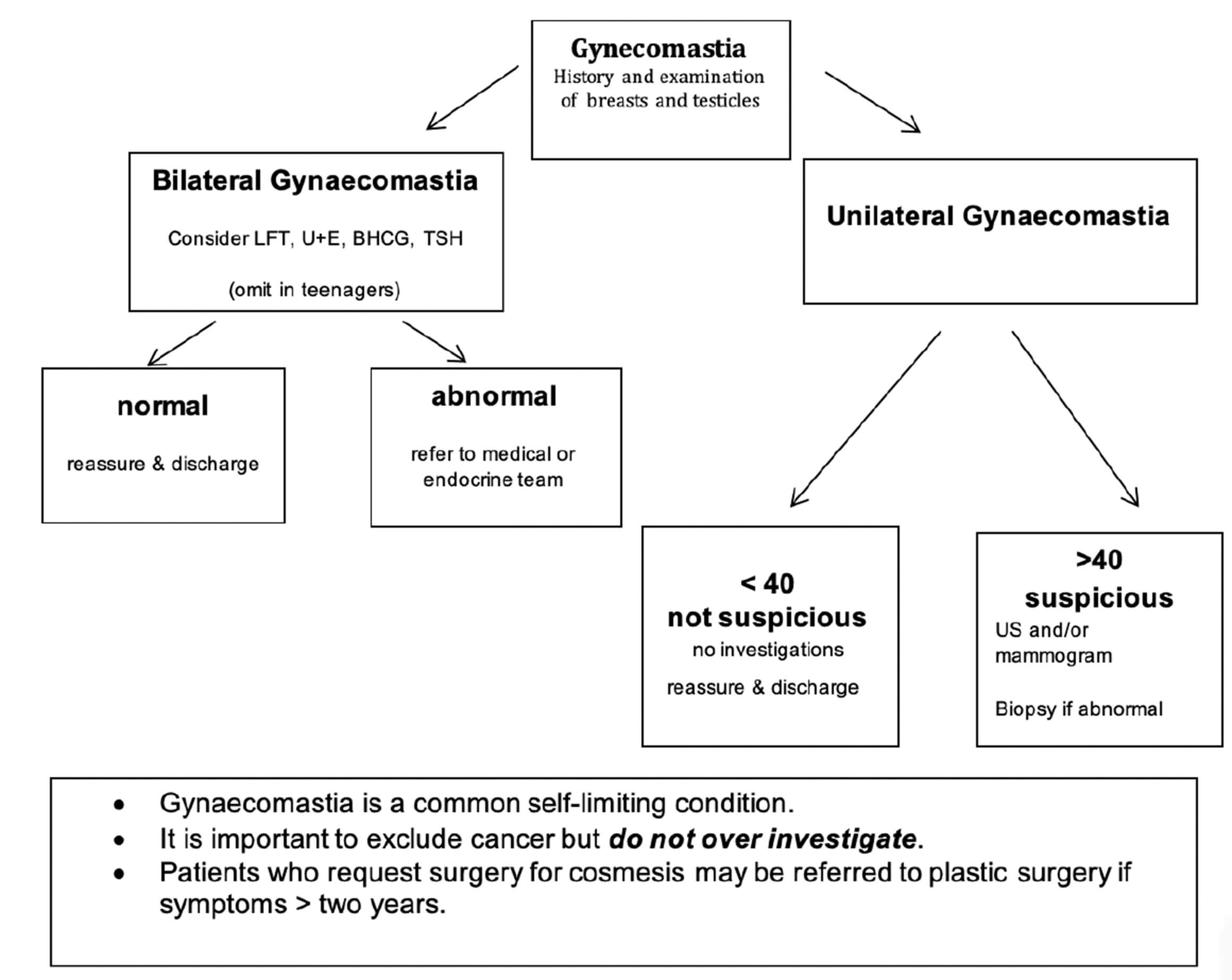
Click on the above image to enlarge
Doctor, I am worried I will get breast cancer - I want to be tested!
Take a thorough breast history and examine fully. Any clinical findings need to be investigated contextually.
Formal analysis of risk can be done using online tools- (IBIS Risk Evaluation is the one we use at our GSH breast clinic). This will issue a report with the likelihood of cancer within the next 5, 10 years and overall lifetime risk. (http://ibis.ikonopedia.com or download file to install on desktop at http://www.ems-trials.org/riskevaluator/)
Most patients grossly over-estimate their risk of developing cancer, and need thoughtful compassionate reassurance.
The following patients should be referred for genetic counselling and testing:
- IBIS lifetime risk estimated >30%
- BRCA mutation risk estimated >10%