An Approach to a Soft Tissue Leg Mass
Doctor, I have a lump on my leg! An approach to a soft tissue mass
Introduction
Differential Diagnosis
Lipoma
Sarcoma
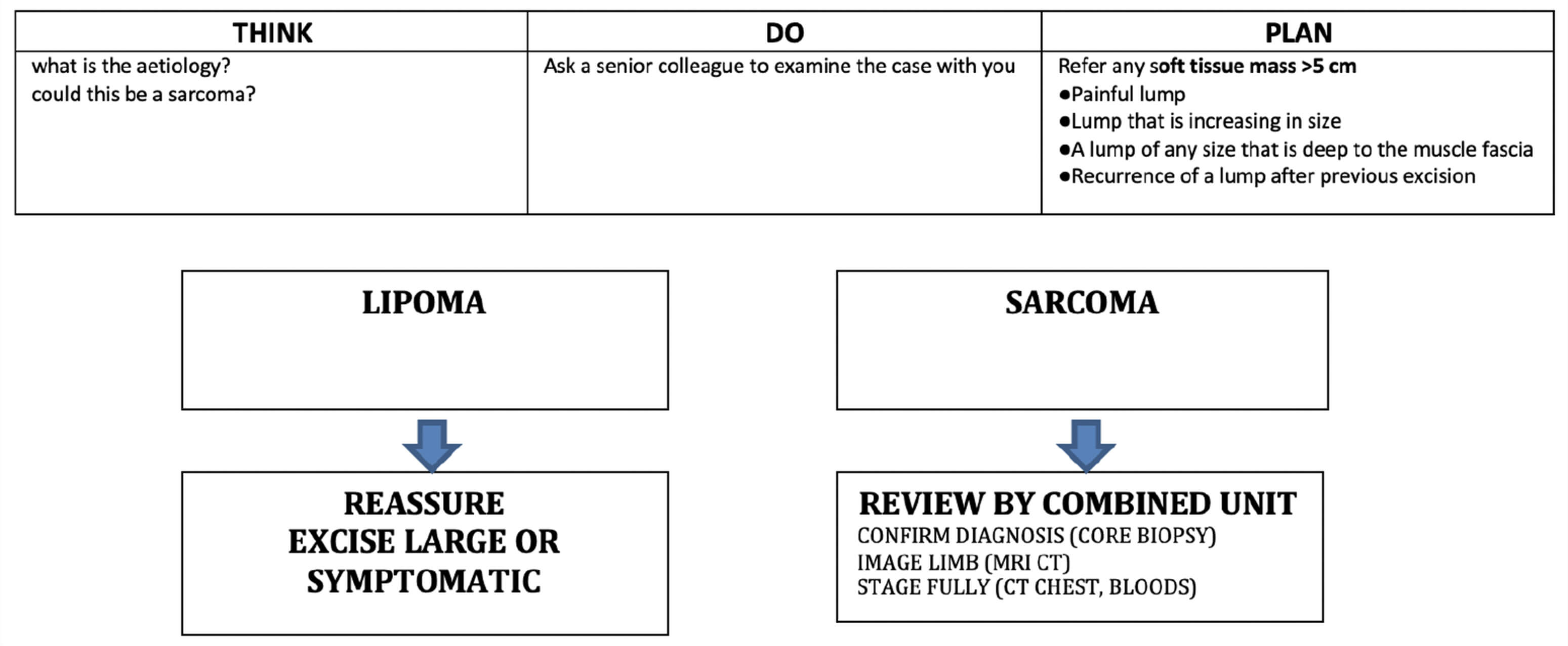
Soft Tissue Lump Algorithm
Context: Outpatients
Introduction
Most of these patients will have a benign soft tissue neoplasm.
Differential Diagnosis
Common | Clues |
Lipoma | Long-standing, painless, minimal change in size |
Uncommon and Weird | Clues |
Heamatoma / Retained foreign body | History of trauma |
False aneurysm | History of penetrating injury in recent past / bruit or thrill on examination |
Seroma | Major soft tissue / degloving injury |
Cold abscess | Medial thigh / frail TB patient / typically painless |
Hydatid | Contains clear fluid only |
Don't Want To Miss | Clues |
Sarcoma | Often has no other symptoms at all Typically faster growing than lipoma, and firm in consistency |
Benign soft tissue masses are at least 100 times more uncommon than sarcomas.
The key clinical question is: is this a lipoma or a sarcoma?
Key Features | Lipoma | Sarcoma |
Duration | Minimal change over years | Increasing size |
Pain | Nil | In late-stage only |
Predisposing Factors | Nil | Neurofibromatosis HIV+ (Kaposi) Previous radiotherapy |
Size | Mostly small | Can be large |
Consistency | Soft "fatty" | Firm |
Location | Superficial | Intramuscular / deep |
Lipoma
These are soft, fatty neoplasms which most typically occur in the subcutaneous tissues. They have an indolent history, and will not cause local symptoms, other than mild discomfort. Management is conservative unless they are large or unsightly, in which case a local excision is curative. The diagnosis can be made on clinical grounds in the majority of cases. If you are not sure, before starting many expensive investigations, ask a senior colleague to examine the patient.
The following patients meet criteria for urgent referral with soft tissue lesion:
- Soft tissue mass >5cm
- Painful lump
- A lump that is increasing in size
- A lump of any size that is deep to the muscle fascia
- Recurrence of a lump after the previous excision
Sarcoma
Diagnosis and staging are best done with the guidance of the surgical oncology unit responsible for the care of the patient, and for practical purposes, a patient with a suspected sarcoma must not have biopsy attempts prior to referral.
Aspiration cytology may be helpful in excluding haematomas and abscesses but it is inadequate in obtaining the finite diagnosis of sarcoma.
The diagnosis can only be made by obtaining a generous amount of tissue for histological evaluation.
Needle core biopsy is the preferred method of diagnosis. On certain occasions, incision or excision biopsy is used, particularly for small (<5cm lesions), but the route must be able to incorporate the final excisional area should the lesion be a sarcoma.
Staging is performed by an MRI scan of the area in question, together with a CT of lungs and liver (a CT of the lungs may reveal metastases not shown on a radiograph). A liver enzyme profile is performed to determine hepatic metastases.
Combined Assessment: Patients are best managed by a multi-disciplinary team comprising of an oncology surgeon, orthopaedic surgeon, pathologist, oncologist and radiologist. Extensive patient counselling is essential.
Real problem:
The biopsy is inadequate:
- Get senior help
- Re-biopsy
- Consider a US-guided biopsy
- Consider open incisional biopsy
Never do a sarcoma operation without histological diagnosis!
Soft tissue lump algorithm
Click on above image to enlarge