Anaesthetic Equipment Decontamination
Management of reusable equipment Definitions and classifications Laryngoscopes Bronchoscopes Magill forceps Nasopharyngeal and rectal temperature probes Suction bowl Suction tubing Oropharyngeal airways Bougies, and intubation guides and stylets Breathing filters and breathing circuits Bag valve mask resuscitators Supraglottic devices Storage of semi-critical items Non-critical medical equipment
Management of reusable equipment
Definitions and classifications
Nasopharyngeal and rectal temperature probes
Bougies, and intubation guides and stylets
Breathing filters and breathing circuits
Storage of semi-critical items
Non-critical medical equipment surfaces
Trans-oesophageal echocardiogram probes (TEE)
Management of reusable equipment
- Reprocessing refers to infection control procedures for removing and inactivating microorganisms on reusable patientcare equipment.
- Reprocessing of reusable patient-care equipment includes cleaning, disinfection, and sterilisation.
- The choice of reprocessing method must be guided by the instrument manufacturer’s recommendations.
- Factors that need to be considered are compatibility among equipment components and materials, chemicals to be used, heat and pressure tolerance of the equipment, and time and temperature requirements of the reprocessing methods.
Definitions and classifications
Spaulding classification12-15
Spaulding developed a classification of instruments into “critical”, “semi-critical” and “non-critical”. This classification is more than 50 years old and is used to determine the level of disinfection for equipment items.
Critical instruments
- Critical instruments have a high risk of contamination.
- This includes those instruments that are in contact with sterile sites within the body or the intravascular system.
- Critical instruments should be subjected to sterilisation such as pressurised steam or low temperature methods such as ethylene oxide gas.
- Examples include surgical instruments, implants and sterile catheters.
- With the advent of multidrug-resistant and aldehyde-resistant organisms there was a call to also include other semi-critical instruments into the category of critical instruments. One such an item is endoscopes. The design is flawed with lots of crevices that may retain microorganisms. A study by Ofstead et al.16 showed that almost 50% of endoscopes retained moisture that can aid in the development of a biofilm. In addition, 71% of endoscopes showed microbial growth and cultured organisms such as Stenotrophomonas maltophilia and Citrobacter freundii.
- Other items that are relevant to the anaesthetist include airway instruments such as laryngoscope blades and handles as well as forceps. Airway instrumentation is often associated with trauma to the mucosa. Several studies have demonstrated occult blood on these items. These instruments thus meet the criteria for critical instruments.17 These items require cleaning and disinfection followed by sterilisation as required and as per manufacturer’s recommendations.
Semi-critical instruments
- Semi-critical instruments have an intermediate risk of contamination.
- These instruments only come into contact with intact mucosa and intact skin.
- Examples given under this category include endoscopes, respiratory equipment and cystoscopes. It is however interesting to note that most of these items are now being considered “critical instruments”.
Non-critical instruments
- Non-critical equipment has a low risk of contamination.
- These instruments only come into contact with intact skin.
- Divided into patient non-critical items such as blood pressure cuffs and oximetry probes and environmental non-critical items such as cot sides, theatre trolleys and uncontaminated floors.
- Cleaning and drying are usually adequate.
Laryngoscopes
Laryngoscope blades14,80
Contaminated anaesthetic equipment has been implicated in the nosocomial transmission of infectious diseases.81 Options for the reprocessing of laryngoscope blades include:
- Use of disposable (single-use) laryngoscope blades (DLBs) (preferred): The metal type only must be used. They should be discarded after single use. DLBs should not be reused, even after sterilisation.
- The sterilisation of reusable laryngoscope blades (RLBs): The light intensity of all RLBs that are steam sterilised should be monitored. Handling and storage are important.
- High-level disinfection (HLD): There are significant concerns about the use of HLD in a South African setting when decontaminating RLBs. Evidence of poor compliance with HLD protocol has been documented, and there is a significant margin for human error in the HLD protocol. If it is used, hospitals must have a specific step-by-step instructional protocol in print that is well understood. Decontamination should occur in a specific designated area, away from patients and other healthcare workers. Frequent in-service training of anaesthesia nurses on HLD must be conducted. The decontamination of RLBs should be monitored and audited for compliance.
- No other method of disinfection, e.g. chlorhexidine and alcohol, should be used to decontaminate RLBs.
Rationale
Laryngoscope blades have been implicated in the transmission of nosocomial disease.82-84 Other studies have identified RLBs as potential vectors for the transmission of methicillin-resistant Staphylococcus aureus, P. aeruginosa, Serratia marcescens and other pathogenic microorganisms.4,85 Further studies have identified the presence of blood (occult and/or visible blood) on RLBs deemed to be ready for use.6,17,86,87 These include a study conducted in the regional, tertiary and central hospitals in a province of South Africa which found blood (visible or occult) and/or visible organic material contamination of RLBs of 80%.6 Staff from 11 of the 15 studied hospitals claimed that the blades were decontaminated by HLD.7
If inanimate objects become contaminated with hepatitis B virus and are not appropriately decontaminated, these objects may contribute to disease transmission for periods of up to one week and possibly longer.88 Hepatitis C has been shown to retain infectivity for several days at room temperature. Wenzel and Edmond89 acknowledged that instruments are sources of pulmonary infections with Gram-negative organisms, such as P. aeruginosa or S. marcescens, reflecting an inanimate environmental reservoir. They concluded that if 1–5% of bronchoscopic procedures were performed on patients with tuberculosis, and if each was followed by a second procedure with the same scope, 460–2 300 patients might become exposed to the pathogen each year if only 10% of the scopes were contaminated. One of the most compelling reasons for re-evaluating the cleaning, disinfection and sterilisation techniques of airway management equipment derives from the report by Agerton et al.90 on outbreaks of tuberculosis following bronchoscopic procedures.
Blood contamination of reusable laryngoscope blades
Traditionally, the RLB was classified by the American Society of Anesthesiologists (ASA) as a “semi-critical” medical device using the Spaulding classification scheme.34,91 According to this scheme, medical devices that directly or indirectly contact mucous membranes or non-intact skin, without ordinarily entering sterile tissue or the vasculature, are classified as semicritical devices, for which this scheme prescribes at least HLD or sterilisation.
Concerns about this classification of RLBs were raised both locally and abroad.6,9 Bleeding in the mouth following routine laryngoscopy has been well described, as well as the contamination of laryngoscope blades with this blood.92 This implies penetration, and not merely contact with the mucosal tissue by the blade. The laryngoscope blade, when used during dental, maxillofacial and otorhinolaryngology surgery, may also become grossly contaminated with blood. The presence of blood contamination from prior patients may facilitate the nosocomial transmission of the hepatitis B and C viruses, HIV and other blood-borne pathogens, the risk of which is difficult to ascertain owing to the paucity of documented cases and ethical constraints in performing such studies.
However, mucosal membrane contact with blood tissue, or other body fluids that are potentially infectious, as well as percutaneous injury, are defined as exposure which places patients at risk of acquiring HIV infection.93 In retrospective case-control and postexposure prophylaxis studies on HIV, an increased risk of HIV infection was associated with exposure to a large quantity of blood from the source, a device visibly contaminated with the patient’s blood, and a deep injury. Other factors included a source patient with AIDS, patients with a high viral load and injury with a hollow-bore needle.93 Furthermore, hepatitis B is approximately 100 times more transmissible than HIV. Contaminated RLBs that may or may not cause a traumatic mucosal breach pose a risk of transmission of hepatitis B, hepatitis C and HIV. Therefore, RLBs should rather be considered to be critical items, rather than semi-critical ones, especially in countries with high endemic levels of HIV and hepatitis B positivity. Several countries in Europe, including Britain and the Netherlands, have changed the decontamination of RLBs from HLD to sterilisation after each use or adopted the use of suitable disposables.9
The rigidity of the high-level disinfection protocol
There are three essential stages in HLD:94,95
- Cleaning: Removal of visible contamination from surfaces with water and friction, e.g. the use of a brush and fluidics, i.e. fluid under pressure, together with enzymatic products. Sequestered organic material poses the greatest risk of crosscontamination for patients as it impedes the effectiveness of these cleaning processes by reacting chemically with the germicide, and/or by forming a protective physical barrier for microorganisms.
- Immersion in a high-level disinfectant: Immersion in a highlevel disinfectant must take place, i.e. ortho-phthalaldehyde (Cidex® OPA) and glutaraldehyde (Cidex® ). The duration of immersion should be in accordance with the manufacturer’s recommendations, with exposure times varying between 8–45 minutes at 20–25 ºC.
- Removal of the disinfectant: Removal of the disinfectant is achieved by adequate rinsing with water.
All three steps are fundamental to the effectiveness and safety of HLD.
The effectiveness and safety of HLD is compromised by:
- Human factors, i.e. owing to complacency and the ignorance of staff, staff shortages and staff turnover.
- Insufficient time allocated to the decontamination process.
- Inadequate numbers of available laryngoscope blades per theatre to allow for compliance with the minimum duration of exposure to the disinfectant in order to achieve effective HLD.
Several of the factors were identified or hypothesised by a study conducted in South Africa, where 60% of the hospitals that claimed to practice HLD were actually noncompliant with the HLD protocol, owing to noncompliance with one or more of the three steps of the process, for example, the omission of cleaning prior to immersion.7
Variations and inconsistencies in reprocessing guidelines and practices can result in ineffective reprocessing, confusion among healthcare staff members, inconsistent and inadequate standards of care, and an increased risk of patient-to-patient disease transmission.96 Moreover, there can be no margin for human error in South African hospitals in this regard because of the risk of transmitting HIV and hepatitis.
The occupational hazard of high-level disinfectants14,95
Commonly used high-level disinfectants in South Africa include ortho-phthalaldehyde (Cidex® OPA) and glutaraldehyde (Cidex®). Ortho-phthalaldehyde (Cidex® OPA) is preferred to glutaraldehyde (Cidex®) for use as a disinfectant. Cidex® OPA has excellent stability over a wide pH range (pH 3–9), is not a known irritant to the eyes and nasal passages, does not require exposure monitoring, has a barely perceptible odour and requires no activation. A potential disadvantage of Cidex® OPA is that it stains proteins grey, including unprotected skin, and thus must be handled with caution with the use of gloves and protective clothing. Cidex® OPA residue remaining on inadequately waterrinsed equipment can stain the patient’s mouth. Allergic reactions have been reported in urology patients undergoing repeated cystoscopies with scopes reprocessed with Cidex® OPA. In April 2004, the manufacturer of OPA alerted the medical community about patients who reportedly experienced an anaphylaxis-like reaction after cystoscopy with scopes repeatedly disinfected with Cidex® OPA.
Glutaraldehyde (Cidex®) causes serious occupational hazard concerns. Cidex® OPA solution should be used in a well-ventilated area and stored in closed containers with tight-fitting lids. Healthcare personnel can be exposed to elevated levels of glutaraldehyde (Cidex®) vapour when equipment is processed in poorly ventilated rooms, when spills occur, when glutaraldehyde solutions are activated or changed, or when open immersion baths are used. Acute or chronic exposure can result in skin irritation or dermatitis, mucous membrane irritation (i.e. the eyes, nose and mouth), or pulmonary symptoms. Epistaxis, allergic contact dermatitis, asthma and rhinitis has also been reported in healthcare workers who had been exposed to glutaraldehyde. Glutaraldehyde exposure needs to be monitored to ensure a safe working environment.
The case for disposable (single-use) laryngoscope blades
Single-use airway equipment is designed to be used once and then discarded. Studies have shown that current techniques for cleaning and decontaminating RLBs are ineffective in removing all remnants of blood. If anaesthetists are not confident that RLBs are appropriately decontaminated, they should use disposable equipment.97
Different DLBs are manufactured with different designs and materials. There may be concern about the quality of some of these devices because they are manufactured at a lower cost to justify their disposal.11 However, metal DLBs offer decreased infection risk with optimal user satisfaction and safety. A multicentre randomised study on more than 1 000 patients for emergency intubation under rapid sequence intubation was published in 2010. It found that single-use metal blades were more efficient than reusable metal blades. Significantly fewer failed first attempts, and there were fewer poor-grade laryngeal views.98
A plastic DLB is reported to be less efficient than a metal reusable blade during rapid sequence induction. This is owing to the increased flexibility of the blade. Jabre et al.99 recommended that conventional RLBs should be available for difficult intubations, and plastic DLBs used for uncomplicated intubations. Therefore, metal DLBs are recommended.
Interestingly, studies have shown that most clinicians would prefer single-use devices to be used on themselves and their families if they were patients.11 The use of DLBs removes concerns about decontamination, and particularly the human factors.
The case for sterilisation of reusable laryngoscope blades95,96
Sterilisation of RLBs has significant advantages over HLD:
- Reliability: Sterilisation has a larger margin of safety, coupled with reliability, consistency and lethality. Sterilisation, in contrast to HLD, significantly removes the “human” element from the process of decontamination.
- Cost-effective: Sterilisation is relatively inexpensive. It decreases the need for high-level disinfectants, with the associated need for storage containers and protective gear.
- Improved efficiency: It links to improved theatre turnover rates, with less time required for decontamination between cases.
- Patient benefits: There is a decreased risk of nosocomial infection and decreased exposure to high-level disinfectants and their residue with sterilisation.
- Healthcare worker benefits: Sterilisation reduces the anaesthesia staff workload. It removes occupational health and safety hazards associated with high-level disinfectants.
The challenge with sterilisation is the progressive decrease in the light intensity of the laryngoscope blades that undergo sterilisation. Therefore, the lifespan of the laryngoscope blade is shorter. Reusable fibreoptic laryngoscope blades have been shown to deteriorate with repeated steam sterilisation, eventually becoming less bright.100-102
Different brands of laryngoscope blades tolerate automated machine cleaning and steam sterilisation differently, and this needs to be kept in mind when making future purchases.
Handling of the contaminated laryngoscope blade
Gadalla and Fong devised a way of improving the handling of the contaminated laryngoscope blade.103 First, the anaesthetist puts on two pairs of clean gloves. Induction is carried out, and then as soon as endotracheal tube placement is complete, the blade of the laryngoscope is held in the gloved hand, and one outer glove is peeled off the hand and inverted over the dirty laryngoscope blade. The other outer glove is also removed. The anaesthetist is then left with a clean pair of gloves. This technique ensures that the used blade never comes into contact with other equipment.
The Association of Anaesthetists of Great Britain and Ireland (AAGBI)9 recommends that the anaesthetist wear gloves during intubation and then place the used blade in a designated receptacle to prevent contamination of surfaces, pillows and drapes.
Laryngoscope handles95,104-106
The laryngoscope handle should be decontaminated after each patient. To minimise laryngoscope handle contamination:
- Remove the blade from the handle immediately after use and place the contaminated blade in a receptacle.
- Do not close the contaminated blade on the handle after intubation.
- Consider covering the handle with a new disposable plastic bag for each patient, as described in the rationale. (This does not change the need for HLD or sterilisation). Consider using the specifically devised double-glove, clean induction technique by Gadalla and Fong.103
- Decontaminate by sterilisation, or HLD or ILD.
Sterilisation
Sterilisation steps are as follows:
- Send the laryngoscope handle to the central sterile supplies department for sterilisation.
- Batteries must be removed in the OT.
- Adequate numbers of handles per OT should be acquired to allow for this.
- The manufacturer should be consulted to determine compatibility with the type of sterilisation. Steam sterilisation is recommended. If the handle is not sterilisation friendly, it should be replaced.
High-level disinfection
HLD steps are as follows:
- HLD should take place after each patient.
- Batteries should be removed in the OT prior to HLD.
There should be a specific step-by-step instructional protocol in print, that can be easily understood.
- HLD must be monitored and audited for compliance.
- Adequate numbers of laryngoscope handles per OT must be acquired to allow for this.
Intermediate-level disinfection
ILD steps are as follows:
- ILD should take place after each patient.
- Chlorhexidine 2% with alcohol 70% should be used.
- HLD or sterilisation must be employed if there is visible blood or organic material contamination.
- Several articles prefer HLD over ILD, so ILD is not the preferred choice.
- After sterilisation or disinfection, the laryngoscope handle and blade should be checked for function and then package into a sealed plastic bag to prevent recontamination.
Rationale
A recent review of laryngoscope blades and handles as a source of infection concluded that the laryngoscope handle should be considered to be a semi-critical item, at least requiring HLD.107,108 It does not come into direct contact with the patient’s oral mucosa. However, it may become contaminated by the tip of the blade when the blade is folded in the “off” position.86,109,110 This “contact point” presents a potential route for the patientto-patient transmission of blood and organisms from the oropharynx. The laryngoscope handle can also be contaminated by the clinician’s gloves, by direct contact with surfaces or other anaesthetic equipment, as well as by indirect contact from splashes or airborne pathogens. Microorganisms can then be transmitted to subsequent patients when the clean blade touches the laryngoscope handle, or when the anaesthesia provider’s gloves touch a contaminated laryngoscope handle. Studies have found high contamination rates of laryngoscope handles, including drug-resistant organisms.87,105,110,111
Traditionally, laryngoscope handles have a knurled surface for good grip. However, the fissures in this surface may provide a protected niche for pathogens109,112 A greater range and heavier growth of bacterial species was found on the knurled, compared with smooth, surfaces of the handle, suggesting that knurled surfaces may harbour more bacteria than smooth surfaces.105 Two recent articles called for the revision of inconsistent guidelines for disinfection and the storage of laryngoscope handles prior to use and recommended at least HLD for laryngoscope handles.96,105
Williams et al.105 isolated one or more species of bacteria from 86% of laryngoscope handles examined, despite the use of low-level disinfection. Isolates included organisms such as Enterococci, methicillin-susceptible S. aureus, Klebsiella spp. and Acinetobacter spp. The Medicines and Healthcare Products Regulatory Agency of the UK reported on the death of a patient who developed sepsis from a contaminated laryngoscope handle.113
Choice of strategies to prevent cross-infection will be influenced by a cost-benefit analysis, but includes the use of newly sterilised laryngoscope handles for each case, disposable laryngoscope handles or HLD of handles between each case. ILD with chlorhexidine 2% with alcohol 70% has been used. However, the trend in the literature is moving away from ILD to at least HLD. If ILD or HLD are used, sterilising handles on a scheduled basis is recommended, especially if C. difficile is encountered.107
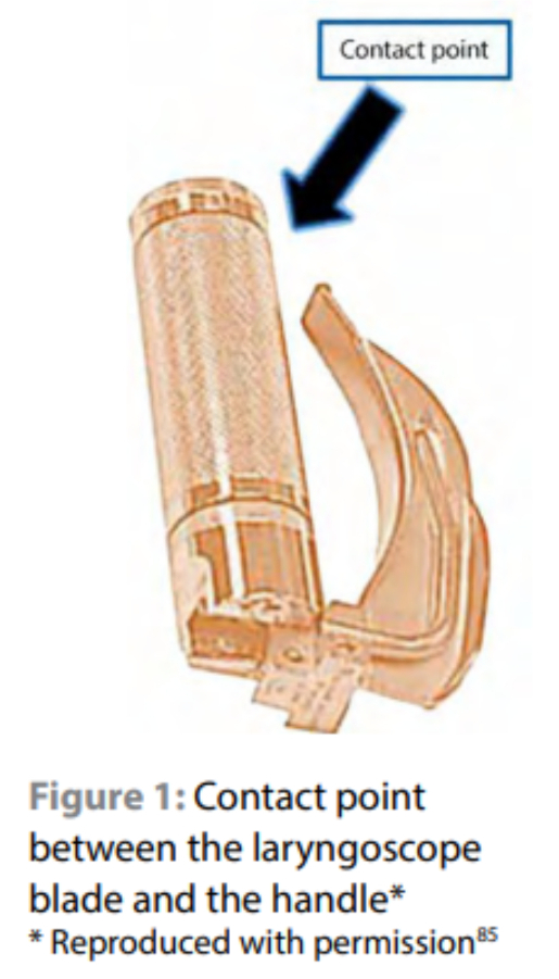
Click on the image to enlarge
A plastic bag or sheath has been used to cover the laryngoscope handle in some cases, but this practice has been criticised for creating a false sense of security. Full sterilisation procedures or HLD of both the laryngoscope handle and the blade have been advocated.9,96
Only one hospital of the 15 studied decontaminated laryngoscope handles after each use in a study that was conducted in regional, tertiary and central hospitals in a province of South Africa.6 It is imperative that laryngoscope handles are decontaminated between use.9,114
Table I: Step-by-step decontamination of laryngoscope blades and handle
| Step 1: Transportation from point of use Transport the laryngoscope blade and handle promptly in a suitable container to the designated decontamination area. Dried debris is both difficult to clean and can impede the effectiveness of disinfection or sterilisation. Therefore avoid delays in reprocessing after use. |
| Step 2: Disassembly in the decontamination area Detach the laryngoscope blade from the handle. Remove the fibreoptic light bundle or the light bulb from the blade if necessary, and/or the batteries or lamp cartridge unit from the handle. |
| Step 3: Cleaning Clean the blade and handle using enzymatic detergent, a brush and fresh, clean water. Soak the entire blade and handle in the detergent solution, unless contraindicated by the manufacturer’s reprocessing instructions. Rinse the blade and handle with a large volume of fresh, clean water. Drying is important as it retards biofilm development. |
| Step 4: Sterilisation or high-level disinfection Steam sterilise the blade and handle, unless contraindicated in the manufacturer’s reprocessing instructions. If steam sterilisation of blade and/or handle is contraindicated, a low-temperature sterilisation process can be considered. Pasteurisation is another recommended method for decontaminating laryngoscope blades and/or handles. If sterilisation of the blade and/or handle is not possible, HLD should be used according to the laryngoscope manufacturer’s instructions. Immersion in a high-level disinfectant: ortho-phthalaldehyde (Cidex® OPA) or glutaraldehyde (Cidex®). The duration of immersion should be in accordance with the manufacturer’s recommendations. After HLD, rinse the blade and/or handle with a large volume of fresh, clean water. Inadequate rinsing may result in instrument damage or injury to the patient’s respiratory mucosa. Do not reuse the rinse water. The blade and/or handle should then be dried with a clean, dry, soft, lint-free cloth. The cloth, dampened with 70% alcohol, may be used to facilitate drying. |
| Step 5: Transportation, storage, handling and care If the laryngoscope is to be stored prior to use, transport it to the storage area, using care to prevent recontamination. The storage area should be clean and dry. To avoid bacterial colonisation, the blade and handle should not be stored in a closed carrying case, container or kit. |
Bronchoscopes
The step-by-step decontamination of bronchoscopes is necessary to prevent both cross-infection between patients and also damage to the scope from debris.
Table II: Step-by-step decontamination of bronchoscopes
| Step 1: Pre-cleaning at the bedside Immediately after use wipe the entire endoscope, including any axillary channels and the insertion tube, with gauze soaked with a detergent solution while at the bedside. Place the insertion tube into the detergent solution and suction the detergent up through the instrument channel for several seconds. Finally, clear the channel by suctioning air. If the patient secretions and enzymatic detergents in endoscopic lumens are allowed to dry, they become difficult to remove. Prompt flushing and wiping prevent this. |
| Step 2: Transporting to the designated decontamination area Used devices must be handled using routine infection prevention and control practices. |
| Step 3: Leak testing Unless otherwise specified by the endoscope manufacturer, leak testing must be performed after each use, prior to cleaning, to verify the integrity of the endoscope. The leak test should be performed according to the endoscope manufacturer’s instructions. The purpose of the leak test is to detect damage to the endoscope. The purpose of the leak test is to minimise damage to parts of the device due to fluid exposure during disinfection. A damaged device must be immediately removed from service and labelled to ensure that it is not used until the device is repaired. |
| Step 4: Cleaning and rinsing Completely immerse and manually clean before disinfection. Manual cleaning should be performed according to specific manufacturer’s instructions for each model. Residual organic material can impede and prevent effective disinfection or sterilisation. Unless otherwise recommended by the manufacturer, the bronchoscope should be completely immersed in a freshly made solution of water and enzymatic detergent that is compatible with the device. The size and diameter of the container/basin should be large enough to prevent undue stress to the bronchoscope. Ensure that all necessary PPE is available for staff. When an enzymatic detergent is used, ensure complete rinsing because the residue may deactivate high-level disinfectants. Brushing and wiping is used to wash debris from the exterior of the scope while it is completely immersed in the detergent solution. The channels and lumens of the endoscope must be flushed and brushed in accordance with the manufacturer’s instructions. The brushes used to clean the lumens should be of an appropriate size and must be inspected before use. Common reprocessing errors are the use of an incorrect brush for a particular channel, or the use of a damaged or contaminated brush. The endoscope, and all removed accessories, must then be thoroughly rinsed with clean, fresh water to remove residual debris and detergent. The rinsing water should be approximately three times the volume of the lumen. Remove excess rinsing water to prevent dilution of the subsequently used liquid chemical disinfectant |
| Step 5: High-level disinfection or sterilisation Bronchoscopes should be sterilised preferably, if the manufacturer’s instructions allow it. (Sterilisable bronchoscopes are available). If sterilisation is not permitted in the instructions, the bronchoscope should receive HLD. If the bronchoscope is not being used on a routine basis, it should be reprocessed prior to use. Manual HLD should be carried out as follows: Completely immerse the previously cleaned and rinsed endoscope and removable parts in a basin of high-level disinfectant. Ensure that the basin is large enough to accommodate the scope without undue coiling and stress, and that it has a tight-fitting lid to contain chemical vapours. Inject the disinfectant into the endoscope channels until the channels are filled with the disinfectant, and that no air pockets remain within them. It is absolutely essential that all of the surfaces be in complete contact with the chemical. Soak the endoscope in the high-level disinfectant for the time and temperature required to achieve HLD. |
| Step 6: Rinsing Rinse with water in accordance with the chemical manufacturer’s instructions to remove chemical residue. If rinsing is performed manually, it must include at least three separate rinses with fresh water each time. When rinsing a lumen, it should be flushed with a volume of water that is at least three times the volume of the lumen. |
| Step 7: Drying Flush lumens with air, followed by 70% isopropyl alcohol until the alcohol can be seen exiting the opposite end of each channel, and followed-up by a second purging of the channels with medical or filtered air to facilitate drying. |
| Step 8: Inspection There must be an effective quality assurance programme for the settings in which the endoscopes are used, with special emphasis on cleaning and HLD or sterilisation. An effective quality assurance programme is fundamental to the delivery of safe and effective patient care. Elements of the quality assurance programme include supervision, including: Visual inspection of the reprocessing procedure and reprocessed device to identify conditions that can affect cleaning and disinfection effectiveness. Training, including additional training and supervised practice each time a new endoscope model or device is introduced. Training when a cleaning or disinfection product or process is changed. Annual competency review of staff responsible for reprocessing. Records indicating that the manufacturer’s recommendations for maintenance schedules of endoscopes are performed |
| Step 9: Storage During storage, endoscopes must hang vertically in a well ventilated dedicated area in a manner that minimises contamination or damage. Other validated methods that ensure dry storage must be used. Endoscopes must not be stored in their transport suitcases. Surfaces should be non-porous and cleanable. Storage cabinets must be cleaned at least twice weekly, using a procedure approved by the infection prevention and control department |
Magill forceps
Magill forceps must be steam sterilised after each use. Adequate numbers of Magill forceps per OT should be acquired to allow for this.
Nasopharyngeal and rectal temperature probes
Nasopharyngeal and rectal temperature probes require sterilisation after each use according to manufacturers’ recommendations. Adequate numbers of nasopharyngeal temperature probes per OT should be acquired to accommodate this.
Suction bowl
The suction bowl is the container that is filled with water that is used to clear anaesthetic suction catheters or Yankauers™. It should be changed to a plastic or metal receptacle that can be replaced after each patient (one suction bowl per patient). The contaminated receiver should be sent for sterilisation.
Rationale
A study was conducted to determine the prevalence of blood (occult or visible) and/or visible organic material contamination of anaesthetic equipment deemed to be ready for use in theatres in regional, tertiary and central hospitals in KwaZulu-Natal. Of the Magill forceps, nasopharyngeal temperature probes and suction bowls that were examined, 50% (0–100%), 80% (0–100%) and 90% (0–100%), respectively, were contaminated with blood (occult or visible) and/or visible organic material.6
Suction bowls are water-filled containers that are used for clearing anaesthetic suction catheters or Yankauers™. Suction bowls do not come into direct contact with the patient’s oral mucosa. The suction bowl and water become contaminated with oral secretions, blood or vomitus each time the anaesthetist dips the suction catheter into the water. Changing the water only is ineffective as the bowl becomes contaminated with blood and secretions with each use and will contaminate the clean new water placed in it. Some hospitals also use these bowls as a common receptacle for used laryngeal mask airways and oropharyngeal airways.
A study that examined the decontamination practices of nasopharyngeal temperature probes and Magill forceps in theatres in regional, tertiary and central hospitals in KwaZuluNatal found that 60% and 53% of the hospitals did not meet the minimum standard required for the reprocessing of nasopharyngeal temperature probes and Magill forceps, respectively.7
Sterilisation is recommended as it has a large margin of safety, coupled with reliability, consistency and lethality.95,115 It also removes the “human” element from the process of decontamination, and is relatively inexpensive.
Suction tubing
Disposable plastic tubing is recommended for suction tubing. The tubing should be replaced after each patient.
Oropharyngeal airways
Single-patient use only is applicable to oropharyngeal airway equipment, which must be discarded after each use.
Bougies, and intubation guides and stylets
A gum-elastic bougie may be disinfected up to five times between patients according to the manufacturer’s recommendations. It should be stored in a sealed packet.9
Alternative single-use intubation aids are preferable to bougie use. Intubation aids and stylets are single-use items. Rigid stylets for use with video laryngoscopes, e.g. GlideRite with the Glidescope, should be sterilised as per manufacturer’s guidelines.116
Rationale
Suction tubing connects the suction catheter or Yankauer™ to the suction apparatus. The danger of not changing suction tubing after each patient is that a clean suction catheter may become contaminated once it is attached to the tubing. Once the suction is turned off, secretions, gastric or bowel contents or blood may track down the tubing owing to gravity and contaminate the clean suction catheters or Yankauers™. Oral and nasal airways are single-use items since they readily become contaminated with transmissible organisms and blood.117
It has been noted that bougies, and intubation guides and stylets have become contaminated with pathogenic bacteria after reuse, and have been associated with cross-infection.118,119
Breathing filters and breathing circuits
Use of a breathing filter must include the following:
- Use a new, high-quality HMEF for every patient. The HMEF must be changed between patients.120
- The filter should be placed on the Y-piece between the endotracheal or tracheostomy tube and the elbow connector or breathing circuit.121-125
- The high-quality HMEF should be above the level of the lungs, with the filter in a vertical position to decrease the risk of contamination from secretion from the patient or condensate from the breathing circuit.126
- The anaesthetist must actively search for complications associated with the use of breathing filters, such as obstruction of the filter with blood or secretions, an increase in airway resistance and possible disconnection.127-136
- The filter should not be placed between the circuit and the absorber as this practice can lead to the desiccation of soda lime, with the resultant risk of carbon monoxide poisoning.120,137-139
- The filter has to be changed when it becomes visibly contaminated with blood or secretions, or with condensate within the breathing system.140
Type of breathing filter
The HMEF must have been tested using the saline test as prescribed in ISO 9360-1:2000 or the European standard norm EN13328-1. The HMEF should have a 99.97% efficiency at a flow rate of 30 l/minute.141 The HMEF should be able to withstand a pressure of 60 hectopascals (≈ 60 cmH2O) without allowing liquid to pass through, or 20 hectopascals above the set pressure limit of the breathing circuit.121,142
The HMEF must have a minimum humidity output of 20 g/m3 in patients ventilated < 10 hours or 33 g/m3 in ICU patients ventilated > 10 hours.141,143,144 When using low flows, the dead space in the filter should be appropriate for the patient’s tidal volume. Ideally, the HMEF should be a hydrophobic pleated filter.121 Electrostatic filters should not be used in cases where there is a high risk of cross-infection as they do not prevent the passage of liquid through the filter.121,126,145-150 Electrostatic filters do not prevent transmission because liquid (carrying viruses and bacteria along) can pass through these filters.
The increase in dead space, increased airway resistance and possible delayed inhalational induction of anaesthesia when using breathing filters/HMEFs, should be considered in children. The lower-weight limit should be a HME of 5 kg and filter of 3 kg.140,151
Breathing circuits
The breathing system consists of the elbow connector or catheter mount, the breathing circuit, the reservoir bag and CO2 absorber.
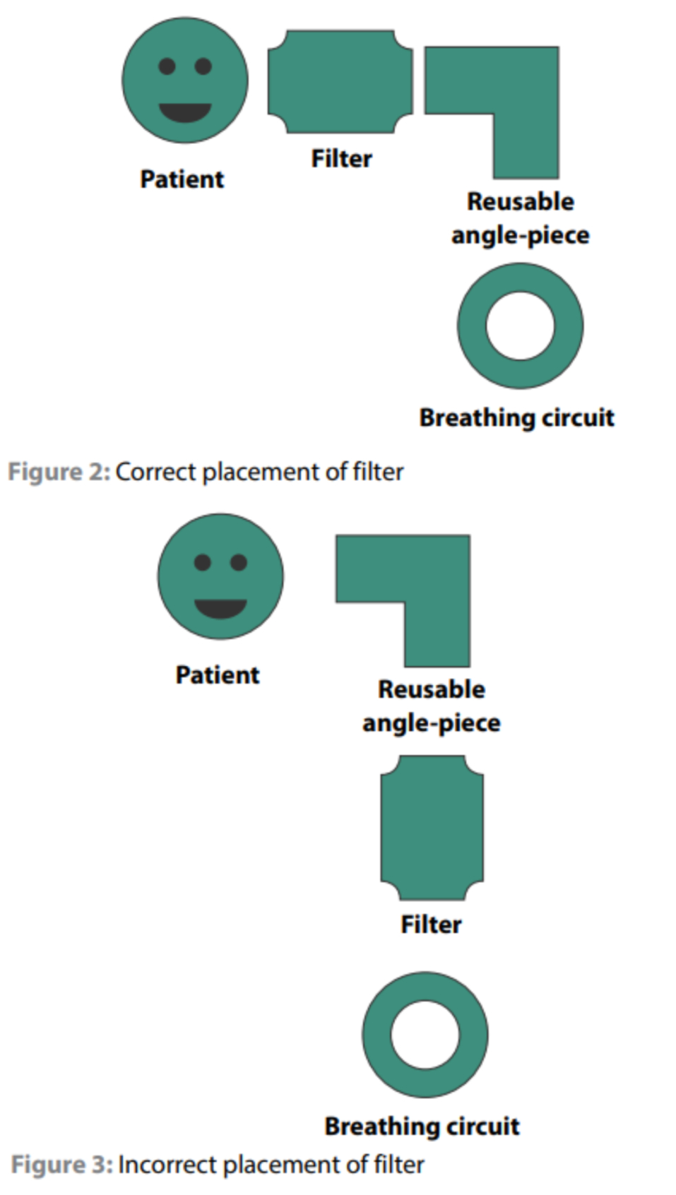
Click on the image to enlarge
The components of the breathing circuit can be reused between cases for up to seven days, provided that:
- A high-efficiency filter has been used.
- There are no defects in the system.
- It has been disinfected according to the manufacturer’s instructions daily.
- It has been cleared by the manufacturer to be used as such.152,153
- The breathing system components are seen as semicritical items and should be disinfected according to the manufacturer’s instructions.154
The CO2-absorber canister should be cleaned every time the absorber material is changed. Disinfection must take place according to manufacturer’s guidelines.154,155
The components of the breathing circuit should be changed immediately in any of the following circumstances:153
- When it is visibly soiled with blood or secretions.
- When used on a patient with a confirmed or potential notifiable infectious disease that involves the risk of transmission via the breathing circuit and reservoir bag, e.g. tuberculosis, acute viral hepatitis, measles, influenza virus, infection and/or colonisation with a multidrug-resistant pathogen and upper or lower respiratory tract infection.
Oxygen tubing, oxygen masks and nasal prongs
These are single-use items and should be discarded after use on a single patient. An area of at least 0.4 m from the mask should be considered to be a potential hazard for aerosolised pathogens.108,157 Patients with high-risk respiratory infections should only be nebulised when necessary and should be isolated during nebulisation in a room with good ventilation.
Rationale
The aim of using breathing filters in anaesthetic circuits is to prevent contamination of the breathing circuit and ventilator with pathogens from the patient. An array of pathogens has been found in contaminated circuits. These include: A. caloaceticus, P. maltophilia, P. aeuroginosa, Flavobacterium meningosepticum, K. pneumoniae, P. mirabilis, Enterobacter cloacae, Citrobacter diversus, E. agglomerans, Candida albicans, Proteus spp. and Streptococcus spp.155,156 It is also to prevent the passage of possibly contaminated liquid out of the circuit back to the patient. In essence, the aim is to prevent cross-contamination. This is particularly important in a country with a high incidence of HIV, tuberculosis and other opportunistic infections. The practice of using viral or bacterial filters allows the reuse of anaesthetic breathing circuits between patients in a cost-constrained environment.
Another benefit of combining a viral or bacterial filter with a HME device is that it prevents the inspissation of secretions, and thus allows for proper airway toilet, therefore decreasing the risk of infection. Dry air also leads to mucosal damage that can become an entry port for microorganisms. Thus, humidification is beneficial in preventing mucosal damage.141,142,158 Different devices can be added to the breathing circuit, and it is important for anaesthetists to appreciate the differences between them.
There is a simple breathing filter, without any HME. Usually, this is a yellow filter. Another HME has no filter and is usually blue. While yet another HME has a built-in breathing filter. This is known as an HMEF. These devices are generally green in colour.141
Other confusion with the terminology derives from the use of the words “electrostatic” and “pleated”. All filters are pleated and have some degree of electrostatic properties. The term “pleated filters” is used to refer to hydrophobic filters.141,159 The aim of pleating the filter material is to increase the surface area and therefore decrease airway resistance, as well as increasing the filtration area.140,159
The efficiency of electrostatic filters decreases over time as the electrostatic charge decreases. It also becomes inefficient when pressurised, so that liquid is able to pass through, and with it, carrying bacteria and viruses. Hartmann et al.145 demonstrated that up to 5.6% of contamination occurs in breathing circuits that had been in use for 72 hours with electrostatic filters.
All of the above filters have to be subjected to quality control tests by being subjected to the international standard, ISO 9360-1:2000, or the European standard, EN 13328-1.141,160 This requires the filters to be challenged with particles in the order of 0.3 μm. This size particle is the size that is most likely to be able to penetrate a filter. Particles below this size are captured by Brownian diffusion, while those that are larger are trapped by inertial impaction and interception.141 Efficiency is measured by the percentage of particles that get trapped by the filter.
Typically, this ranges from 95–99.97%. Thus, efficiency is always reported as a percentage. Other symbols commonly found in the terminology are the letters “N”, “P” or “R” in front of the efficiency percentage, e.g. N95. This refers to the ability of the mask to withstand oil. “N” means not oil proof, “R” means resistant to oil and “P” means oil proof. The “N” masks are the most commonly used masks in the medical field.
There has been a longstanding debate on whether or not a new circuit should be used for every patient, or whether a filter can be used to avoid contamination and then the circuits reused among patients. Some studies have shown that filters placed at the Y-connection were able to prevent the contamination of breathing systems.122-125 It is also known that filters reduce airborne microbes when tested in vitro.146-148,161,162 Evidence by Von Hassel showed that the risk of acquiring a lower respiratory tract infection from shared breathing circuits without using filters was very low at 0.1–0.2%. He subjected the circuits to HLD and pasteurisation and only used filters in HIV-reactive patients and those with tuberculosis. However, low-flow anaesthesia was not used, and thus, it is possible that the high gas flows had washed any contaminants out of the circuit. The American Thoracic Society164 noted that although HMEFs reduce circuit colonisation, they do not significantly reduce the incidence of ventilator-associated pneumonia and cannot be regarded as a tool to be used to totally prevent ventilator-associated pneumonia.
What is clear is the risk of complications associated with the use of filters.127-139 There have been case reports of patients suffering fatal events when breathing filters became blocked with blood or secretions. This led to an increase in airway resistance and could have contributed to an increase in dead space. HMEFs may also be associated with a risk of carbon monoxide poisoning. In particular, this is problematic when the HMEF is placed between the circuit and the absorber. This can lead to the desiccation of soda lime or bara lime, with the resultant production of carbon monoxide. Carbon monoxide poisoning might not be easily detected as the oxygen saturation monitor will give a false reassuringly high reading. The risk is also higher when higher gas flows are used.
This has led to North American societies recommending the single use of a breathing circuit per patient without the use of an airway filter. European societies have taken a different stance in view of cost constraints, and in their guidelines have recommended that breathing circuits can be reused, as long as a high-efficiency HMEF is utilised at the Y-piece and changed between patients, or when it becomes visibly soiled. In addition, the filters have to be able to withstand a pressure of 60 hectopascals in order to prevent liquid from condensation within the circuit passing back through the filter, carrying bacteria and viruses with it.121,142 The British Society for Antimicrobial Chemotherapy120 recommends the use of HMEs rather than heated humidifiers to reduce the incidence of ventilatorassociated pneumonia.
When circuits are to be reused, it is recommended that a high efficiency filter is used. Chant et al.165 reported on the possible patient-to-patient transmission of hepatitis C. A case series was described in which five of the patients on the elective minor operations list developed hepatitis C approximately seven weeks after surgery. All of the viruses where typed to be the same strain. A second such case was also reported.166
Various pathogens are, or have, the potential to be airborne over long distances (> 1 m). These pathogens pose a high risk of contamination of the breathing surface and ventilator, as well as the surrounding environment. Tuberculosis and influenza viruses are examples of such pathogens.
When reusing circuits between patients, it must be ensured that the circuit is classified as such by the manufacturer. Reusing circuits that are classified as “single-use” makes the anaesthetist liable.167 The manufacturers of multi-use circuits have specific instructions on the disinfection and cleaning of such circuits, and these have to be followed carefully to ensure continued performance.
Masks and tubing used for oxygen therapy are contaminated with secretions from the patient’s upper airways. They should never be reused between patients. Ip et al.156 demonstrated that aerosolised droplets could be detected as far as 0.4 m from a simple face mask during normal breathing. This distance can increase dramatically when the patient sneezes, coughs or vocalises. Patients with high-risk infections should only be nebulised when necessary, and in rooms that are isolated and have good ventilation. The risk is that nebulisation increases the velocity and distance in which aerosolised particles can travel. Nebulisation was thought to be the main cause that led to an outbreak of severe acute respiratory syndrome in Hong Kong in 2003.168
Bag valve mask resuscitators
All resuscitators should be fitted with a high-efficiency breathing filter between the valve and the mask before being used on a patient. Resuscitators used on the same patient should be capped at the patient connection port when not in use. Resuscitators should be cleaned and disinfected according to the manufacturers’ instructions. The resuscitator should be disassembled and all the parts washed thoroughly, using clean water and mild detergent. It is necessary to ensure that the detergent is suitable for the material. Do not disassemble the pressure release valve and the positive end expiratory pressure valve. All of the parts should be rinsed in clean water to remove the detergent. All of the parts should be allowed to dry in a clean controlled environment, where the risk for recontamination is low. The components should then be subjected to one of the following decontamination techniques:14
- Pasteurisation for 30 minutes (not the oxygen reservoir bag).
- Autoclaving not to exceed 132 °C (not the oxygen reservoir bag).
- Ethylene oxide gas (all parts are suitable).
- Liquid sterilisation (all parts are suitable) with Cidex® OPA or sodium hypochlorite. Wash thoroughly to remove any excess disinfectant.
Manual resuscitator should be sterilised:
- For first-time use.
- Between patients.
- When visibly contaminated.
- Every 24 hours of use in the same patient.
Rationale
Manual resuscitators are classified as semi-critical items and should be decontaminated using HLD or sterilisation. It is important to follow the instructions of manufacturers in this regard as certain parts of the resuscitator might not be compatible with the chosen method of sterilisation. It has been shown that bacteria can be cultured from resuscitators which were macroscopically “clean”. Paediatric Ambu® bags in the obstetric OT are used more frequently than adult ones and have been linked with disease transmission.
Supraglottic devices
Single-use (disposable) SADs are preferred to reusable SADs.
If reusable SADs, e.g. LMA Classic™, are used, they should be sterilised in an audited sterile service department and not more often than recommended by the manufacturer, e.g. 40 times for LMA Classic™. Do not decontaminate and reuse single-use SADs.
Rationale
Reasons for the preferred use of disposable SADs as opposed to reusable SADs include:
- The potential for the iatrogenic spread of protein-born prion diseases: The main concern in the reuse of the laryngeal mask airway (LMA) is the potential for the iatrogenic spread of protein-born prion diseases. Prions are proteins which become distinctive infectious agents that can reproduce without nucleic acids, and can cause transmissible spongiform encephalopathies in humans, including Kuru and Creutzfeldt– Jakob disease, under certain conditions.169 Variant Creutzfeldt– Jakob disease, a transmissible prion disease, was identified in humans in Great Britain in 1996.170 Recently, large numbers of prion proteins were detected in human tonsillar tissue, raising significant concern in several countries about reusable anaesthetic and surgical equipment.171 Concerns were raised in Europe about the SAD owing to its close proximity to tonsillar tissue. Prions are extremely resistant to inactivation by disinfectant chemicals and heat. Accordingly, the cleaning and autoclaving of reusable SADs may not be adequate to prevent the iatrogenic spread of prions. Several studies have found significant protein contamination on reusable SADs, even after cleaning and autoclaving.117,172-174 The risk to patients is as yet unquantified owing to several challenges, such as the long incubation period between exposure to prions and the development of clinical features, combined with an unknown number of carriers, undetermined exposure times and incomplete penetration. However, investigators have estimated that the number of carriers of variant Creutzfeldt–Jakob disease could range from thousands, to as many as millions of people worldwide.175 Moreover, the risk to anaesthesia staff during both the use and decontamination of the LMA has not been considered.
- Monitoring of reuse: Tracking the number of times that the reusable SAD is reused is essential. However, this is often not carried out. The original LMAs were designed for use up to 40 times.
- Affordability: Studies have shown that the cost of a disposable SAD compares favourably with the cost per use of a reusable SAD, even when staff time and cleaning costs were excluded.176
- Efficiency: There is no need to allocate time to the cleaning and sterilisation of the LMAs.
- When surveyed, the majority of clinicians would want singleuse devices to be used on their families and themselves if they were patients.11
Storage of semi-critical items
Semi-critical items should be packaged and stored in a way that prevents recontamination. Suggested compliant storage methods include a peel pouch or a closed plastic bag. These items should not be left unwrapped in or on top of anaesthesia workstations and trolleys.
Non-critical medical equipment surfaces
Non-critical medical equipment surfaces extend to blood pressure cuffs, stethoscopes, and frequently used control mechanisms, e.g. pop-off knobs, flow controls and vaporisers. It is necessary to disinfect with a low- or intermediate-level disinfectant after each patient. Medical equipment surfaces can become contaminated with blood and infectious agents and contribute to the spread of healthcare-associated infection.177,178 Loftus et al.179 recently reported that multidrug-resistant bacterial transmission to the anaesthesia work area occurred during the practice of general anaesthesia.
Disposable devices
“It seems to me that reusing disposable devices has an element of poetic justice ingrained, if one can become poetic about economics.” —VW Greene
A single-use medical device is to be used on a single patient during one procedure.113 The reuse of disposable or single use devices started in the 1970s and is a growing and common practice worldwide, especially in resource-limited settings.113,180-182 Informed consent should be obtained from the patients if an item is to be reused on them. Certain issues exist, including the decontamination technique and the risk of crosscontamination, material alteration, a clear limit to the number of times that an item can be reused, mechanical failure of the device, exposure risks to health care workers and ethical and legal implications.80,183 Although the reuse of single-use devices is strongly discouraged, the suggested practice from the ISID80 and the WHO184 are as follows:
- A facility should be committed to the reuse of single-use devices and have an institution-specific policy with clear guidelines.
- The disposable devices should be classified and reprocessed as per their intrinsic risk: critical, semi-critical and non-critical.
- Functionality and integrity of the device should be maintained.
- The package labelling and manufacturer’s guidelines should be followed.
- Reprocessing of disposable devices should be cost justified and performed by a licensed reprocessor.
- Both the physician and the patient should be informed that a device being used is a reprocessed single-use device.
- Any person who reuses a single use device takes full responsibility for its safety and effectiveness.
- A platform for reporting any adverse events from the reuse of single-use devices should be available. The Association of Perioperative Registered Nurses (AORN) has recommended that the sterility, integrity and functionality of a reprocessed single-use device must be documented as safe for patient care and/or equal to the original device specifications.
Trans-oesophageal echocardiogram probes (TEE)
A semi-critical device with increasing usage in anaesthetic practice that has the potential for cross-contamination, especially if the probes are damaged.12 A sheath should preferentially be used but does not eliminate the risk of contamination and does not exclude the probe from HLD. The recommended basic principles for reprocessing TEE probes are:12
- Clean the probe shaft and tip either with immersion or with a detergent-moistened wipe or enzymatic cleaner to remove gross contamination.
- Use a second wipe to wipe the proximal non-immersible parts such as the handles.
- Ensure that there is no structural damage to the probe.
- Use HLD to disinfect the probe tip and flexible shaft.
- Thoroughly rinse and dry before storage.
- The manufacturers’ instructions regarding chemical disinfectants should be followed.
South African Society of Anaesthesiologists (SASA) Guidelines for Infection Control and Prevention in Anaesthesia in South Africa 2021