GuidelinesOther guideline
Ankle Dislocation
Ankle Dislocation
Clinical
Posterior
Anterior
Lateral
Superior
Reduction
Ankle dislocations without fracture occur when significant force applied to the joint results in loss of opposition of the articular surfaces. Because of the large amount of force required and the inherent stability of the tibiotalar joint, dislocation of the ankle joint is rarely seen without an associated fracture.
Associated fractures are the rule rather than the exception with ankle dislocations.
- Neurovascular injury is the principal concern, as with any dislocation.
- Vascular compromise may result in avascular compromise of the talus, permanent sensation or nerve damage, and lower extremity tissue necrosis, and gangrene if not promptly reduced.
- Tented skin may be subject to ischemic necrosis.
Clinical
Four types of dislocations are seen around the ankle joint: posterior, anterior, lateral, and superior:
1. Posterior
- A posterior dislocation in the most common type of ankle dislocation. The talus moves in a posterior direction in relation to the distal tibia as force drives the foot backward. The wider anterior talus wedges back, resulting in forced widening of the joint.
- This must be accompanied by either a disruption if the tibiofibular syndesmosis or a fracture if the lateral malleolus. This occurs most commonly when the ankle is plantar flexed.
2. Anterior
- Anterior dislocations result from the foot being forced anteriorly at the ankle joint.
- Typically, this occurs with the foot fixed and a posterior force applied to the tibia or with forced dorsiflexion.
3. Lateral
- These dislocations result from forced inversion, eversion, or external or internal rotation of the ankle.
- They are associated uniformly with fractures of either or both the malleoli or the distal fibula.
4. Superior
- Diastasis occurs when a force drives the talus upward into the mortise. These dislocations usually are the result of a fall from a height.
- In such cases, the patient should be evaluated carefully for concomitant spine injury and fracture of the calcaneus.
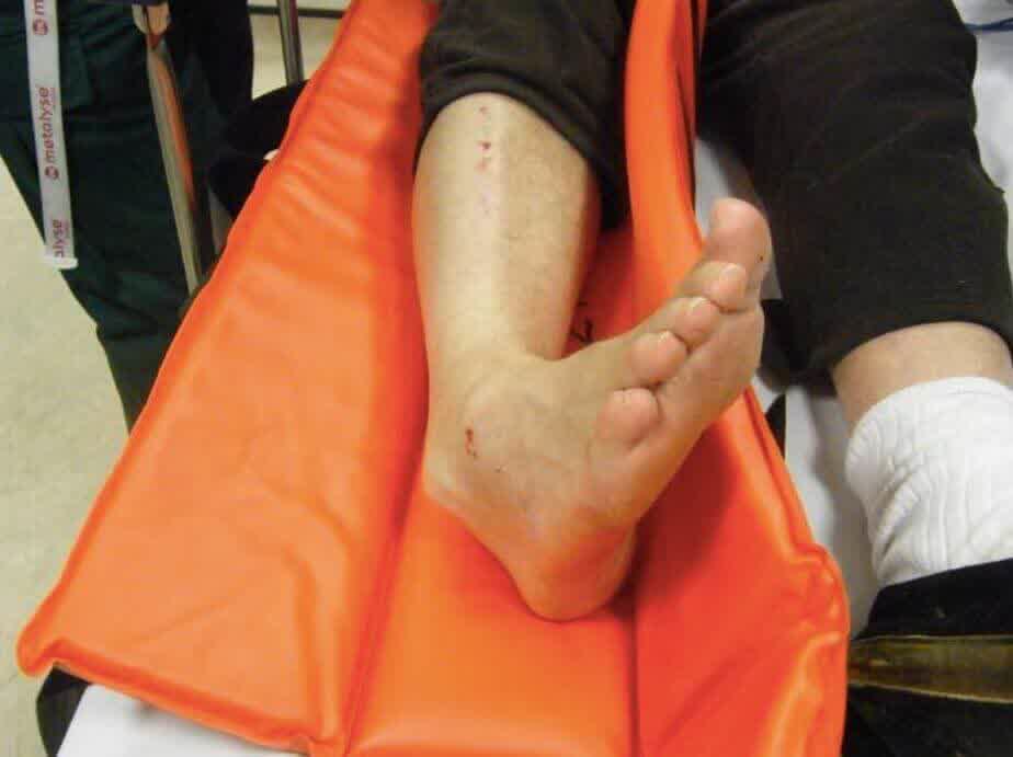
5. Reduction
- Early reduction is essential since delay may increase risk of neurovascular compromise or damage to articular cartilage. In patients with vascular compromise, perform reduction prior to radiologic examination.
- Dislocations should be reduced as soon as possible under conscious sedation.
- The different types of dislocations require different techniques the joint should be aligned to a neutral position and immobilised in a back slab with elevation post reduction.
- PEARL – Making the dislocation “worse” by applying traction and then aligning the joint is often successful.
References
- Rockwood CA Jr, Green DP, Bucholz RW, eds. Rockwood and Green's Fractures in Adults. 4th ed. Philadelphia, Pa: Lippincott Williams & Wilkins; 1996:971-85.