Ankle Injuries - Anatomy
Ankle injuries - Anatomy
Anatomy revisited
A few take home points on ankle anatomy
Bones
Movement
Ligaments
PEARL
Anatomy revisited
The talocrural joint commonly referred to as the ankle joint is the articulation between the tibia, fibula, and talus.
It is a synovial-type joint that permits movement predominantly in the sagittal plane.
This “hinge”-type joint allows approximately
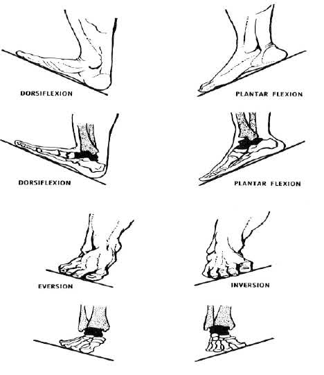
- 20º of dorsiflexion
- 30º-50º of plantar flexion
- 25º of inversion/ eversion
This complex motion is achieved by a relatively simple arrangement of bone, articular cartilage, synovial fluid, ligaments and tendons
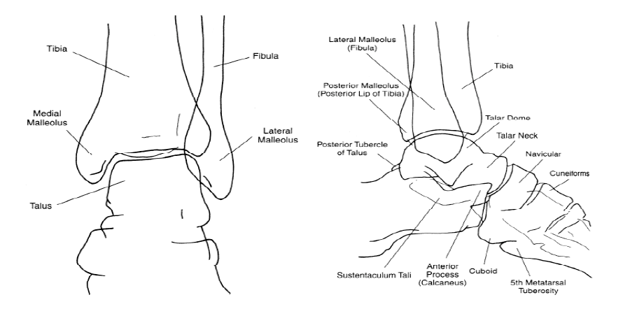
A few take home points on ankle anatomy
Bones :
The dome of the talus (trochlea) sits in the mortise, a socket created by the distal articulation of the tibia and fibula. Bony prominences (termed malleoli) form the medial and lateral aspects of the mortise, respectively, and the inferior end of the tibia (plafond) forms its roof.
Movement :
Adduction (inversion), abduction (eversion), internal rotation, external rotation, dorsiflexion, and plantar flexion describe the motion of the talus within the mortise.
Internal and external rotation occur when the talus rotates either inward (internal rotation) or outward (external rotation) around the long axis of the leg. Dorsiflexion and plantar flexion occur as the talus rocks either posteriorly (dorsiflexion) or anteriorly (plantar flexion) within the mortise.
Ligaments
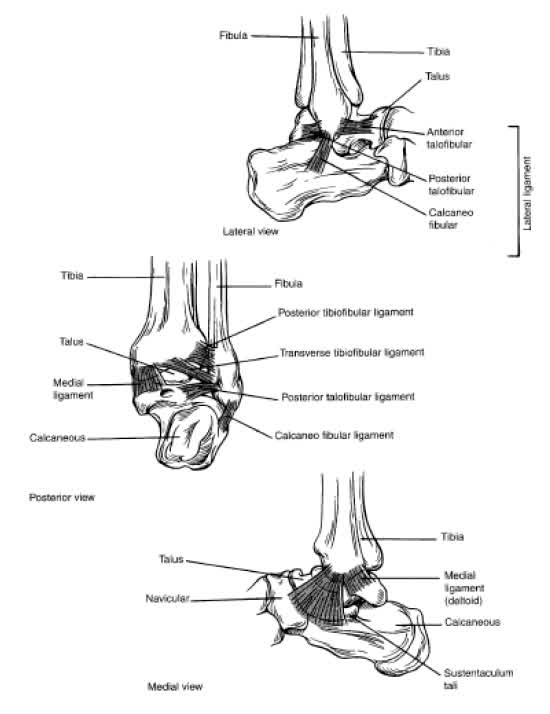
The talocrural ligaments provide stability to the ankle.
Three groups of ligaments connect the ankle bones on all four sides, forming a ring.
- The deltoid or medial collateral ligament stabilises the joint medially
- The lateral collateral ligaments steady the joint laterally
- Syndesmotic ligaments maintain anterior and posterior alignment.
PEARL
Anatomic stability depends on the number and location of breaks in the ring.
A single break in the ring (isolated ligament rupture or undisplaced single malleolar fracture) is usually a stable injury. More than one break (of either a ligament or bone) involving opposite sides of the ring disrupts the proper anatomic and functional relationship of the talus within the mortise and results in an unstable injury. Unstable injuries include such patterns as a lateral ligament tear and medial malleolar fracture or bi-malleolar fractures.