Ankle Injuries - Imaging
Ankle injuries - Imaging
Should all ankles be imaged?
Ottawa Ankle Rules
Plain films of the ankle are one of the most frequently ordered radiographic series in EC patients with musculoskeletal trauma, second only to cervical spine films. Only 15% of plain films show a significant fracture.
The question of when to order ankle x-rays has stimulated more than four decades of clinical debate. In the early 1990s, a Canadian group developed clinical decision rules (CDRs) for ankle radiography.
These rules attempt to
- Reduce costs by eliminating unnecessary ankle x-rays.
- Radiation exposure
- Decreasing time spent in the emergency centre
The Ottawa Ankle Rules are simply :
An ankle film should be ordered if there is post traumatic ankle pain with :
- Unable to weight bare at time of injury and on presentation for 2 steps on the injured ankle.
- Point tenderness upon palpation of the tip or distal 6cm of the posterior aspect of either malleolus.
Foot film is indicated if the patient complains about mid foot pain has either of the following :
- Unable to ambulate four steps at either the scene or in the emergency centre.
- Point tenderness over either the proximal 5th metatarsal or navicular bone.
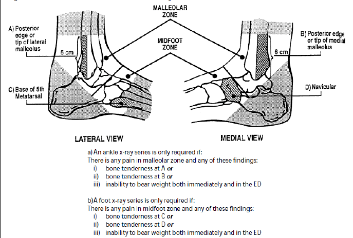
The big question to the public and clinician is how many fractures a missed? If one read the literature the initial study claimed sensitivities of nearly 100% and that the few insignificant avulsion fractures that are missed can be safely be treated as ankle sprains with no negative impact to the client.
In the setting where follow up can be problematic and the clinical suspicion is still high I would suggest doing a plain film.
If a fracture is still suspected even with no radiological evidence a back slab while non-weight bearing with orthopaedic follow up is suggested at a fracture clinic.
XR Views
- AP - This view demonstrates the medial and lateral malleoli and an oblique view of the talar dome it does reveal the entire talo-crural joint.
- Lateral - lateral view visualises the anterior and posterior elements of the distal tibia, including the posterior malleolus, as well as the base of the fifth metatarsal.
- Mortise - The mortise view is filmed like an AP, except the leg is internally rotated 15º-20º. This positioning provides a true AP view of the talus, revealing the entire joint space, both malleoli, and the breadth of the talar dome. The width of the joint space should be uniform, and tibia and fibula should overlap.