Atrial Fibrillation
Atrial Fibrillation
Introduction
Identification
Severity
Management based on severity
Relevant history, examination
Investigations
Dose charts
Referral/discharge criteria
Treatment Algorithm
Introduction/ description
AF is the most common sustained cardiac rhythm disturbance in adults. It is a risk factor for thromboembolism and congestive heart failure (CHF), and it causes symptoms such as chest pain and shortness of breath. The incidence and prevalence of AF is increasing. Lifetime risk over the age of 40 years is approximately 25%
Identification/ recognition/ diagnosis of problem
Clinical presentation ranges from asymptomatic to cardiogenic shock and stroke. The majority of patients are largely asymptomatic with only palpitations, dyspnoea, fatigue and dizziness. Usually identified by a fast, irregular rhythm and volume pulse and confirmed on ECG.
Differential diagnosis
- Atrial Flutter
- Atrial Tachycardia
- Atrioventricular Nodal Re-entry Tachycardia (AVNRT)
- Multifocal Atrial Tachycardia
- Paroxysmal Supraventricular Tachycardia
- Wolff-Parkinson-White Syndrome
Severity assessment (triage)
- Unstable AF (resulting in chest pain, hypotension or haemodynamic compromise)
- Stable AF - Rate vs Rhythm control
- < 48 hours
- > 48 hours
Management based on severity
Immediate action
- Stabilise using ABCs, perform blood glucose, apply oxygen if hypoxic and establish IV access.
- Decide whether stable or unstable (hypotension, chest pain, cardiac failure, decreased conscious level)
- Unstable: The majority of unstable AF patients will have a fast rate, although in a small cohort patients will present with a slow rate. In these patients consider high degree AV block (pacing might be needed), electrolyte abnormalities (hypokalaemia/ - magnesaemia), iatrogenic (Digoxin toxicity) or hypothermia.
- Aggressively pursue cause: AF is a nearly always a symptom and that the underlying cause requires treatment in order manage the patient optimally
Causes of AF include:
- Ischaemic heart disease
- Hypertension (Left ventricular hypertrophy with left atrial dilation)
- Valvular heart disease (esp. mitral stenosis / regurgitation)
- Thyrotoxicosis
- Acute infections
- Electrolyte disturbance (hypokalaemia, hypomagnesaemia)
- Drugs (e.g. sympathomimetics)
- Pulmonary embolus
- Pericardial disease
- Pre-excitation syndromes (WPW)
- Cardiomyopathies: dilated or hypertrophic
- Phaeochromocytoma
Non-drug treatment
- Obtain an ECG on presentation.
ECG Features of Atrial Fibrillation
Irregularly irregular rhythm, absence of P waves and an isoelectric baseline with a variable ventricular rate
QRS complexes usually < 120 ms unless pre-existing bundle branch block, accessory pathway, or rate related aberrant conduction
Fibrillatory waves may be present and can be either fine (amplitude < 0.5mm) or coarse (amplitude >0.5mm)
Fibrillatory waves may mimic P waves leading to misdiagnosis
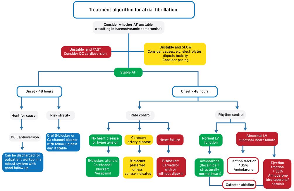
- For unstable AF with a fast ventricular response rate immediate direct current (DC) cardioversion is required.
- Stable patients with an onset of less than 48 hours at presentation can also be safely DC cardioverted.
General principles for DC Cardioversion
Monitored area (Resus preferred) as for procedural sedation
Separate providers managing airway and cardioversion
Sedation recommendation: Etomidate (0.1mg/kg) and Fentanyl
Deliver synchronised 150J biphasic, repeated if required
Repeat 12 Lead ECG following DC Cardioversion
Additional considerations in AF
Pre-cardioversion use of halve dose Amiodarone 150mg IV if time allows
Anticoagulation with Enoxaparin 1-1.5mg/kg SC prior to cardioversion
Drug treatment
- DC cardioversion pre-treatment: anti-arrhythmic drugs may be used to pre-treat patients before electrical cardioversion in ED in order to decrease early recurrence of AF and to enhance cardioversion: Amiodarone 150 mg can be given over 10 minutes prior to DC cardioversion
- Stable AF with onset < 48 hours: Rate-slowing agents alone are acceptable while awaiting spontaneous conversion in certain patients: age less than 65, no co-morbidities and hemodynamically stable
- Stable AF with onset > 48 hours:
- Rate control: Drugs include β-blockers to Ca-channel blockers with an aim of a target resting heart-rate ≤ 80 BPM and during moderate physical activity ≤ 110 BPM. In the EC an aim of 80- 110 BPM is acceptable
- Rhythm control: Drugs include Amiodarone, although Flecainide (where available) can also be used provided preserved left ventricular function.
- Anticoagulation/ prevention of thromboembolism
With a rhythm control strategy in AF onset ≥ 48 hours (or uncertain duration), patients should receive therapeutic oral anticoagulants (OAC) therapy (warfarin or dabigatran) for 3 weeks before and at least 4 weeks postcardioversion.
With a rate control strategy in AF onset ≥ 48 hours (or uncertain duration), a risk assessment should be made using the CHA2DS2-VASc score (end of section) to guide anti-coagulation needs.
In all cases the benefit of using anti-coagulation should be weighed up against the risk of bleeding. The HAS-BLED score (end of section) can be used to guide decision making.
Relevant history, examination
- Specifics in the history include checking for stimulants, medication (e.g. Theophylline), chronic medical conditions, etc.
- Specifics in the physical examination include checking for goitre and features of heart failure
Investigations
In addition to the ECG also obtain:
- Bloods: FBC, U&E, TSH, cardiac enzymes (the latter only if specifically indicated; troponin often raised due to fast rate-related ischaemia necessitates a repeat measurement in 3 to 6 hours)
- CXR: straight left heart border suggests left atrial enlargement, right atrial enlargement with hyperinflation.
- Bedside Cardiac Ultrasound: provides clues to ejection fraction, valvular pathology and chamber size.
Dose charts
β blockers
β blockers are more effective at rate control than Ca-channel blockers. Avoid in hypotension, acute decompensated heart failure or asthma. The IV route is preferred in the acute setting.
- Atenolol (cardio selective): IV 5 mg over 5 minutes, repeated if no effect in 10 minutes
- Oral (largely asymptomatic patients): 50 mg stat, then 50- 100 mg OD
- Metoprolol (cardio-selective): IV 2- 5 mg at 5 minute intervals until desired effect (max of 15 mg)
- Oral (largely asymptomatic patients): 50 mg stat, then 50- 150 mg in 2- 3 divided doses
- Carvedilol has α and β blocking activity: Oral 3.125 mg BD, increased to effect to max 25 mg BD
Ca-channel blockers
Avoid in hypotension, broad complex tachycardia, acute decompensated heart failure or severe left ventricular dysfunction. The IV route is preferred in the acute setting.
- Verapamil: IV 2.5- 5 mg over 2 minutes, repeated at 5- 10mg every 15- 30 Minutes (max of 20 mg)
Oral (largely asymptomatic patients): 40- 80 mg TDS
Digoxin*
Digoxin is a cardiac glycoside that rate controls as well as increases inotropy and can therefore be used in heart failure. The oral route is preferred. It is often used in combination with β blockers (e.g. Carvedilol). Due to its narrow therapeutic index, the β blocker is usually titrated to effect rather than the digoxin
- Digoxin: Oral 500 µg load (if digoxin naive) and then 62.5 µg to 125 µg OD
IV (rarely used, requires monitoring and normal renal function): 500 µg over 30 minutes
Amiodarone
Amiodarone is likely to cardiovert AF to sinus rhythm. Please ensure that rhythm control is desired
- Initial 150 mg Amiodarone infusion: add 150 mg to 100 mL 5% Dextrose and infuse over 10- 60 minutes
- Followed by giving a 900 mg maintenance infusion as follow:
- Add Amiodarone 900 mg to 500 mL 5% Dextrose
- Infuse Amiodarone 360 mg (200 mL of mixed solution) over 6 hours (or 33 mL/h for 6 hours)
- Infuse the remaining Amiodarone 540 mg (300 mL of mixed solution) over 18 hours (or 17 mL/h for 18 hours)
Pill in pocket
The pill in the pocket regiment rhythm control treatment strategy is acceptable as initial therapy for patients who are asymptomatic with new onset paroxysmal AF and no structural heart disease. This strategy involves a larger, one-time dose of a class I antiarrhythmic (propafenone or flecainide). A fast acting atrioventricular nodal blocking agent such as metoprolol should be used in conjunction to prevent concealed conduction that can progress to ventricular tachycardia or fibrillation. It is recommended that this approach be followed in consultation with a cardiologist.
Enoxaparin*
Enoxaparin1 mg/kg BD
Warfarin
Start with 5- 10 mg daily for 2- 5 days until desired prothrombin activity is reached. Warfarin has significant drug interactions which should be checked prior to commencement. Monitoring of INR is required throughout treatment on warfarin.
*Reduce dose in renal failure, see SAMF
Referral/ discharge criteria
Hospital admission is recommended for highly symptomatic patients with decompensated heart failure or myocardial ischemia. Hospital admission should be limited to highly symptomatic patients in whom adequate rate control cannot be achieved, or where an outpatient workup cannot be achieved. After conversion to sinus rhythm has been achieved, the decision to introduce antiarrhythmic drug therapy should be based on the estimated probability of recurrence and the symptoms during AF. Long-term therapy will need to be determined by an appropriate outpatient consultation.
CHA2DS2-VAScscore
| Risk factor | Score |
| CHF or LVEF < 40 % | 1 |
| Hypertension | 1 |
| Age > 75 | 2 |
| Diabetes | 1 |
| Stroke/ TIA/ Thromboembolism | 2 |
| Vascular disease | 1 |
| Age 65- 74 | 1 |
| Female | 1 |
| Risk guidance | |
| Score 0 | Low risk: consider use of Aspirin |
| Score 1 | Intermediate risk: consider use of either Aspirin or Warfarin/ Dabigatran |
| Score ≥ 2 | High risk: Strongly consider use of Warfarin/ Dabigatran |
HAS-BLED score
| Risk factor | Score |
| Hypertension | 1 |
| Abnormal renal/ hepatic function | 1 (each) |
| Stroke | 1 |
| Bleeding | 1 |
| Labile INRs | 1 |
| Age ≥ 65 | 1 |
| Drug or alcohol abuse | 1 (each) |
| Risk guidance | Bleeding rate (%/ year) |
| Score 0 | 1.13 |
| Score 1 | 1.02 |
| Score 2 | 1.88 |
| Score 3 | 3.74 |
| Score 4 | 8.70 |
| Score ≥ 5 | Insufficient data |
Treatment algorithm for atrial fibrillation
References
Dorian P. Rate Control in Atrial Fibrillation. N Engl J Med 2010; 362:1439
Gheorghiade M, van Veldhuisen DJ, Colucci WS. Contemporary use of digoxin in the management of cardiovascular disorders. Circulation. 2006;113(21):2556
Rossiter D, Blockman M (eds.). South African Medicines Formulary. 11th Ed. Cape Town: Health and Medical Publishing Group; 2014
Cameron P, Jelinek G, Kelly A, et al. Textbook of Adult Emergency Medicine. 4th Ed. Elsevier; 2015