Breast Diseases
Breast Diseases
Physical Examination
Imaging
Tissue diagnosis
Indications for excision biopsy
Impalpable breast cancer
Indications for mammography at GSH breast clinic and Metro West facilities
Guidelines to Special Investigation
The core function of a diagnostic breast clinic is to exclude or confirm with confidence a breast carcinoma, and secondarily not to over manage benign breast disease.
The diagnosis of breast cancer is based on the triple assessment and comprises:-
- Physical examination
- Imaging - mammography and/or ultrasonography
- Biopsy - fine needle aspiration biopsy (FNAB) and/or core needle biopsy
The three modalities are complementary, enabling the diagnosis of breast cancer most cases.
Physical Examination
Breast cancer may present as a lump or increased tissue thickening in the breast, with skin or nipple changes, nipple discharge or may be incidentally discovered by radiological imaging.
The examination of the breast should proceed with the patient in the upright sitting position with inspection for obvious masses, asymmetry, skin or nipple retraction and skin changes. Raising the arms above the head or tensing the pectoralis muscles may accentuate asymmetry or skin dimpling. Then, with the patient supine, and the hands placed above the head, the breasts should be gently palpated against the chest wall, feeling for any masses or thickening. Next, the axillae are palpated for any nodal involvement while supporting the patient’s arm.
A non-tender breast lump is the most common finding in women with breast cancer. Common causes of benign breast lumps are fibroadenomas, cysts and fat necrosis. The differential diagnosis is strongly influenced by the patient’s age.
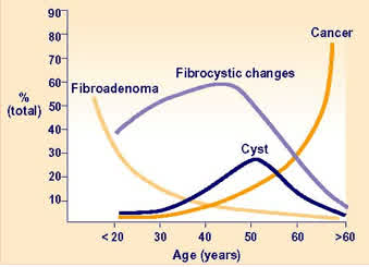
Figure 1: age distribution of benign and malignant breast lesions in patients presenting with a palpable breast lump
Breast lumps should be characterized according to their size, consistency and location. Carcinomas are typically firm and less well circumscribed, and their movement produces a drag of the adjacent tissue. Cysts and fibroadenomas are typically firm, but well circumscribed and mobile.
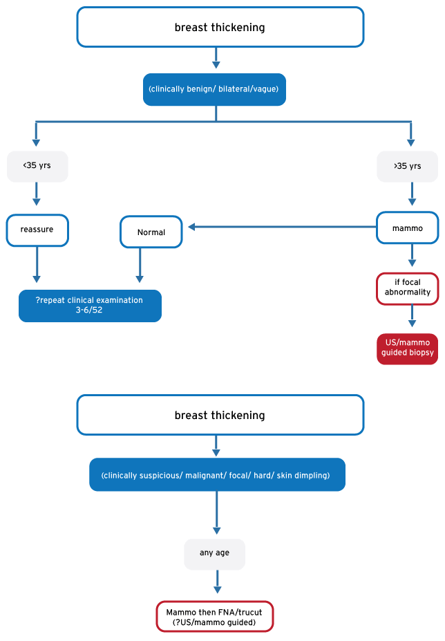
Some carcinomas may not necessarily present with a well-defined discreet lump on examination, but rather an area of focal breast thickening, with or without overlying skin dimpling. In the majority of cases, these clinical findings reflect fibrocystic changes, but it is important to consider that some carcinomas, particularly lobular in type, may present with similar subtle clinical findings. An area of focal breast thickening should be evaluated thoroughly with breast imaging and directed needle biopsy.
Oedema of the skin produces a clinical sign known as peau d`orange (skin of the orange) and, when associated with breast cancer, is a sign of locally advanced disease. When combined with erythema, warmth and tenderness, these signs are the hallmark of inflammatory carcinoma, and may be mistaken for mastitis or breast abscess.
Nipple discharge is common and rarely associated with an underlying breast cancer. Common causes include physiological discharge, intraductal papilloma, duct ectasia and periductal mastitis. A spontaneous unilateral non-milky discharge from a single duct orifice is significant and warrants further investigation, although in the absence of a palpable mass or suspicious mammogram, this symptom is usually not associated with cancer. This clinical scenario is best investigated with a microdochectomy (excision of breast duct).
An eczematous nipple reaction is highly suggestive of Paget`s disease. Histologically, this condition is produced by intraductal carcinoma occurring in the large lactiferous sinuses just under the nipple.
Carcinoma cells invade into the epidermal layer of skin of the nipple, and may then spread radially onto the areola. By contrast, benign skin conditions such as eczema usually originate on the areola. Paget`s disease may or may not be associated with an underlying mass. A punch biopsy of the affected skin is diagnostic.
Please record your clinical findings on the drawing in the clerking sheets accurately, and state your clinical diagnosis PRIOR to knowing the cytology or mammographic report. Biopsy can occasionally cause haematomas and make later evaluation of clinical stage less accurate.
Imaging
Mammography is the first-line radiological test. The classic appearance of carcinoma is a hyper-dense, spiculated mass, although architectural distortion, asymmetry, stellate lesions and calcification may also indicate carcinoma. It is important to remember that mammography has a false negative rate of approximately 15%. A normal mammogram, therefore, does not exclude breast cancer, and clinical signs should not be ignored.
U/S has a sensitivity of about 75% for the diagnosis of breast cancer. It is seldom used in isolation, but it is useful in assessing a breast mass where mammography is non-specific, particularly in young women with dense breasts. In our clinic it mostly done at the discretion of the reporting radiologist .
Tissue diagnosis
Fine-needle Aspiration (FNAB) should preferably be performed after mammography. It is carried out using a 22 G needle on a 10 ml syringe. Leaving 2 ml of air in the syringe, negative pressure is applied while making multiple passes through the lump. Thin smears of aspirate are prepared on 3 slides, 2 of which are sprayed with a fixative, and one which is air dried. The cytology tech will report on the slide on site, and if a cancer is diagnosed the patient will be immediately seen by the attending Radiation Oncology registrar. If the report shows an inadequate aspirate, it can be repeated immediately.
Our unit does not recommend definitive treatment based on cytological assessment only, since false positive diagnoses are rare (<0.5%), but well documented. Therefore almost all patients will have a core biopsy at the same time. This is then reported in time for Wednesday’s combined clinic. A definitive diagnosis of breast cancer can be based on cytology alone, only if the “malignant” cytological report is concordant with clinical and mammographic opinions. If any of these evaluations fall short of a confident “malignant” report (ie are merely indeterminate or suspicious) cytology is not sufficient to guide cancer decisions.
Core Biopsy: establishes a histological diagnosis. Using a cutting needle device (e.g. Trucut), it is carried out immediately after FNAB. A core biopsy can establish cell type, differentiate between in situ and invasive cancer, and will often establish receptor status and tumour grade. Core biopsy is more technically difficult than FNAB but, when positive, establishes an unequivocal diagnosis of carcinoma.
Both needle techniques have a false negative rate, mostly as a result of technical error or geographic mis-sampling.
Excision biopsy: the diagnosis of carcinoma is made without excision biopsy. However approximately 5% of cases of breast cancer are not confidently diagnosed with a triple assessment, and require formal excision. For small lesions, radiologically guided biopsies may greatly simplify the diagnostic process.
Indications for excision biopsy
- Discordant results of needle biopsy / mammography, i.e. Cytology report suspicious or malignant but mammogram and core biopsy inconclusive / benign or mammogram suspicious / malignant, cytology and core biopsy inconclusive / benign
- Patient request
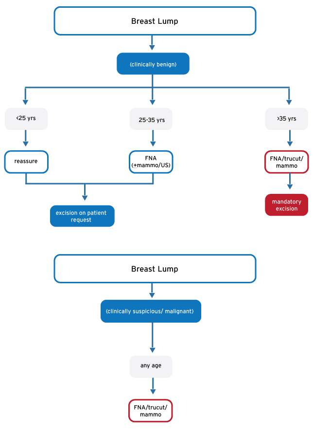
- Symptomatic discreet mass in patient older than 35 years
Impalpable breast cancer
With the use of imaging modalities an increasing number of breast cancers are identified before they present with any clinical signs. In this context the diagnosis may be made by US guided core biopsies, or by guided excision biopsy, using radioactive tracers (radio-guided occult lesion localisation-R.O.L.L.).
Indications for mammography at GSH breast clinic and Metro West facilities
Mammogram: Currently available daily at GSH, and on Mondays at MPH via Pink Drive. It is a limited resource.
Diagnostic: for the evaluation of a clinical breast complaint. This is the commonest indication.
- Mammography is mandatory in any clinical case where there is suspicion of malignancy.
- Every palpable breast lump in a patent older than 30 must be evaluated by means of mammography AND tissue biopsy.
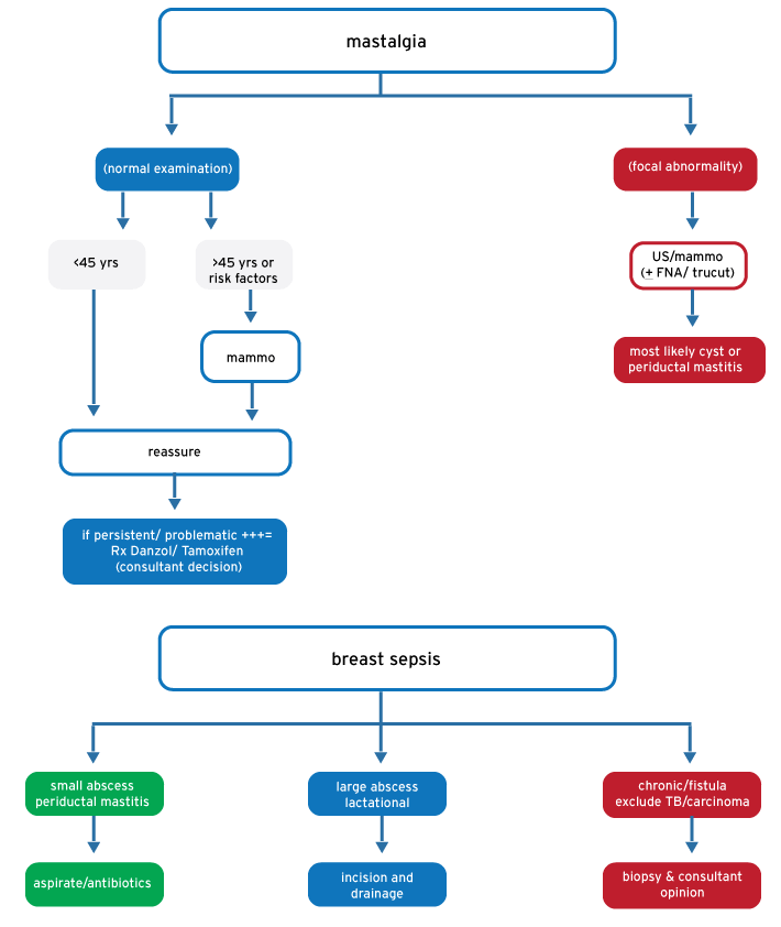
- Mammography is not indicated in patients presenting with unequivocally benign breast complaints (i.e. mastalgia, periductal mastitis, normal clinical examination)
Screening: population based screening is not feasible. Screening is supported only in limited specific situations. In such circumstances it is to be performed annually.
High risk patients:
Risk evaluation: the follow subgroups merit regular annual screening by means of clinical review and mammography
- Known high risk genetic mutations- BRACA1,2 + others
- Previous high risk breast pathology, such as LCIS, atypical hyperplasia, DCIS
- Previous breast carcinoma, with breast conserving surgery, yearly x 5 years
- Lifetime breast cancer risk > 20% (http://www.cancer.gov/bcrisktool/) or download Ibis risk evaluator (http://www.ems-trials.org/riskevaluator/) to evaluate family pedigree in depth. Programs can be downloaded on to OPD computers free of charge or carried on smart phone/ tablet)
Moderate risk patients:
- Prior to commencing HRT
- Yearly until HRT is stopped
- HRT not to be continued for >5 years
Opportunistic screening: patients presenting to the breast clinic requesting mammographic screening, will be offered a once off investigation. They are not to be screened on an ongoing basis, unless they meet risk criteria listed above. Patient requests are to be treated sympathetically, but not prioritised.
Guidelines to Special Investigation