Bronchiolitis
Bronchiolitis
Summary algorithm
Introduction
History
Examination
Differential diagnosis
Investigations
Management
Admission criteria
Indications for ICU admission
Discharge criteria
Advice for caregivers
Appendices
Summary algorithm
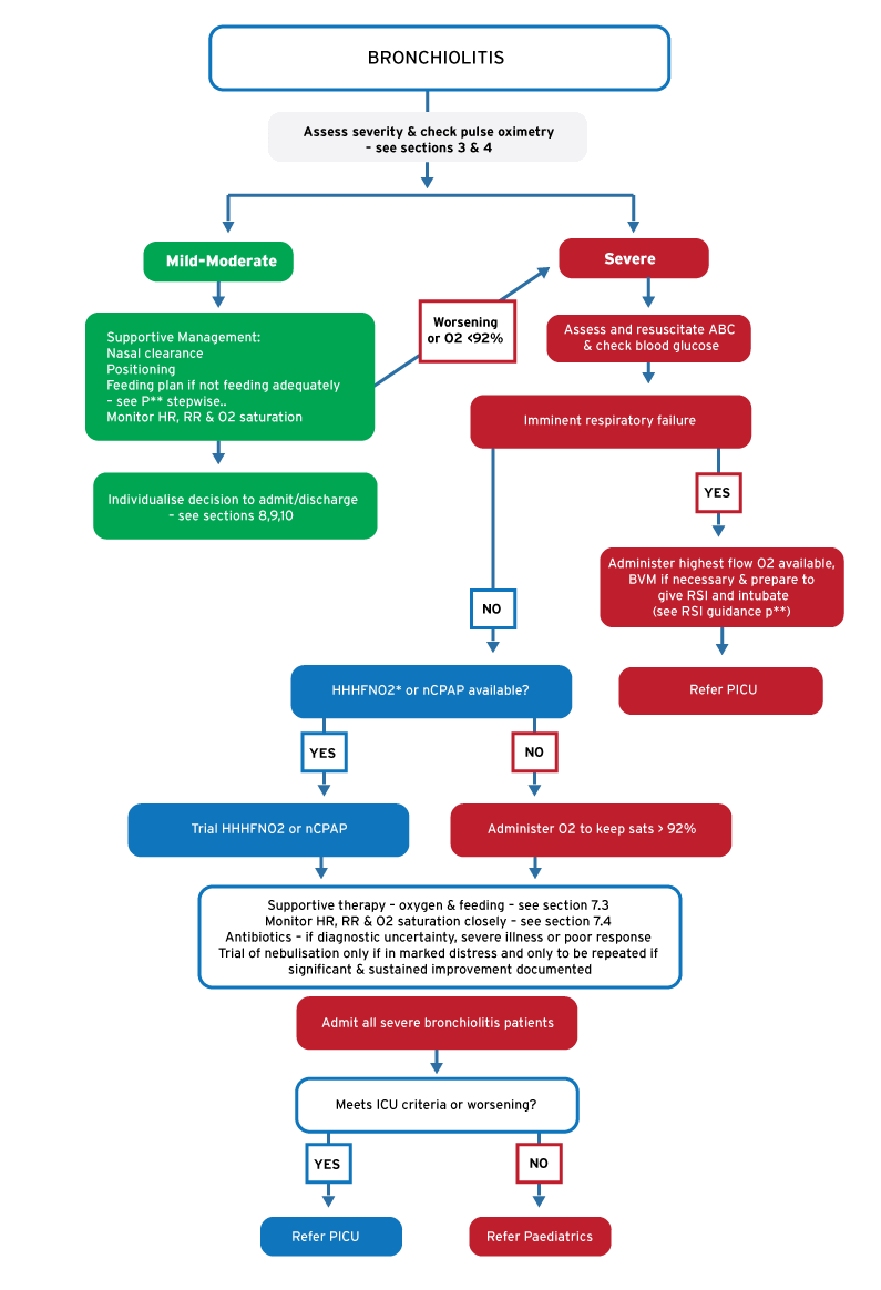
Click to enlarge
*Heated humidified high flow nasal cannula oxygen (HHHFNO2)
Introduction
Acute Bronchiolitis is a viral-induced inflammation of the smallest airways (bronchioles). The clinical manifestations result from airway inflammation, oedema and mucous plugging in the bronchioles leading to air-trapping. Worst affected are small babies <1 year of age as their airways get easily blocked.
Respiratory syncytial virus (RSV) is the most common viral pathogen. Other viruses include rhino, para-influenza, influenza, adeno and human metapneumovirus & measles. There is a seasonal tendency.
Bronchiolitis is generally self-limiting, with a brief coryzal period followed by a duration of 2-5 days; but the wheeze may persist a week or longer (post-viral wheeze). Of note the illness often worsens for 1-3 days after the onset of fast breathing. Secondary bacterial infection is uncommon if immune competent.
History: make the diagnosis
Based on History and Clinical examination, not Special investigations)
- Children younger than 2 years with constellation of mild URTI signs, low-grade fever, increased respiratory effort and wheeze.
- Often feed poorly – ask approx. what % of the normal intake the baby is managing
- Neonates & young babies may present with apnoea only (mechanism not known)
RISK FACTORS for SEVERE BRONCHIOLITIS | |
| Infants < 3 months Ex-premature infants Immunosuppression/unimmunised Lack of breastfeeding | Congenital heart disease (CHD) Chronic lung disease (CLD) Neurological disease Overcrowding, passive smoke exposure |
Examination
DETERMINE THE SEVERITY AND NEED FOR EMERGENCY INTERVENTIONS
MILD/MODERATE DISEASE | SEVERE DISEASE | |
LOOK FOR CLINICAL SIGNS of Respiratory Distress and Dehydration | Mild-moderate increase in work of breathing: -Tachypnoea, Hyperinflation -Lower chest wall indrawing -Expiratory wheeze -Crepitations | Severe respiratory distress: Grunting, nasal flare, use of auxiliary muscles, head nodding, central cyanosis (hypoxic), inability to feed & marked increase in work of breathing NOTE: Premature/very young babies may present with apnoea as 1st sign of bronchiolitis + have a high risk of further episodes |
DO PULSE OXIMETRY in all | If saturation < 92% in air give O2 and treat as severe disease | Oxygen if saturation < 92% in room air Assess need for ventilatory support (see below) |
Differential Diagnosis
- Pneumonia
- Viral induced asthma/wheeze
- Foreign Body Aspiration
- Chronic lung disease
- Cardiac cause (don’t be caught out by cardiac failure!)
- Gastro esophageal reflux with/without aspiration
- Vascular ring, Tracheo-oesophageal fistula
Investigations
CXRs and blood tests generally unhelpful and not necessary to make the diagnosis
MILD/MODERATE DISEASE | SEVERE DISEASE | |
Blood tests | None needed routinely | FBC + Differential count, Blood culture, CRP, Glucose and Electrolytes, Arterial blood gas |
Imaging (CXR) | Not routinely done | May show areas of atelectasis and consolidation |
Viral Tests | Not routinely done | Viral etiology (testing) does not predict severity or improve acute care. May be done for cohorting purposes or in severe cases when diagnosis unclear |
Management
Antibiotics and bronchodilators
ANTIBIOTICS
No Antibiotics for uncomplicated mild-moderate bronchiolitis
However, have a low threshold for starting antibiotics in children with risk factors or those requiring hospitalisation with suspected concomitant bacterial infection (see CAP guidelines). It is almost impossible to distinguish bacterial from viral lower respiratory tract infections on clinical or laboratory grounds and therefore antibiotics can be given if deemed beneficial by the clinician. Oral antibiotics can be given if the child is relatively well.
BRONCHODILATORS
Bronchodilators are NOT effective in the routine management of bronchiolitis
There is no supportive evidence for a trial or regular use of Adrenaline, ß2 agonists, or Ipratropium bromide. Nebulised hypertonic (3%) saline may be administered to hospitalised infants (weak evidence for use) as it may decrease in-patient length of stay. There is no supportive evidence for the use of Montelukast, chest physiotherapy or corticosteroids (systemic, nebulised or inhaled). Palivizumab is only indicated for infants with chronic lung/cardiac disease and prematurity and must be prescribed by sub-speciality clinicians.
SUPPORTIVE TREATMENT
Oxygen Therapy
- Nasal prongs are recommended at a flow rate of 2L/min (1L/min in infants) which would provide approx. 28%-35% inspired oxygen (FiO2).
- Simple facemask (FM) oxygen can deliver 30-50% at flow rate 6-10L/min only IF FM IS TIGHT FITTING
- Venturi mask system typically delivers 28-40-% FiO2 depending on the flow rate and FM fitting
- Non-rebreather(NRB) FM (reservoir + one-way flow valve) can deliver FiO2 up to 90% WITH TIGHT FM
- O2 should be discontinued when the child is improving clinically & saturations remain > 92% in room air.
Feeding & IV fluid therapy – see Stepwise assisted feeding for children with respiratory distress
- Encourage children to eat and drink normally.
- Enteral feeds through a nasogastric tube should be started if the child is too distressed to feed.
- Intravenous fluids are only required in children who are severely ill; use cautiously.
- Electrolytes should be measured as the syndrome of inappropriate antidiuretic hormone secretion (SIADH) is common. Restrict IV fluids to 40-60% of normal requirements after correction of hypovolaemia. If hyponatraemia is a problem, fluids containing half normal saline should be used.
MONITORING
- The frequency of monitoring will depend on the child’s condition.
- Children on oxygen therapy should have observations done 4 hourly including temperature, pulse, respiratory rate and patterns, and oxygen saturation.
Failure to improve after 48 hrs of treatment should prompt a review of therapy and a search for complications.
It is important to consider whether the child:
- is receiving the prescribed medications
- is tolerating the treatment e.g. vomiting
- has dual pathology or pathogens and/or has a resistant organism
has developed a complication of the disease (pneumothorax, pneumonia, effusion, empyema, lung abscess, or distal disease secondary to bacteraemia)
Admission criteria
- RR>50/min or Severe respiratory distress
- O2 saturations <95% in room air – but only need O2 if O2 sats <92%
- Unable to feed at least 50% of usual feeds – see assisted feeding plan
- Apnoea (even just a history of apnoea from the carer)
- Lethargy
- Carer not able to understand &/or comply with discharge instructions
- Carer unable to return to hospital in the event of worsening condition of baby
- Other high risk factors (as outlined above in section 3.1)
- Early in course of illness (e.g. Day 1-3) in small or at risk baby that is likely to worsen
Indications for ICU admission:
- Failure to maintain saturations of >92% on FiO2 of ≥70% (i.e. partial or full non-rebreather facemask (NRB FM)) – if no NRB FM’s then administer highest FiO2 possible
- Apnoea (not history alone – ongoing likelihood of apnoea and/or marked distress)
- Hypercarbia with resulting acidaemia (pH <7.25)
- Exhaustion (clinical judgement)
Discharge criteria:
- Feeding at least 50% of normal feeds
- Maintaining O2 saturation >92% in room air and not needing oxygen in past 6 hours
- Only minimal or no respiratory distress
- No high risk factors (see section 3.1)
- Parent able to understand and comply with discharge instructions
- Parent/carer able to bring the baby back if condition worsens
Advice for caregivers
- Nasal clearance: feeding may be hampered by nasal secretions – show carer how to administer saline nasal drops. Give about 10 minutes before each feed and then again just before feeding. Gently remove any secretions that are sneezed out.
- Feeding: baby may not be able to take the full amount as normal. Therefore may need to offer smaller amounts but more often. E.g. instead of 120ml 3 hrly offer 80ml 2 hrly or 40ml hrly.
Return if any of the following occur:
- The work of breathing increases. (Describe to the carer what you mean both visually & in words include e.g. nasal flaring, grunting, fast breathing, chest indrawing between-above-below ribs etc.)
- Pale or blue lips/tongue
- Exhaustion: getting very tired or weak
- Not able to feed at least 50% of feeds or vomiting repeatedly
- Parental concern that the baby is ‘not right’
Appendices
See separate guideline for Stepwise Assisted Feeding Plan for Bronchiolitis.
References
- Fernandes RM, Hartling L. Glucocorticoids for Acute Viral Bronchiolitis in Infants and Young Children. JAMA Clinical Evidence Synopsis 2014;311(1):87-88
- Gadomski AM, Scribani MB. Bronchodilators for bronchiolitis. Cochrane Database of Systemic Reviews 2014; issue 6. Art No.: CD001266. DOI:10.1002/14651858.CD001266.pub4
- Ralston SL, Lieberthal AS, Meissner HC, Alverson BK, Baley JE, Gadomski AM, Johnson DW, Light MJ, Maraqa NF, Mendonca EA, Phelan KJ, Rosenblum E, Sayles S, Hernandez-Cancio S.
- Clinical Practice Guidelines: The Diagnosis, management, and Prevention of Bronchiolitis. Pediatrics 2014;134:e1473-1502
- Schroeder AR, Mansbach JM. Recent evidence on the management of Bronchiolitis. Current opinion 2014;26(3):328-333
- Stollar F, Alcoba G, Gervaiz A, Argiroffo CB. Virologic testing in bronchiolitis: Does it change management decisions and predict outcome? Eur J Pediatr 2014;173:1429-1435