Burns
Introduction Background Classifying a burn Initial management Discussion criteria Ongoing management SETTING Primary and Secondary Healthcare Institutions in the Western Cape which refer to Red Cross War Memorial Children’s Hospital (RCWMCH) FOR STAFF Primary and Secondary Doctors and Nurses who refer patients to RCWMCH PATIENTS Patients under the age of 13 years old with burns I
SETTING
Primary and Secondary Healthcare Institutions in the Western Cape which refer to Red Cross War Memorial Children’s Hospital (RCWMCH)
FOR STAFF
Primary and Secondary Doctors and Nurses who refer patients to RCWMCH
PATIENTS
Patients under the age of 13 years old with burns
Introduction
In the paediatric population, hot liquid burns are far more common than flame burns. This is because children are far more vulnerable to significant injury from hot fluids than adults are. They burn faster and at a lower temperature than adults, so what would be a minor scald in an adult, has the potential to be a significant burn requiring emergency care in a child.
The context that many of our paediatric patients live in exposes them to increased risk. Overcrowding, informal plumbing systems and insufficient supervision increases the chance that a young child will sustain a significant injury.
Given the high incidence and severity of burns in the paediatric population in South Africa, it is important that primary care clinicians and nurses have a good knowledge of the initial management of severe burns and the skills to safely manage smaller minor burns independently.
The purpose of this guideline is to assist clinicians in identifying where a burn can be safely managed at the primary level or if discussion with RCWMCH is required. Guidelines on the initial management are provided.
Background
Your skin is the largest and most exposed organ in your body. When skin is damaged it loses its ability to fulfil its role of thermoregulation, preventing fluid loss and providing a mechanical barrier against pathogens, trauma and the sun. Part of the management of a burn is assisting the patient with the functions of the skin which have been lost due to damage to the skin.
The clinician managing a major burn needs to manage the massive inflammatory cascade, other possible trauma and severe pain associated with burns as well as compensate for the loss of function of the skin.
Classifying a burn
Classification of burns is important to predict where they should be managed and what can be expected in terms of the rate of healing. Three factors are taken into consideration when classifying a burn: Type, depth and surface area.
Type of burn: the mechanism of the burn.
- Flame: Caused by thermal energy of fire.
- Hot liquid: caused by contact with or submersion in hot liquids, such as water, tea, etc.
- Inhalation: burns to the airway caused by breathing in hot gasses (and sometimes fluid). This is more commonly supraglottic (above the cords), although infra-glottic injuries can also occur.
- Chemical: burns caused by contact with chemicals. These burns can be ongoing until the chemical is removed from the skin.
- Electrical: injured due to electrical energy passing through the tissues. The visible burn is often much less impressive than the underlying damage to muscle, bone and other tissues.
- Contact: burns because of touching a hot surface such as a stove top or hot sand.
- Friction: abrasive injuries because of being dragged or rubbed against a rough surface.
- Cold: severe cold exposure is rare in South Africa but will also cause tissue damage.
Depth of burn: the layer of skin which has been damaged.
| Depth | Superficial | Partial thickness (Superficial) | Partial thickness (Deep) | Full thickness |
| Appearance | Dry and erythematous. No blistering. | Blistering with moist erythematous surface after blister ruptures. Blanches with quick capillary refill. |
Blistering with moist white or mottled erythematous surface after blisters rupture. Blanches and slow capillary refill. |
Dry, charred, white. Not blanching. |
| Layers involved | Limited to epidermis. | Superficial dermis | Deep dermis | Beyond dermis. |
| Pain sensation | Painful | Painful | Less painful | Painless |
| Healing pattern | Rapid, less than 1 week | 1-3 weeks | 3-4 weeks, may require grafting | Needs grafting to heal. |
Surface area as a percentage of the patient’s total body surface area (TBSA).
It is essential that the patient is washed prior to assessing the surface area of the burn, as loose blistered skin needs to be removed prior to making this assessment. Burns along the hairline necessitate that the hair is shaved to beyond the edge of the burn to delineate this edge correctly.
There are several different charts used to calculate the percentage total body surface area of a burn. Below is the Lund and Browder chart, which is more accurate in children than the Rule of 9s commonly used in adult burns. Please note that different values are used for certain areas for different age groups.
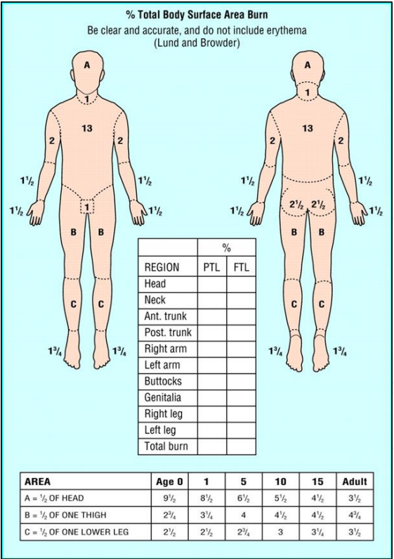
Click on the image to enlarge.
Another useful tool to use when calculating %TBSA burnt is to use the surface area of the patient’s own flat palm with the fingers together as a template. This will represent 1% of the patients TBSA and can be used to measure smaller burns. It is also useful to use the measure islands of unburnt skin within a larger burnt area such as the back or thigh. This can then be subtracted from the surface area calculated using the charts above.
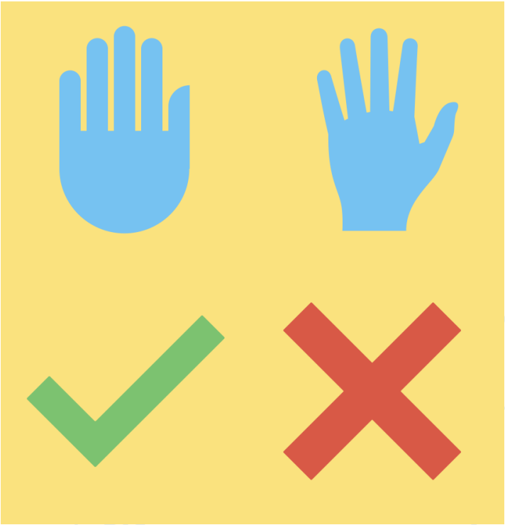
Click on the image to enlarge.
Note that the fingers are included in the surface area of this measurement and that they should be arranged flat, and next to each other – not spaced out.
Initial management
First Aid
Get the patient to safety and the stop ongoing burning by running the area under cool water for 20 minutes. Once the burns have been cooled, cover them with cling film/gladwrap and keep the patient warm.
Commercial products, such as Burnshield®, can also be used if available. Please note that Burnshield® is a first aid measure to cool the wound and is not intended to be left on the wound as a dressing.
Resuscitation
Standard ATLS approach to trauma resuscitation is relevant here as always.
Special consideration of the needs of burns patients will be discussed.
Airway:
Strongly consider inhalational injury in patients with facial burns, soot in the mouth and nose, singed eyebrows or nasal hairs. Inhalational injuries will lead to increased facial swelling and a threatened airway. Early intubation is indicated prior to this swelling. Signs of stridor and difficulty breathing are evident once the airway is already threatened – aim to intubate before these are evident.
Breathing:
Inhalational injury will cause injury to the lungs in 2 ways:
- Thermal injury which predominantly affects the upper airway (requires early intubation)
- Inhalation of the products of combustion which cause chemical pneumonitis and systemic toxicity (requires ventilation support)
Management is predominantly supportive:
- Providing supplemental oxygen
- Facilitating adequate ventilation through intubation and ventilation as needed
- Avoiding tight, circumferential dressings
- Escharotomies of full thickness burns.
All patients with a risk of CO poisoning (from being in an enclosed space) should be on 100% O2.
Circulation:
All large burns (>10% in children) require 2 large bore access points (Preferably into healthy skin) Early use of intraosseous access is appropriate if needed.
Patients presenting in shock should receive fluid resuscitation bolus of 10-20ml/kg with continual reassessment. This should be done independently of the Parkland’s Fluid resuscitation.
Patients should then be started on continuous fluid as per the Modified Parkland’s formula:
𝑤𝑒𝑖𝑔ℎ𝑡 (𝑘𝑔) 𝑥 𝑝𝑒𝑟𝑐𝑒𝑛𝑡𝑎𝑔𝑒 𝑇𝐵𝑆𝐴 𝑏𝑢𝑟𝑛 𝑥 3𝑚𝑙
This is given ivi over 24 hrs and should be on top of standard maintenance fluids which can be given ivi or orally depending on the size of the burn (see below).
(In flame burns the Parklands formula fluid is divided with ½ being administered in the first 8 hours post burn and the second half going in over the next 16 hrs. )
Disability:
Consider the mechanism of the burn and exclude any possible head injury, hypoxia, toxicity (CO poisoning and cyanide) and hypoglycaemia.
Analgesia:
Ensure the patient receives adequate analgesia. Ketamine is recommended for the initial scrub and dressing due to it’s analgesic and sedative properties and safety profile.
Washing:
This is essential management to enable wound assessment, reduce infection and remove debris. This is the most important part of the assessment of the burn and should be done as soon as possible and prior to estimating the final %TBSA burn.
If possible, this can be done under sedation. Use soap and warm water to do the washing.
Photos of the wounds should be taken after washing and a careful assessment of the TBSA should be done after fully exposing the wounds.
The following picture illustrates the importance of washing prior to formally assessing the percentage TBSA burnt – note the significant difference in the apparent size of the burn:
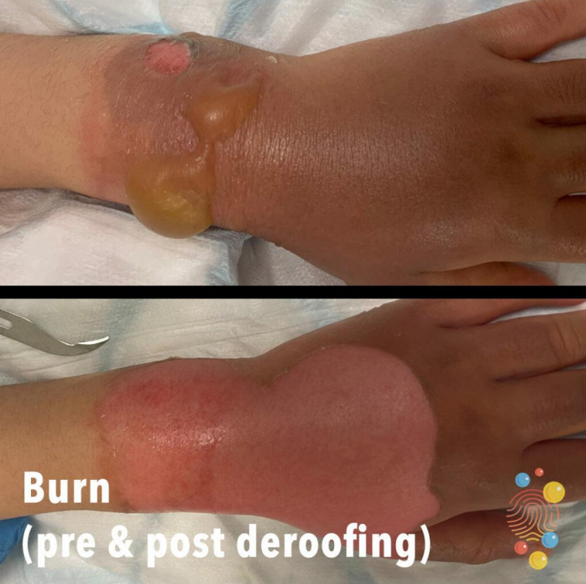
Click on the image to enlarge.
A note on escharotomies:
Escharotomies are a life saving procedure. They should be done on the torso or limbs when circumferential full thickness burns are observed. This is because the thickened, stiff skin causes compression of the underlying tissues which inhibits ventilation in the case of chest burns, and inhibits distal limb perfusion in circumferential limb burns.
Doing an escharotomy can be an intimidating procedure if you have not seen one before but waiting for an experienced clinician may cost the patient their limb or life. Reference charts are available, showing you where to make your escharotomy incision for each body part – do not hesitate to look it up if you are unsure.
Although this procedure is often discussed alongside the procedure of a fasciotomy, please be reassured that they are quite different procedures and the escharotomy is far simpler, easier to do and less likely to cause bleeding or nerve injury.
In an escharotomy the goal is to cut through the burnt skin to break the ring of compression. The skin is already dead so will not have sensation and will not bleed. The incision needs to be deep enough to allow the tissues underneath to expand and does not need to pass beyond the subcutaneous fat. As the incision runs along tissue it will reach areas that are not full thickness: aim to have an incision from the top to the bottom of the burn without cutting into healthy tissue along the way. This does not require anaesthesia and can be done at the bedside.
Incisions should be in the longitudinal pain, deep enough to reach the subcutaneous fat and reach just into the normal skin above and below the burn.
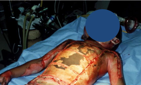
Click on the image to enlarge.
The image above shows a child with chest and arm burns. Note that the incisions run along the chest and upper parts of the arms. The incision are deep enough to have allowed the sheets of burnt skin to separate but there is minimal bleeding coming from the subcutaneous fat.
Discussion criteria
The outcome of the discussion will be influenced by your centre’s capacity for burns care.
ALL referrals need images to allow for assessment. Ideally all images should be taken AFTER the wounds have been washed.
Not all discussions require transfer.
We may ask you to discuss with a centre closer to you for a review if you are from far.
Any burn likely to need surgical involvement needs to be managed at a centre with capacity for paediatric burns care.
Cases that should be discussed
- Burns ≥ 10% TBSA
- Full thickness burns ≥ 5% TBSA
- Suspicion of inhalational injury
- Electrical burns
- Chemical burns
- Special area burns (face, hands, feet, perineum, over major joints)
- Septic burns
- Circumferential full thickness burns (limbs, neck or chest)
- Neonates or infants
- Polytrauma
- All cases where non-accidental injury is suspected
Feedback will be one of the following
- Acute transfer
- Transfer to OPD
- Wound care at your facility
- Discuss with a larger centre closer to you
Ongoing management
| Intervention | 10-14% TBSA | 15-19% TBSA | 20-29% TBSA | 30+% TBSA |
| 2 large bore ivi line | Yes | Yes | Yes | Yes |
| Bloods (FBC, U&E, CMP, albumin) | Yes | Yes | Yes | Yes |
| NGT | Yes | Yes | ||
| NJT | Consider | Yes | ||
| Urinary catheter and monitoring | Yes | Yes | Yes | |
| Nasal prong O2 | Yes | Yes | Consider Intubation | |
| ICU admission | Neonates >20% | Yes | ||
| Supplemental vitamins | Yes | Yes | Yes | |
| Tetanus | Yes | Yes | Yes | Yes |
| 6 hrly HGT | Yes | Yes | Yes |
Children's Surgery Network, Western Cape Government, Red Cross War Memorial Children’s Hospital (RCWMCH), Clinical Guideline_Version 1_2025
Burns