Burns (Adult) - Approach
Developed by Prof H Rhode, Dr Albie van der Merwe, Dr Stephan Fourie & Prof A Millar & released as Circular H62 of 2011 (& updated 2016 by Dr S Cox, Dr R Martinez & Prof H Rhode)
Developed by Prof H Rhode, Dr Albie van der Merwe, Dr Stephan Fourie & Prof A Millar & released as Circular H62 of 2011
(& updated 2016 by Dr S Cox, Dr R Martinez & Prof H Rhode)
Assess airway/breathing
Assess the neurological state of the patient (AVPU)
Circulation
Remove any sources of heat or chemicals
Estimate the percentage of total body surface area
Fluid resuscitation requirements
Assess urine output (this is the best guide to resuscitation)
Insert a nasogastric tube
Decompression incisions (Escharotomy)
Medications
Local wound care
Assess airway/breathing
- Careful airway assessment must be done where there are flame or scald burns of the face and neck or the circumstances indicate a ‘closed space’ mechanism of injury.
- Secure the C –spine with an inline stabilising collar, when the mechanism of injury could indicate additional trauma (MVA, fall from high tension wire)
- Look for signs of inhalational burn- history of hot gas, smoke, steam. Indications for airway assessment include
- Carbonaceous sputum
- Burns to nose, mouth, pharynx
- presence of pharyngeal burns
- air hunger
- stridor
- Change of voice/ hoarseness
- Tracheal tug
- Singed nasal hair
- Restlessness
- Hypoxia
- Chin lift/jaw thrust to open airway.
- Adrenaline nebulisation with 3ml adrenaline could relief symptoms, but should not delay intubation in severe cases.
- INTUBATE if significant airway obstruction present or WORSENING symptoms. Intubation is necessary in the case of unconscious patients, hypoxic patients with severe smoke inhalation, or patients with flame or flash burns involving the face and neck if there is evidence of compromised airway patency. Oedema of the upper airways often develops within hours after resuscitation. It may however take 24 hours in some cases for the swelling of the airway to manifest, so close monitoring is essential during the first 24-48 hours
- Keep in mind that the emergency room evaluation may show normal X-ray of the chest and blood gasses.
- All patients with major burns must receive high-flow oxygen for 24 hours.
- Patients with carbon monoxide poisoning may have the following symptoms:
- 15-20% - headache, confusion, restlessness
- 20-40% - nausea, disorientation, irritability
- 40-60% - hallucination, ataxia, syncope, convulsions, coma
- >60% - death
- Carboxyhaemoglobin half- life is 250 minutes. On 100% oxygen the half-life is 40 minutes.
- Administer humidified oxygen at 8 litres per minute via non- rebreathing mask. Ventilation if intubated.
- Useful laboratory test: Blood gases including carboxyhaemoglobin level.
- If breathing seems to be compromised because of tight circumferential trunk burns, consult with burn centre surgeons immediately. Urgent escharotomies may be required to facilitate chest expansion
Asses the neurological state of the patient (AVPU)
- Alert
- React to Voice
- React to Pain from stimulus
- Unresponsive
Take appropriate action based on the patient’s neurological state. Consider trauma, hypoxia & carbon monoxide poisoning as possible causes.
Circulation
- Stop any external bleeding.
- Identify potential sources of internal bleeding.
- Establish large-bore intravenous (IV) lines and provide resuscitation bolus fluid as required in all compromised patients, using standard ATLS® protocols. Perfusion of potentially viable burn wounds is critical. Consider size of burn and fluid requirements when determining bore size. Often the extremities are burnt and then a CVP line needs to be placed.
Reminder: IV lines may be placed through the burned area if necessary (suture to secure). Avoid the saphenous vein if at all possible, and avoid cut-downs through unburned skin if possible.
Attach cardiac monitor for all electrical burns for the first 24 hours, to identify life-threatening dysrhythmias.
Remove any sources of heat or chemicals
- Remove any clothing that may be burned, covered with chemicals or that is constricting.
- Melted plastic and tar can be removed with the topical application of liquid paraffin solution
- Cool any burns less than 3 hours old with cold tap water for at least 30 minutes and then dry the patient. Ice deepens the burn.
- Cover the patient with a clean dry sheet or blanket to prevent hypothermia.
- Use of melaleuca alternifolia (Burnshield®) is a very effective means of cooling and dressing the injury for the first 12 hours.
- Rings and constricting garments must be removed.
ESTIMATE THE PERCENTAGE TOTAL BODY SURFACE AREA
Initially, use the Rule of Nines. In the case of all paediatric patients and for a more accurate assessment, use the Berkow diagram or the patient’s open hand representing 1% of TBSA.
Reminder: Accurate estimation of burn size is critical to ongoing fluid replacement and management.
Shave the hair to inspect burn % in hairline or your estimation will be wrong.
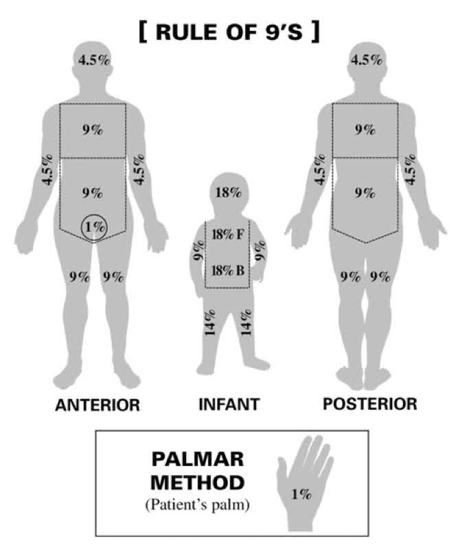
(Palmar method only for small burns)
FLUID RESUSCITATION REQUIREMENTS
- If resuscitation is delayed for more than 2 hours or if the patient presents with hypovolaemic shock, start with immediate fluid boluses according to ATLS principles
- Patients with < 10% TBSA burns can be resuscitated orally (unless the patient has an electrical injury or associated trauma). This needs ongoing evaluation and the patient may still require an IV line.
- For patients with burns 10 - 40% TBSA, secure a large-bore IV line; add a second line if transportation will take longer than 45 minutes.
- Burns > 40% TBSA require 2 large-bore IV lines.
- If the transfer will take less than 30 minutes from the time of call attempt a line, but don’t delay transfer for an IV line.
- Initiate fluids for ongoing resuscitation and fluid losses using the modified Parkland formula: (3 ml Ringer’s lactate) x (kg of body weight) x (% burn) = ml in first 24 hours.
- Example: In the case of a patient weighing 70 kg with a 50% TBSA burn: (4 x 70 x 50) = 14000 ml needed in the first 24 hours.
- Inhalation injury: Add 2ml/kg/% burn to this regime.
- Associated trauma with blood loss, consider blood at 10 ml/kg.
- Electrical burns may require more fluid than expected because of non-visualised deep seated injury, and may need diuresis
- Daily maintenance requirements are in excess of volumes calculated above
Assess urine output (this is the best guide to resuscitation)
- Insert a Foley catheter in patients with burns >15% TBSA. Adequate urine output is 0.5 to 1ml/kg/h in adults
- Reminder: Lasix and other diuretics are never given to improve urine output; fluid rates are adjusted to increase urine output.
- Observe urine for burgundy colour (myoglobinuria) seen with massive injuries or electrical burns. There is a high incidence of renal failure associated with these injuries, requiring prompt and aggressive intervention. Increase urine output to 2 ml/kg/hr. Sodium Bicarbonate 25meq/litre in resuscitation fluid to alkalinise the urine. Mannitol 12,5gram/litre fluid
- Reminder: If the urine is red or brown consult a burn centre.
- REASSESS. RE-EVALUATE frequently. Check pulse rate, capillary refill (<3secs), urine output (0.5-1ml/kg/hr), alertness. Lowered BP indicates DECOMPENSATED shock and all measures should be taken to avoid reaching this point.
- Fluid bolus for inadequate perfusion as evidenced by low urine output or tachycardia: WARM 20ml/kg Ringers lactate.
- Reduce fluids if urine output is more than 2ml/kg/hr, to avoid fluid creep.
- In the event of pulmonary oedema, secondary to fluid overload, treat with furosemide 0,5mg/kg/intravenous dose. Consider inotropes if myocardial failure.
- Beware of hyponatraemia with major fluid shifts.
Insert a nasogastric tube
Insert a nasogastric tube in any patient with burns > 30% TBSA, or any patient who is unresponsive, shocked or with burns > 20% if preparing for air or long-distance transportation.
Decompression incisions (Escharotomy)
Assess for circumferential full-thickness burns of extremities or trunk. Elevate burned extremities on pillows above level of the heart to reduce the development of oedema. If transfer will be delayed, discuss with a burn surgeon indications and methods for decompression incisions (escharotomies). If an escharotomy is required, do not delay the procedure.
Medications
Give tetanus toxoid immunisation.
After fluid resuscitation has been started, pain medication must be titrated or given in small intravenous doses (not intramuscular). As an initial dose of 0.1 mg per kilogram body weight of morphine can be provided. Blood pressure, pulse, respiratory rate & state of consciousness should be assessed after each increment of IV morphine & then continuously assessed.
Local wound care
- The burn wound is carefully assessed for extent and depth and then treated topically with an antiseptic/antibacterial agent as a prophylactic or therapeutic measure against burn wound colonisation and burn wound infection.
- The initial burn wounds can be washed with soap and water or 1% Chlorhexidine or can be debrided under anaesthesia where facilities exist The burn wound is washed with water and soap or with chlorhexidine solution and then covered with the selected topical agent.
- Grease & road contamination can be removed by the application of petroleum jelly. A closed burn wound technique is used and the topical agents chosen according to burn unit protocol.
- Frequently used topical agents include silver sulphudiazine (changed daily), providing iodine (changed daily), Mupirocin with or without Chlorhexidine (changed daily) or a nano-crystalline dressing changed every 3 days.