C-spine - Clinical exam and Injury Patterns
C-spine - Clinical exam and injury patterns
Physical exam
Essential of the neuro examination
Classification of C-spine injuries
Flexion injuries
Extension injuries
Axial load injuries
Rotational injuries
Physical Exam
After the primary ABCD’s has been stabilised the physician can then focus on the secondary survey.
Important steps should be performed and documented :
- An assessment of the patient’s mental status is critical, as this will influence the decision to obtain radiographs. Many minimally brain-injured patients (GCS 14) appear to have a normal mental status on superficial examination. When there is any doubt about the patient’s mental status, the physician should have a low threshold to image the cervical spine.
- The physician must inspect and palpate the entire spine for evidence of ecchymosis, deformity, or focal tenderness. Determine whether the patient has focal vertebral tenderness as opposed to para-vertebral muscle tenderness.
- Obtain radiographs if the patient has point tenderness over the bone, or if this distinction cannot be made.
- Finally, the physician will have to decide whether a distracting painful injury is present. As is above this remains an contentious issue.
Essentials Of The Neuro Examination
- A detailed and precise neurological examination is critical and should be documented I suggest using an injury data form.
Classification of C-spine injuries
The cervical spine can be anatomically divided into anterior and posterior columns. The posterior longitudinal ligament acts as a fulcrum between the two.
Cervical spine injuries are described according to the mechanism of injury and resultant radiographic alterations of these columns. So pay close attention to the handover from EMS staff.
- Flexion
- Extension
- Axial load
- Rotational mechanisms
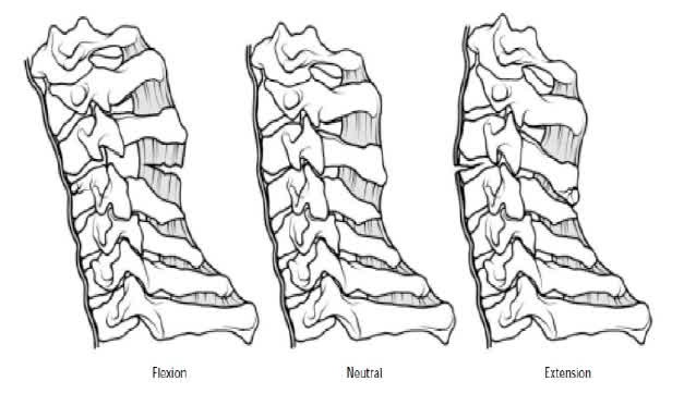
Each predispose patients to particular fracture patterns.
Look for injury patterns!
Flexion Injuries
Flexion injuries cause compression of the anterior column and distraction of the posterior column.
This results in varying degrees of crush to the anterior aspect of the vertebral body, and ligamentous disruption and widening of the posterior column (“crushed in the front and wide in the back”).
- Simple wedge fractures,
- flexion teardrop fractures,
- unilateral facet dislocations,
- and bilateral facet dislocations
represent a progression of severity (and instability) of flexion injuries.
Extension Injuries
Extension injuries cause distraction of the anterior column and compression of the posterior column. There is variable disruption and widening of the anterior ligaments and varying degrees of crush of the posterior elements (“wide in the front and crushed in the back”).
Extension injuries include the hangman’s fracture and extension teardrop fractures.
Axial Load Injuries
Axial load injuries cause compression of both the anterior and posterior columns.
These are the result of forces from above (skull) or below (pelvis) that are applied to the vertebral column in neutral position at the time of impact.
Adjacent vertebral bodies are forced against each other with overwhelming force. The resultant vectors cause the vertebral body to shatter outward, resulting in a “burst fracture.”
A burst fracture is usually stable because the ligamentous structures of the vertebral column remain intact.
One unstable burst fracture involves C1 (Jefferson fracture).
- It occurs when vertical forces are applied to the skull and transmitted through the occipital condyles to the lateral masses of the atlas.
- This results in disruption of the anterior and posterior arches of C1.
- On the lateral view, look for a widened pre-dental space (between the dens and the anterior arch of C1) and soft-tissue swelling anterior to C2.
- On the odontoid view, the lateral masses of C1 extend beyond, and overhang, the lateral masses of C2.
This extremely unstable injury requires immediate orthopaedic / neurosurgical consultation.
Rotational Injuries
Rotational injuries cause varying degrees of rotation of both the anterior and posterior columns, usually around a facet dislocation. This occurs when one of the facets acts as a fulcrum, and rotation plus flexion allows the contralateral facet to dislocate.
The superior facet jumps above and anterior to the inferior facet. Thus the superior facet comes to rest in the intervertebral foramen, resulting in a “locked in” position and a stable injury (even though the posterior ligaments are somewhat disrupted).
- The AP radiograph will reveal the spinous process above the injury angled away from the midline in the direction of the rotational injury.
- On the lateral projection, a subluxation of the vertebra above the facet dislocation will be present.
- In addition, the jumped facet will be blurred compared to the sharp line that defines the other facets.