C-Spine - Evaluation of Plain Films/Imaging
C-Spine - Evaluation of plain films/imaging
Lateral view
Anteroposterior view
Open-mouth odontoid view
A few key points on interpreting C-spine plain films
PEARLS
How many C-spine views? Lateral good enough – standard 3 views or 5 views.
In a resource limited setting strong recommendation is to get the following 3 views :
A. Lateral view—is the film adequate?
- Alignment: anterior, middle and posterior arcs
- Bones: vertebrae and spinous processes uniformity and height
- Cartilage: intervertebral disk space height and length
- Soft tissue: pre-vertebral soft tissue width
B. Anteroposterior view
- Alignment of spinous processes
- Distance between spinous processes
- Uniformity and height of vertebrae
C. Open-Mouth Odontoid View
- Spacing of dens and lateral masses
- Lateral alignment of C1 and C2
- Uniformity of bones
The lateral view alone to clear a c-spine prior to intubation has two distinct pitfalls
- it may delay definitive airway management
- it does not exclude cervical injury, and thus may provide the operator with a false sense of security
- there is no evidence to support an increased risk of neurologic injury during endotracheal intubation, provided effective immobilisation is maintained at all times.
A few key points on interpreting C-spine Plain Films
Make a point to examine the patient if you are interpreting the plain film it helps to direct the eye to areas you suspect might have a fracture.
Lateral View
Because approximately 70% of detectable abnormalities will be visible on the lateral view, this radiograph should be inspected first.
An adequate lateral film must demonstrate all seven cervical vertebrae as well as the top of the first thoracic vertebral. To obtain an adequate view of the lower cervical spine a swimmers view can be requested.
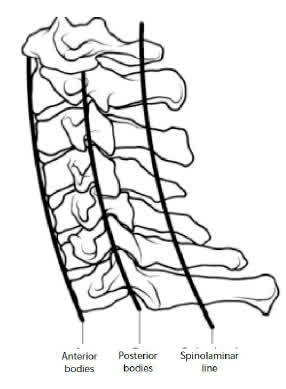
If the film is adequate, use the mnemonic “ABCS” to examine the film:
- Alignment : Three arcs should be easily traced on the lateral radiograph. The first is composed of the anterior margins of the vertebral bodies. The second is defined by the posterior margins of the vertebral bodies. The third is along the bases of the spinous processes (the spino-laminar line). These arcs should be traced as smooth, unbroken lines. If there is a step-off in any of these arcs, suspect a fracture or ligamentous disruption. The only exception to this rule is that occasionally the line along the bases of the spinous processes appears to have a posterior step-off at the C2 level.
- Bones: Inspect all of the vertebral bodies, which should have a uniform square or rectangular shape from C2 and below. Any small fragment may represent a fracture. Examine all laminae and spinous processes carefully for uniformity and smooth edges. Any loss of height or wedging either anteriorly or posteriorly may be a clue to a compression fracture. The anterior height should be no less than 3 mm shorter than the posterior height. Carefully examine C2, especially the odontoid, which articulates with C1. The odontoid should form a smooth arch just behind the anterior portion of C1 and should be closely applied to the posterior portion of C1. This space between the anterior dens and the anterior ring of C1 is the predental space and should be 3 mm or less in adults and 5 mm in children. There are a number of radiographic signs distinct to C2. Oblique fractures of the second cervical vertebra below the odontoid may cause the body of C2 to appear enlarged or “fat” compared to C3 (known, not surprisingly, as the “fat” C2 sign) There is also a radiographic “ring” on C2 best seen on the cross-table lateral view. If this ring is broken, suspect a type III odontoid fracture.
- Cartilage : The next step is to examine the cartilaginous structures. The intervertebral disc spaces should be uniform in height and length. Narrowing of a disc space may be a clue to acute disc herniation or an adjacent vertebra fracture. Widening of a disk space may suggest rupture of the annulus fibrosis or longitudinal ligamentous structures. Give careful attention to the spaces between the spinous processes. Any widening or “fanning” of these spaces could represent significant disruption to the posterior ligamentous complex.
- Soft Tissues: Finally, examine the soft-tissue spaces of the lateral radiograph. Abnormal swelling of the prevertebral soft tissue suggests a vertebral fracture. The soft tissue immediately anterior to C1-C4 should be 7 mm or less and, for C5-T1, 22 mm or less. Any widening of these tissue planes should prompt careful scrutiny for subtle fracture and may suggest the need for consultation or CT scan of the involved area.
http://www.trauma.org/archive/spine/lateral-cspine.html is an excellent reference.
Anteroposterior View
An AP radiograph is necessary to evaluate the cervical spine. This view should include C3 to T1 due to the fact that the mandible obscures C1 and C2.
The alignment of the vertebrae and spinous processes is crucial to interpreting the film :
- The spinous processes should form a straight line down the middle of the vertebral body.
- If one spinous process appears to be out of line compared to the rest consider a facet joint injury.
- The distance between the spinous processes should be roughly equal; none of the spaces should be 50% wider than the one immediately above or below.
- Abnormalities in alignment or spacing could be an indication of unifacet dislocation or fracture of the lateral articulating surface.
These findings should prompt you to carefully scrutinise the lateral view and perhaps order oblique films. CT scanning may be helpful when an abnormality is seen.
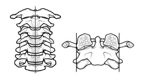
Odontoid View
This view identifies fractures involving C1 and the odontoid process of C2. This radiograph can be difficult to interpret due to the overlapping shadows and densities from the occiput and central incisors.
A few tips when scrutinising this view :
- The dens should be parallel with the lateral masses of C1.
- The space on each side of the dens (between the dens and lateral masses) should be equal. If this space is asymmetric, it may indicate a fracture.
- Also inspect the lateral alignment of C1 and C2 if alignment abnormal suspect a fracture.
PEARLS
- Know your anatomy, working in a busy emergency centre there is no excuse to miss a potential fracture with cursory glance at a plain film
- Never settle for inadequate films
- Never ignore subtle findings
- Always be suspicious in the patient with persistent significant neck pain and “normal” plain films.
A pertinent question in a setting where CT is not available at most of the facilities dealing with trauma patients on a daily basis one approach is for transferring a patient to a trauma centre is:
- A suspected fracture on plain film to get adequate images of the bony anatomy and identify the level of injury.
- High clinical suspicion where inadequate images are obtained of the upper and lower C-spine. the upper or lower cervical spine cannot be adequately visualised
- Decrease GCS in trauma patients
- Patients with persistent pain and/or neurologic deficit despite normal plain films. (Make sure a senior clinician / radiologist has checked the films).