Chest Trauma - Penetrating
Penetrating Chest Trauma
Assessment
Management
Disposition
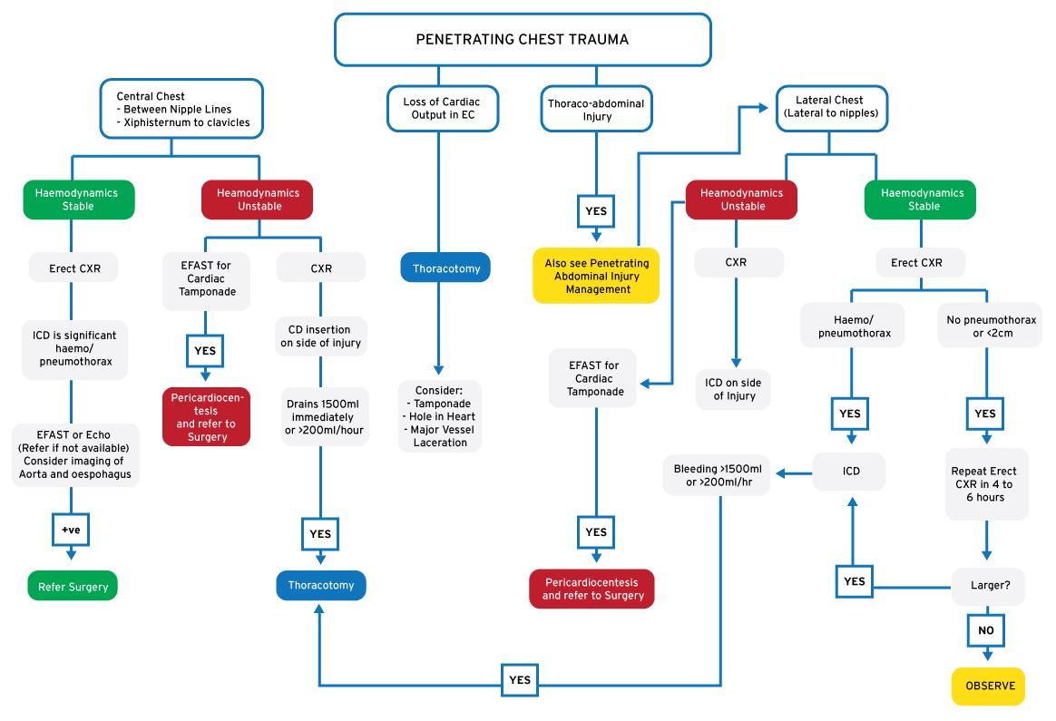 Assessment
- Cardiac Injury
- Vascular Injury
- Lung Injury
- Diaphragmatic Rupture
Early Surgical Referral
Penetrating injury to the chest with any of the following:
- Haemodynamic Instability
- Vascular Injury
- Peritoneal Signs
- Immediate drainage of > 1500 ml blood in chest drain
- > 200ml blood per hour in chest drain
Chest injuries frequently accompany multiple other injuries: ALWAYS look for co-existing injuries
Use a standard ATLS approach
Assess – Intervene – Reassess: Assessment and Management occur in tandem
The goal is to determine whether there is significant intra-abdominal trauma
DMIST Handover from Ambulance
- Triage patient using SATS
- Measure and Record Vital Signs
- Fingerprick Haemoglobin
Examination
Primary: Assess ABC’s then secondary Head to Toe examination
Specifically for Penetrating Chest Trauma
- Check haemodynamic status and Hb
- Look for potential sites of blood loss (front and back)
- Screen for external bleeding (Chest; Neck; Abdomen)
- Screen for internal bleeding (Chest; Abdomen; Neck)
- Chest Examination: Chest movement; Discolouration; Tenderness; Air Entry; Trachea; Surgical Emphysema
- Assess wound tract carefully but do NOT do a blind finger exploration
Be aware of tracts extending to the neck and abdomen.
Take a SAMPLE History
- Signs and Symptoms: Shortness of Breath; Haemoptysis
- Allergies
- Medications
- Past Medical history
- Last Meal
- Events: Mechanism and Type of Injury
Investigations
- Hb and Glucose
- Oxygen Saturation
- Bloods: Formal Hb, XMatch, Arterial Blood Gas may be indicated
- Chest X-Ray (erect is best, lateral decubitus for suspected small haemothorax)
- Other X-Rays as Indicated
Point of Care EFAST scan
- Look for intra-abdominal, pleural and pericardial fluid
- Look for pneumothorax
- If stable, consider CT scan of chest
Management
Stabilise the ABC’s
Airway:
- Injury extending to neck may cause pressure on airway
- Tension pneumothorax may compromise trachea
Breathing:
- Treat haemo/pneumothorax
- Decompress and treat tension pneumothorax
- Cover sucking chest wounds
Circulation:
- Insert at least one large bore (14G preferable) on the opposite side of the injury
- Thoracotomy indicated in the event of massive haemo-thorax (emergency) or no cardiac output (emergency center)
- Consider cardiac injury in the absence of significant visible bleeding
Monitor the Patient:
- Attach monitors (ECG, SpO2, BP)
- Urinary catheter
- Nasogastric tube (not with BOS fracture)
IV Analgesia:
Morphine Sulphate
Constitute 10mg in 10 ml of sterile water (10 ml total)
Loading dose: 0.1 to 0.15 mg/kg (use less for elderly and frail) then bolus 0.05 mg/kg every
15 minutes. Titrate to effect.
Beware drop in BP and respiration.
Disposition
Discharge:
- Wound judged to be non-penetrating and
- Normal vital signs after 4 hours and
- Repeat CXR normal after 4 hours
- No admission criteria
Discharge all patients with a Chest Injury Discharge Advice form.
Admit:
Level 2:
- Any penetrating chest injury OR
- Needs admission for other injuries
Provided no criteria for Level 3 hospital admission
Level 3:
(See Direct Trauma Referral Criteria)
- Poly-trauma
- Pregnant
- Haemophilia
- Injuries requiring advanced imaging (CT scan/Fluoroscopy/Bronchoscopy)
- Injuries requiring specialist care (e.g cardio-thoracic surgery)