GuidelinesOther guideline
Detection of Papilloedema
Detection of Papilloedema
Key points
Key examination signs
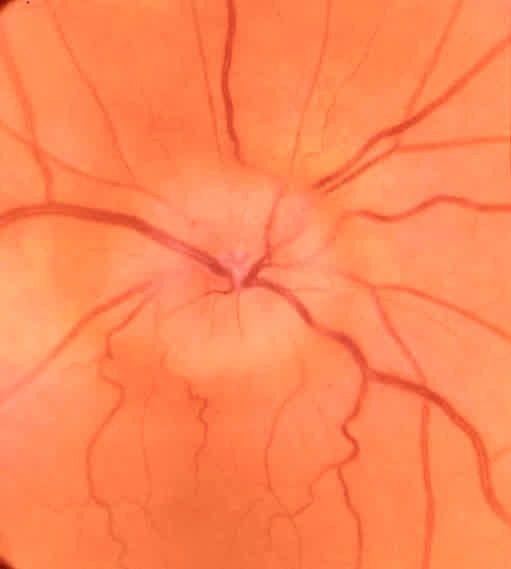
Papilloedema is swelling of the optic nerve head (optic disc) due to raised intracranial pressure. See Figure below.
Key points
- It may take several hours to develop after a sudden elevation of the intracranial pressure and longer with a gradual elevation.
- It is usually bilateral (but can be unilateral)
- Ophthalmoscopic detection is difficult and it is recommended that it is only excluded after a confident examination with a dilated pupil.
- Visual loss is rare and if present usually limited to brief loss of vision lasting a few seconds (visual obscurations).
- The presence of venous pulsations on the discs suggest there is not raised intracranial pressure but this is a difficult sign to see and they are absent in about 10% of the normal population.
Key examination signs
5 mechanical and 5 vascular signs
Mechanical:
- Elevation of the nerve (can look like a champagne cork pushing into the eye)
- Loss of the cup
- The margin of the disc becomes obscured
- Retinal folds
- Hard exudates
Vascular:
- Hyperaemia of the disc
- Engorgement and tortuosity of the veins
- Cotton wool spots
- Haemorrhages around the disc
- Loss of vessels as they cross the margin of the disc