Diagnosis and Management of Asthma
Symptoms and signs of asthma The characteristic symptoms of asthma are wheeze, shortness of breath, tightness of the chest and cough that vary over time and in intensity. Patterns of symptoms that suggest asthma are: variability: day and night, day to day, seasonal precipitation by a range of factors including environmental allergens (house dust mite, grass pollens, animal dander, occ
Symptoms and signs of asthma
The characteristic symptoms of asthma are wheeze, shortness of breath, tightness of the chest and cough that vary over time and in intensity.
Patterns of symptoms that suggest asthma are:
- variability: day and night, day to day, seasonal
- precipitation by a range of factors including environmental allergens (house dust mite, grass pollens, animal dander, occupational exposures), non-specific irritants (smoke, dust and fumes), cold weather and exercise
- symptomatic response to a bronchodilator and an ICS.
An expiratory wheeze is a cardinal sign of asthma but may be absent at the time of consultation. Airway constriction is variable, does not always result in detectable signs, and may not be present at the time of the consultation. A personal history of other atopic disorders such as allergic rhinitis, allergic conjunctivitis and eczema is supportive evidence that the respiratory symptoms are due to asthma. A positive family history of asthma and other atopic disorders are helpful supportive evidence. However, not all people with asthma are atopic, particularly late-onset asthma.
A history of current or past cigarette smoke exposure is important as cigarette smoking is the primary cause of chronic obstructive pulmonary disease (COPD), which needs to be differentiated from asthma. Furthermore, smoking affects asthma control and response to an ICS. Patients with asthma may have features of COPD and vice versa, which may be referred to as asthma-COPD overlap (ACO).[7]
Lung function testing
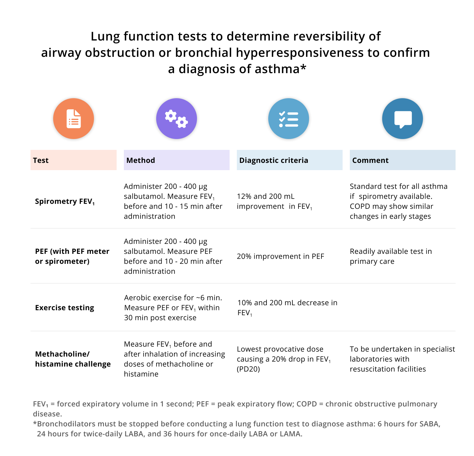
Table 1: Click on image to enlarge
Spirometric lung function tests, including measurement of peak expiratory flow, are key to the diagnosis, assessment of severity, management and monitoring of asthma. Normal lung function does not exclude a diagnosis, especially in well-controlled asthmatics, and particular note should be paid to patients with significant signs and symptoms but normal spirometry.
Reduction in forced expiratory volume in 1 second (FEV1) and peak expiratory flow (PEF) are the hallmark of asthma but may be seen in other diseases. A ratio of FEV1 to forced vital capacity (FVC) below 70% or the lower limit of normal (LLN) is characteristic of obstructive airways disease. Significant bronchodilator responsiveness (reversibility) with normalisation of the airway obstruction is the major physiological characteristic of asthma. The standardised criteria for bronchodilator responsiveness are an increase in FEV1 of >12% and 200 mL, 10 - 15 minutes following the inhalation of 200 - 400 μg of salbutamol, or a 20% improvement in PEF from baseline. The degree of reduction of FEV1 is generally related to severity of the airway obstruction. Asthma improvement is usually mirrored by an improvement in FEV1 and PEF.
Not all asthma patients will exhibit bronchodilator responsiveness at presentation; this could be due to recent bronchodilator treatment and patients should be advised to withhold their SABA for 6 hours, twice-daily LABA for 24 hours, and once-daily LABA for 36 hours prior to testing. The test may need to be repeated at different visits to detect variability in lung function. Alternatively, demonstrating a diurnal variation in PEF may be of value. An average of more than 10% variation over 2 weeks is indicative of asthma. This can also be used to identify environmental (including occupational) causes of asthma symptoms by monitoring PEF 2 - 4 times each day for at least 2 weeks. To measure diurnal variation, PEF is measured first thing in the morning and in the evening. There are several methods of calculating diurnal PEF variability. A useful method is the difference between the maximum and the minimum value for the day, expressed as a percentage of the mean daily PEF value, and averaged over 2 weeks.
Asthma can also be confirmed by demonstrating increased hyperresponsiveness to bronchoconstrictor stimuli. This is usually only of value in subjects with near-normal spirometry. A methacholine/ histamine challenge test should only be undertaken in a specialist lung function laboratory. Alternatively, airway hyperresponsiveness may be demonstrated using an exercise test for exercise-induced bronchoconstriction. A fall of 20% in PEF (or decrease >10% and 200 mL in FEV1) from baseline within 30 minutes after a 6-minute run and having achieved >80% of the predicted heart rate for age, is supportive of a diagnosis of asthma.[8] Allergy testing may be supportive evidence of asthma but is not specific and not all people with asthma are atopic. Measurement of exhaled nitric oxide (FeNO) is not routinely recommended for the diagnosis of asthma in adults.[1] Lung function testing options are summarised in Table 1.
Patients who are on anti-inflammatory treatment (ICS) for asthma may not demonstrate features of asthma with any of the lung function tests. If it is essential to confirm a diagnosis of asthma, it may be necessary to reduce the dose of treatment gradually and repeat the tests after 4 weeks of a dose reduction. SABA for symptom relief should be used in the interim. Use of a SABA must be avoided for at least 6 hours prior to repeat testing.
Laboratory testing
Routine laboratory tests are generally not required to make a diagnosis of asthma. A full blood count with a differential white cell count may demonstrate eosinophilia. Serum immunoglobulin E (IgE) may be raised, and serum allergen testing may be indicative of allergic sensitisation. In severe and difficult-to-control asthma, phenotyping is important, and the above tests should be performed. Measurement of FeNO is a surrogate marker of allergic inflammation but may be raised in many other conditions, and is not recommended in routine practice.[9]
Differential diagnosis
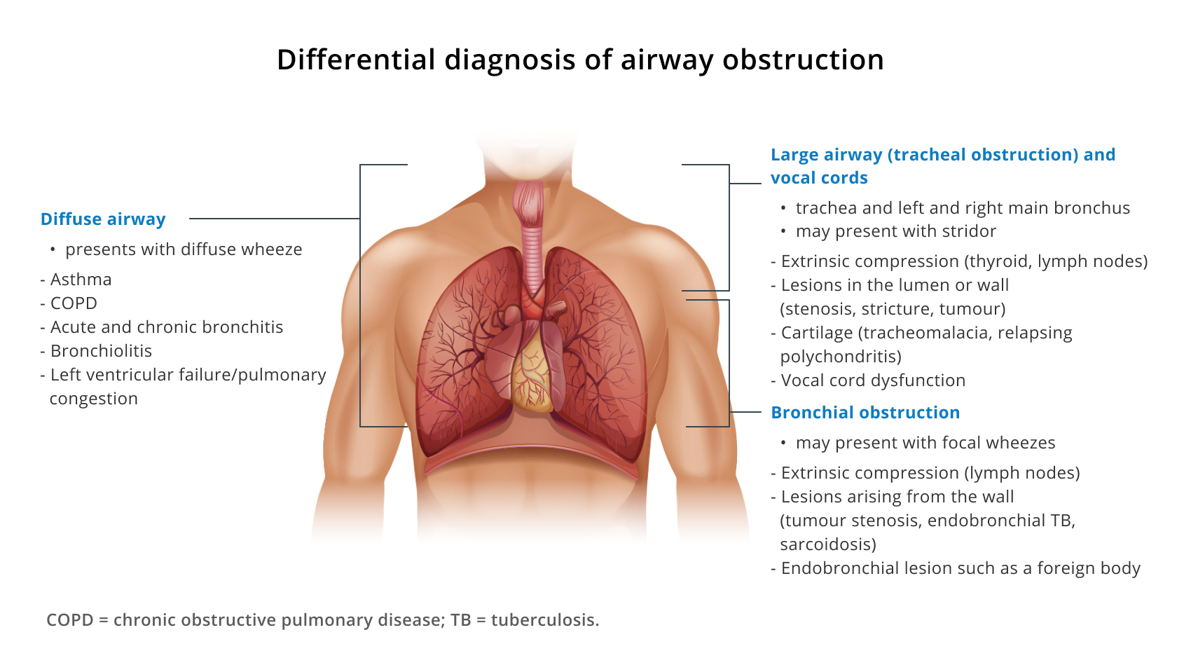
Image 1: Click on image to enlarge
Many conditions may present with symptoms similar to asthma. These must be excluded with a careful history and examination and particularly if the criteria for a diagnosis of asthma are not fulfilled. Image 1 summarises the conditions that frequently mimic asthma and asthma symptoms, using an anatomical approach.
Cough as an isolated symptom is common in clinical practice and is also a common symptom of asthma. Upper respiratory tract infections, usually caused by viruses may cause a cough without wheeze, and usually remits within 6 weeks. Chronic rhinitis and sinusitis with a postnasal drip may also present with a chronic cough and may be the first manifestation of atopy with asthma developing later.
Upper airway obstruction is a serious condition and requires urgent investigation and management. It may present with stridor that is often mistaken for a wheeze unless one auscultates carefully and times the ‘wheeze’ which occurs during inspiration. A localised wheeze must prompt investigations for focal lung disease, including endobronchial obstruction. Diffuse wheezes are present with acute and chronic bronchitis, COPD, left ventricular failure, and bronchiolitis.
It is important to distinguish asthma from COPD because of the different treatment approach and prognosis. Many people with asthma who smoke develop COPD, and patients with COPD may develop asthma. This overlap has received much attention as the coexistence of both asthma and COPD frequently makes the control of symptoms more difficult. There are no formal diagnostic criteria but patients with features of both conditions could be labelled as ACO if for no other reason than to identify the potential challenge in treatment and to default to an asthma-led treatment approach.
Assessment of asthma severity or control
The evaluation of asthma ‘severity’ has recently undergone a paradigm shift with the recognition that so-called ‘mild’ asthma may be associated with significant mortality, even though symptoms are infrequent or ‘mild’.[1] Additionally, in patients who have a diagnosis of asthma and are already on asthma treatment, the diagnosis of severity is based on intensity of treatment that is required to control symptoms (and exacerbations) rather than the frequency of symptoms, which simply reflect ‘control’. For those potentially with truly severe asthma characterised by ongoing symptoms despite at least medium-dose ICS-LABA combination therapy, a specific workup should be conducted to confirm the need for high-dose corticosteroids or biologic therapy. (Further details are provided in the section below on special considerations in asthma management.)
The principles of asthma therapy are to achieve asthma control, which may be defined as: no night-time waking or limitation in daily activities, and symptoms occurring no more than twice a week or requirement for SABA reliever usage less than twice a week.[1] The ACT may also be used to evaluate level of control with a score >20/25 considered to be indicative of well-controlled asthma. [10]
Guidelines for the management of asthma in adults and adolescents, South African Thoracic Society, 2021