Epistaxis
Epistaxis
Basics
Evaluating the patient
Acute management
Step 1: Direct pressure
Step 2: Identify and cauterise the source
Step 3: Nasal Packing
Inserting a posterior pack
Further care
Hypertension and epistaxis
Anticoagulant therapy and epistaxis
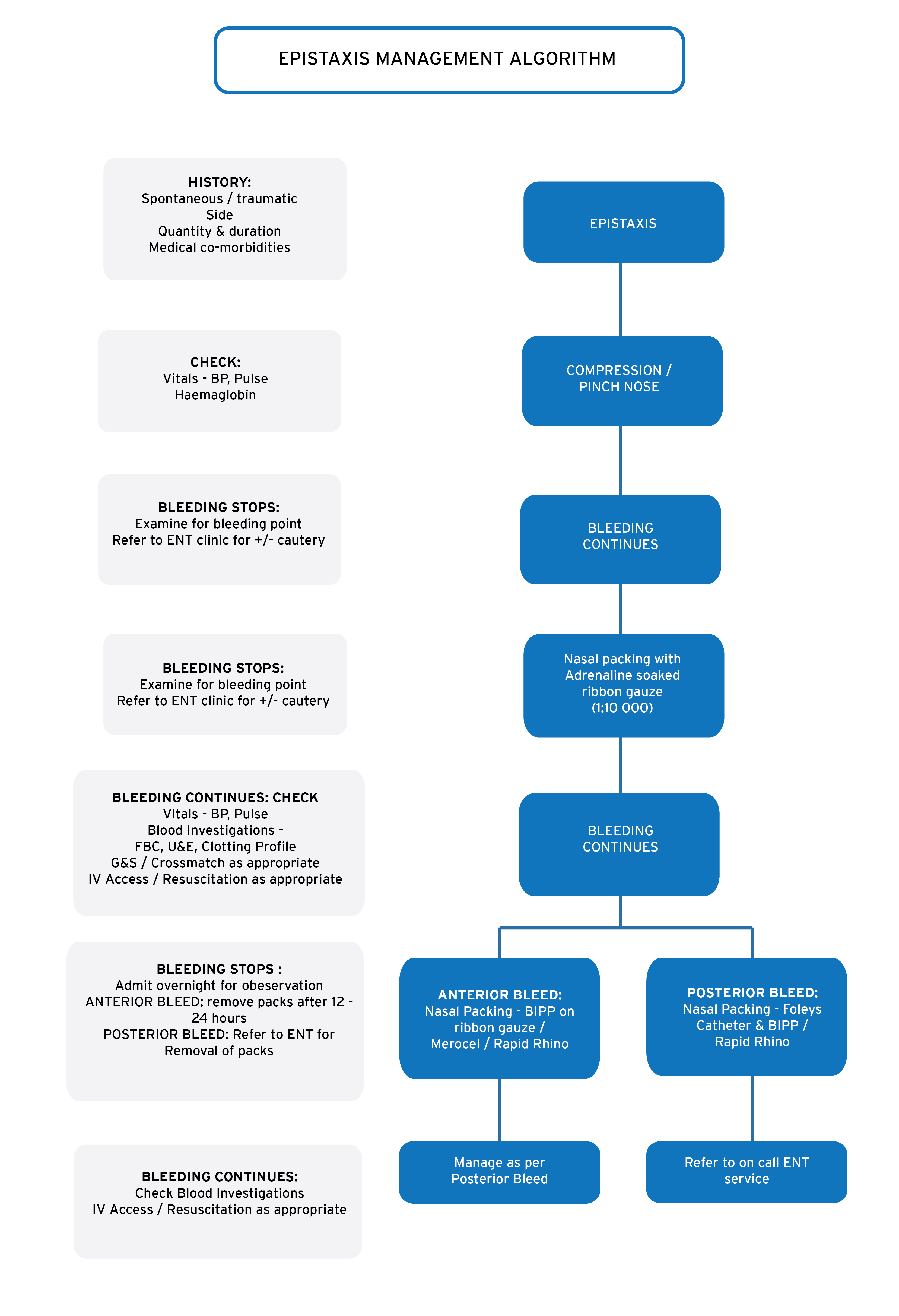
Epistaxis can be defined as bleeding from the nostril, nasal cavity or nasopharynx. In true epistaxis the source of bleeding is the nose or nasopharynx. Bleeding from other sources, like the mouth or upper GI tract may exit the nose, mimicking epistaxis.
Epistaxis is the most common ENT emergency and may range from minor and innocent to severe bleeding with the potential for airway and hemodynamic compromise.
Basics
The nasal mucosa is delicate and highly vascular. The origin of the bleeding is almost always (90%) anterior at Kiesselbach’s plexus.
The most common causes of epistaxis are minor trauma and upper respiratory tract infections.
One should consider the possibility of a secondary cause of epistaxis, such as coagulopathy or a nasal tumour, in all patients. Patients presenting with recurrent or severe nose bleeds should prompt a more complete evaluation.
Common Causes of 2⁰ epistaxis
- Anticoagulant medication
- Other coagulopathies
- Benign and malignant tumours
- Vascular malformations
- Intra-nasal foreign bodies
Evaluating the Patient
The initial survey should focus on three important questions:
- Is there compromise of the airway?
- Has the patient lost enough blood to cause hemodynamic compromise (shock)?
- Has the patient lost enough blood to necessitate a blood transfusion?
If the answers to any of these questions are yes, initial management should include stabilisation of the patient’s condition using a structured approach to life threatening emergencies, such as ACLS® or ATLS®. These patients should all have good vascular access. Send a cross match sample to the blood bank if the bleeding is severe.
Once the patient is stable you should stop the bleeding and then consider the cause.
Acute Management
- Arrest of haemorrhage
- Determination of site and cause
As the patient is being stabilised it is important to stop the on-going bleeding. Most nose bleeds can be managed with simple skills and equipment.
Management should follow a step-wise approach, progressing to the next step once the previous step fails. Make sure to adhere to universal precautions by protecting your hands and face. You and the patient should both gown up.
Step one: Direct pressure
Ask the patient to sit forward to prevent blood from trickling down the throat. The patient should gently blow any clots from the nose, then apply direct finger pressure to the nose in a pincer-like motion. Note that the pressure is applied to the anterior, cartilaginous portion of the nose, not the bony base. You could consider using a commercially available nose clip, or improvise one from a washing pin.
The pressure should be maintained for > 10 minutes before a check is made if the bleeding has been arrested. If not, repeat this up to three times. Consider applying pressure for ≤30 minutes at a time.
Step 2: Identify and Cauterise the Source
Ask the patient to gently blow out any clot. Apply a local aesthetic (i.e. lignocaine) and vasoconstrictor (i.e. oxymetazoline) spray to the nose. Re-apply pressure for a few minutes.
Now carefully examine the nose and identify the cause of bleeding. Cauterise the bleed point with a silver nitrate stick. First wet the tip and then roll over the bleed point for 5 to 10 seconds or until a gray eschar forms.
Step 3: Nasal Packing
If cautery fails to arrest the bleeding, pack the nose. There are three groups of packing solutions:
- Traditional ribbon gauze packing. The gauze should either be soaked in an adrenaline solution or covered with BIPP (bismuth iodine paraffin paste).
- Intra nasal sponges and tampons
- Intra nasal balloon devices
Traditional packing works very well if done correctly. Grasp the gauze ribbon about 6 inches from its end with a bayonet forceps. Place it in the nasal cavity as far back as possible ensuring that the free end protrudes from the nose. On the first pass, the gauze is pressed onto the floor of nasopharynx with closed bayonets.
Next, grasp the ribbon about 4-5 inches from the nasal alae, and re-position the nasal speculum so that the lower blade holds the ribbon against lower border of the nasal alae. Bring a second strip into the nose, and press downward.
Continue this process, layering the gauze from inferior to superior until the naris is completely packed. Both ends of ribbon must protrude from the naris and should be secured with tape. If this measure does not stop the bleeding, consider bilateral nasal packing.
There are many commercial nasal sponges (i.e. Merocel) and balloon devices (i.e. Rapid Rhino) available. Sponge devices are usually lubricated and then inserted into the nose. The sponge expands and tamponades the bleeding. Ballon devices are inserted in a similar fashion. A balloon is then inflated (much like with an ET tube or urinary catheter) that tamponades the bleeding.
If all the above steps fail to control the bleeding you should consider that the bleeding point may be in the posterior nose or nasopharynx. Insert a posterior pack and discuss the patient with an ENT surgeon.
Inserting a Posterior Pack
The simplest way to insert a posterior pack is to use a size 12 or 14 Foleys catheter. Lubricate the catheter and gently insert it into the nose until the tip is entirely in the nasopharynx. Inflate the balloon with 5 to 10ml of saline and pull the catheter back so that the balloon sits snugly against the posterior nasal choana. Now also insert an anterior pack. Secure the catheter in place with an umbilical catheter or instrument clamp.
Further Care
Most patients can be discharged once the bleeding stops. Prescribe a nasal moisturiaer like Vaseline to reduce the chance of re-bleeding. Patients should avoid nose blowing, straining, bending over and sports for a few days. They should sneeze with an open mouth.
Patients with anterior packs can be discharged if stable. They should receive antibiotics like Amoxil and be reviewed in 24 to 48 hours. At that time, gently remove the pack and observe the patient for 1 to 2 hours.
The following patients should be admitted and discussed with an ENT surgeon:
Admit the following:
- Airway compromise
- Hemodynamic compromise
- Patient requires a transfusion
- Bilateral anterior packs
- Bilateral anterior packs
- All posterior packs
- Secondary epistaxis
- Recurrent or severe epistaxis
- Unwell patients
- Patients on anticoagulant therapy
Hypertension and Epistaxis
Up to date no studies have shown a direct correlation between hypertension and epistaxis. No anti-hypertensive has been advocated, and the elevated blood pressures in these patients are usually attributed to the anxiety and invasive methods of stopping the bleeding.
Long standing hypertension and the atherosclerosis are deemed risk factors for epistaxis.
Anticoagulant therapy and Epistaxis
Warfarin should not be reduced, stopped, or reversed without careful consideration of the medical history and the severity of the epistaxis should be taken into account. If the INR is within the therapeutic range and the bleeding is controlled, it may be safe to continue the warfarin.
Proposed protocol for anticoagulants and epistaxis is as follows:
All patients should have a full blood count and platelet count checked, with an INR included if they are on warfarin.
Metal valve patient on warfarin: continue warfarin if INR levels are therapeutic, and withhold if over- anti-coagulated and then recommence once INR levels are therapeutic. Discuss with cardiologist if life-threatening bleed occurs.
Patients on aspirin should have their medication continued. If a life-threatening bleed occurs, then, in cases of coronary stenting, it should be discussed with cardiologist, who may recommend platelet transfusions.
All other patients should have warfarin continued if within therapeutic range and withheld only until back within range if over anti-coagulated.
Patients on aspirin or clopidogrel should have their medication continued, unless a life-threatening bleed occurs, when a platelet transfusion may be considered on the advice of a cardiologist.