Focused Cardiac Ultrasound
Partners In Health Ultrasound Series: Focused Cardiac Ultrasound
Goals
Views
Technique and normal findings
Parasternal long axis view (oblique sagittal plane)
Parasternal short axis view
Sub-xiphoid view
Abnormal findings
Pericardial effusion
Cardiac tamponade
LV systolic dysfunction
LV diastolic dysfunction
RV dysfunction
Pearls and pitfalls
Goals
In the rapid evaluation of the hypotensive, hypoxic or dyspneic patient:
- Identify a pericardial effusion
- Recognise signs of cardiac tamponade
- Evaluate left ventricular systolic and diastolic function
- Assess for right ventricular dysfunction
Probe & Mode: phased array probe, use the “Cardiac” mode or exam type. This will switch the screen dot to the right side of the screen putting the scan into cardiology orientation.
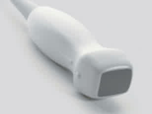
Patient positioning: supine or left lateral decubitus
Views
- Parasternal long axis (PLAX)
- Parasternal short axis (PSAX)
- Sub-xiphoid
Technique and Normal Findings
Parasternal Long Axis View (Oblique Sagittal Plane)
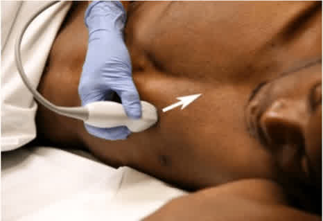
- Place the probe just lateral to the left sternal margin between the 4th and 5th intercostal spaces with the probe marker pointed toward the patient’s right shoulder.
- Adjust the probe (slide up and down a rib space or rotate the transducer slightly) to optimise your image quality.
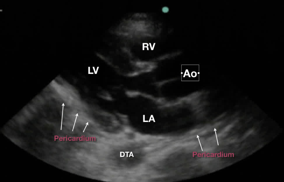
- Normal Anatomy: RV=Right Ventricle seen superficially, close to the probe. LV = Left Ventricle. Ao=Aortic Outflow tract including aortic valve. LA= Left Atrium.
- The fibrous pericardium is the hyperechoic (bright) structure that forms the heart’s border near the bottom of the screen, shown here in arrows.
- The descending thoracic aorta (DTA), seen in cross-section, is the posterior most structure.
- The apex of the heart should not be visible on this view.
- The RV, LVOT and the LA are approximately equivalent in size and the RV is less than 2/3 of the LV’s diameter.
Parasternal Short Axis View
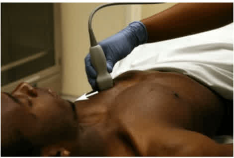
- After obtaining the PLAX view, rotate the probe 90° to the patient’s left shoulder.
- Tilt the probe toward the heart’s apex until the papillary muscles of the LV are visualised just below the mitral valve.
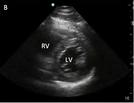
- In this view, the heart should appear as a symmetrically contracting circular LV with a smaller crescent-shaped right ventricle (RV). The inter-ventricular septum should be slightly bowed/curved into the RV.
Sub-xiphoid View
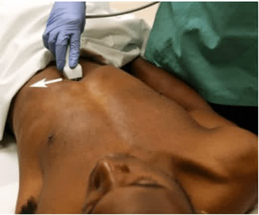
- Place the probe 1-2 cm below the xiphoid process with the probe marker and the transducer face pointing toward the patient’s left shoulder. The probe should lay quite flat, and be pushed up into the patient’s subcostal area with plenty of gel to avoid artifact.
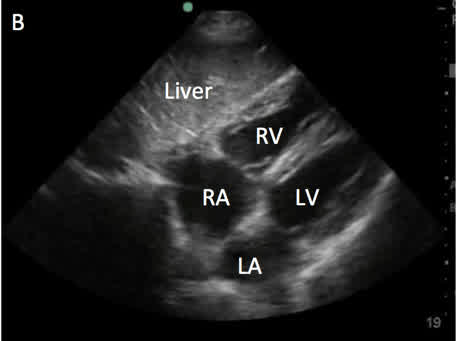
- All four chambers of the heart should be visualised.
- The atria are toward the left of the screen and the ventricles are toward the right.
Abnormal Findings
Pericardial Effusion
- A pericardial effusion can be identified on all views as a hypoechoic (dark) band between the bright pericardium and the grey myocardium.
- It is most easily visualised on the PLAX view behind the posterior LV wall at the bottom of the screen.
- The DTA is a useful landmark on the PLAX view to differentiate between a pericardial and pleural effusion. If the effusion is above the DTA, it is pericardial. If it is below the DTA, it is a pleural effusion.
- Pericardial effusions should be described in terms of whether they are circumferential and how large (centimetre measure between myocardium and pericardium).
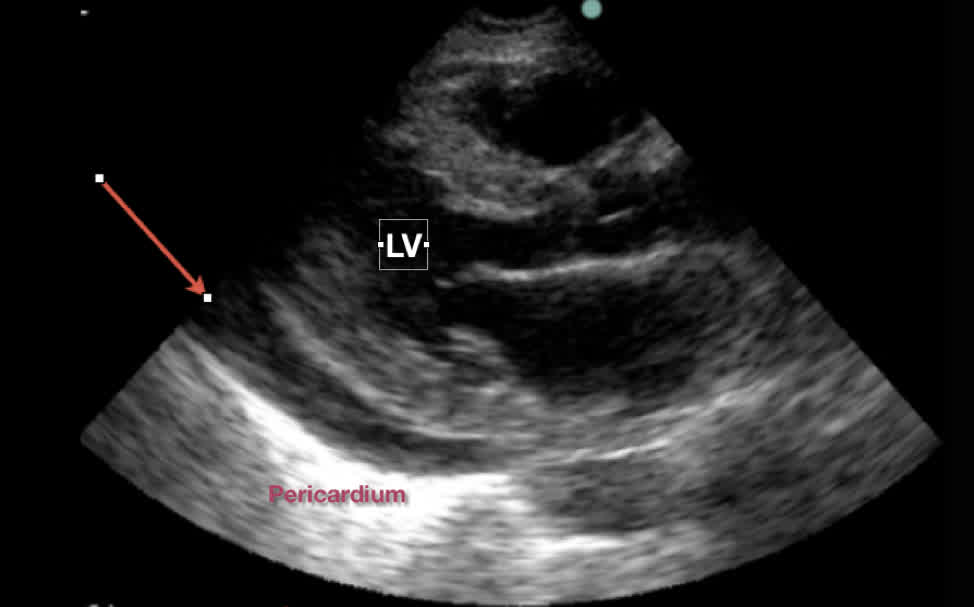
- A pericardial effusion on the PLAX view – the hypoechoic band (arrow) is above the DTA.
Cardiac Tamponade
- Cardiac tamponade is the result of a pericardial effusion that compresses the ventricular walls, prevents normal filling and results in hemodynamic collapse.
- When pressure in the pericardial space exceeds ventricular pressures, the RV with its compliant wall is the first to show signs of impaired filling during diastole.
- To assess for RV diastolic collapse (early sign of cardiac tamponade), obtain a PLAX view and observe the RV free wall as the heart beats
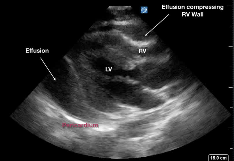
- If the RV free wall caves in or scallops (arrow) during the time when the mitral valve is open (diastole), this suggests cardiac tamponade.
LV Systolic Dysfunction
- Two methods to assess LV systolic function are the visual estimation of gross LV function by clinician impression and the EPSS technique.
- Normal LV function is identified when the anterior leaflet of the mitral valve (ALMV) is observed to nearly touch the septum and the entire ventricle contracts vigorously and symmetrically.
- When the LVEF is depressed, the total volume of the LV does not change significantly between systole and diastole, and the ALMV does not reach beyond the middle of the LV cavity.
- EPSS stands for E-point septal separation and is done using M-mode on the PLAX view – this technique objectively quantifies how close the ALMV comes to the septum.
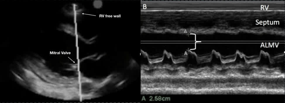
- Place M-mode along the tip of the ALMV.
- On the tracing measure the distance between the septum and the ALMV.
- If this distance is <7mm, LVEF is normal. If EPSS is >10mm, LVEF is depressed.
LV Diastolic Dysfunction
- Two findings can be visually estimated to determine the presence or absence of significant diastolic dysfunction: LV hypertrophy (LVH) and LA enlargement (LAE).
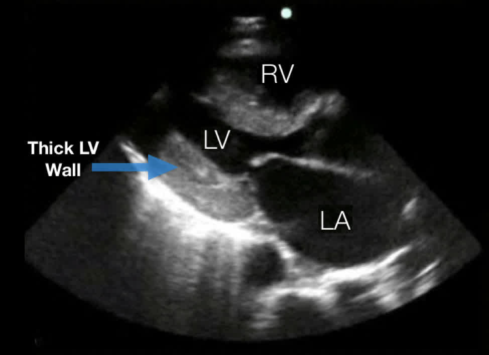
- In cases of dramatic LVH, a massive pathologic LV can be easily distinguished from a normal sized chamber.
- In more subtle cases, measure the LV free wall in PLAX view. If the wall thickness is >10mm, this suggests LVH.
- LAE is suggested if the LA appears grossly enlarged on multiple views.
- On the PLAX view, the LA is larger relative to the LVOT and the RV.
RV Dysfunction
- The dilated/pressure overloaded RV can be identified on multiple views.
- In the right clinical setting, signs of RV dysfunction can prompt further workup of pulmonary embolism as a cause of acute dyspnea.
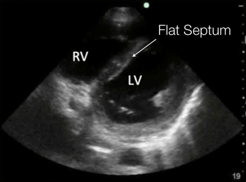
- On the PSAX view, a strained RV compensates for an increase in pressure by ballooning out its free wall. As a result, the septal wall flattens causing a D-shaped appearance of the LV instead of its normal circular appearance.
- In any view, if the RV diameter appears to be larger or equal to the LV this suggests RV high pressure.
- If the RV wall is thickened, it suggests chronic RV high pressures (eg. Pulmonary hypertension), while if the RV wall is thin this suggests an acute process creating high Right Heart pressure (eg. Pulmonary embolism)
Pearls and Pitfalls
- Stabilise your hand while scanning – subtle movements of the transducer can have a significant impact on the quality of images. Rest your hand on the patient’s chest wall – this allows you to make more controlled movements and obtain a more optimal study.
- Obtain as many views as possible – a single view may be misleading especially in the case of assessing chamber size or identifying a loculated pericardial effusion not visible on all views.
- The epicardial fat pad is a benign finding of a small hypoechoic region above the RV free wall at the top of the screen – it can sometimes be difficult to distinguish from a loculated effusion.
- Studies suggest an experienced sonographer’s visual impression of the gross systolic function or squeeze of the LV is as accurate as quantitative methods such as ejection fraction calculation.
- The EPSS technique relies on the assumption of a normal mitral valve and should not be used in cases of suspected or known valvular disease, eg. Rheumatic valvular disease with thickened, immobile leaflets.
- In the hypertensive dyspneic patient with a grossly normal EF, identifying LVH/LAE with other sonographic signs of congestive heart failure suggests decompensated diastolic heart failure.
- Bedside cardiac ultrasound is not sensitive or specific in diagnosing pulmonary embolism (PE). It can only identify the presence of RV dysfunction while ruling out decompensated heart failure in the assessment of the acutely dyspneic patient.
- Patients with acute sub massive PEs can have unremarkable bedside cardiac ultrasounds.