Gastronomy
Introduction Background Anatomy of a gastrostomy Important details on history Important details on examination Basic care Trouble shooting Using a foley’s catheter as a temporary gastrostomy SETTING Primary and Secondary Healthcare Institutions in the Western Cape which refer to Red Cross War Memorial Children’s Hospital (RCWMCH) FOR STAFF Primary and Secondary Doctors and Nurses who refer patien
Important details on examination
Using a foley’s catheter as a temporary gastrostomy
SETTING
Primary and Secondary Healthcare Institutions in the Western Cape which refer to Red Cross War Memorial Children’s Hospital (RCWMCH)
FOR STAFF
Primary and Secondary Doctors and Nurses who refer patients to RCWMCH
PATIENTS
Patients under the age of 13 years old with a gastronomy
Introduction
Gastrostomies are a common medical device used to assist with long-term tube feeding in patients who are unable to tolerate enough oral intake safely to maintain their nutritional needs. These devices classical replace a nasogastric tube and can be used to instil liquid food into the stomach or to decompress gas and fluid out of the stomach when needed.
There are several issues that are commonly seen in patients with gastrostomy tubes in situ. These range from minor leaks and granulomas to more serious complications such as tube displacement into the peritoneum. Most concerns involving gastrostomies do not need specialist surgical input and can be managed at primary level with supplementary guidance from gastrostomy nurses.
The purpose of this guideline is to equip primary care clinicians and nurses to manage the minor complications seen with gastrostomy tubes and to highlight scenarios when referral to a paediatric surgeon in indicated.
Background
The 2 most important things to know about gastrostomies are:
- If the tube is left out for too long, then the gastrostomy hole will close (this can happen in the matter 20 minutes).
Because of this, it is always important to replace the tube when it falls out. You can use the same tube, a new tube or even a foley’s catheter to do this.
- If a gastrostomy is new and you replace the tube before it has had a chance to heal, then the tube can end up outside of the stomach, instead of in the lumen of the stomach. If you feed through the tube under these circumstances then the patient will have food in the abdominal cavity which will cause severe sepsis and can lead to death.
To prevent this from happening you need to check when the gastrostomy was created before you replace the tube. If the gastrostomy is less than 6 weeks old then the chance of the gastrostomy not yet having fully healed is high. You should still attempt to replace the tube if it goes easily but DO NOT use the tube to feed the patient until it has been checked with a contrast study (which might need to happen at RCWMCH if you can’t do it at your facility). The advantage to attempting to put the tube in gently, even though you will not use it to feed, is that you will stop the hole from closing and might save the patient another operation in the future.
Anatomy of a gastrostomy
The primary concept with a gastrostomy tube is that there is a tube that passes through the patient’s abdominal wall directly into the stomach lumen. This allows fluid to be instilled into the stomach and gas and fluid to be aspirated out.
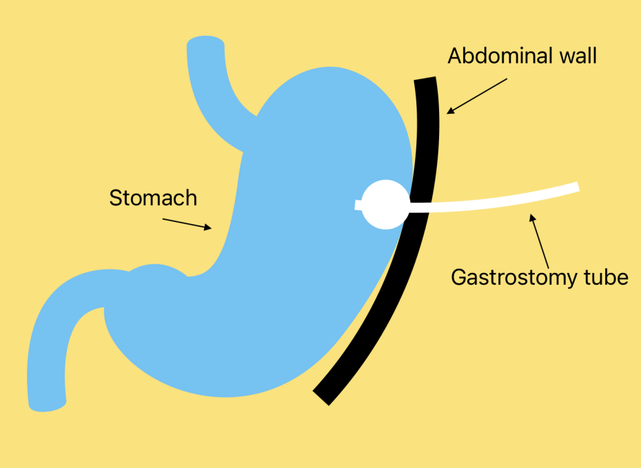
Click on the image to enlarge.
There are a few different tubes that are used as gastrostomies. The differences between the tubes are mainly to do with how they are kept in position:
The main types of tubes that we use at RCWMCH are the balloon gastrostomy, percutaneous gastrostomy tubes and the button gastrostomy.
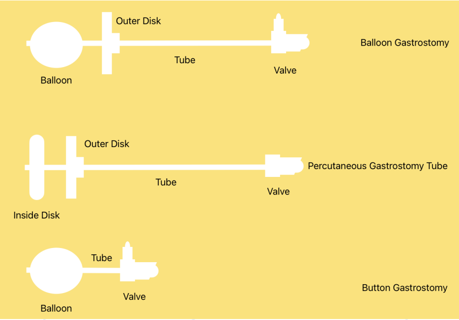
Click on the image to enlarge.
On the outside part of the tube there is usually a disk (outer disk) that stops the tube going in too deep. When the tube was inserted the distance at which this disk should be was measured. Button gastrostomies are a little different: there is no disk on the outside, but the valve where the feeds are instilled acts as a stopper on the outside. These button gastrostomies are measured to fit the patient exactly and they have an extra tube that needs to be attached during feeds because the gastrostomy buttons are very short.
On the piece of the tube which is inside the stomach there is always either a balloon or a disk (inside disk) that stops the tube from falling out. When tubes have a balloon this means the tube can be removed and replaced by letting the fluid in the balloon out – similarly to a foley’s catheter. When they have a disk, you have to cut the tube to separate it from the disk (usually surgeons would use a gastroscope to retrieve the piece they cut off – although it will usually pass in the stool if left in the bowel lumen).
You can tell if a tube is a balloon gastrostomy by looking at the valve where the feeds attach. In a balloon tube there is a valve for the balloon to be inflated but in a tube that has a disk there is only a port to instil feeds through.
Important details on history
- When was the gastrostomy created?
- What was the reason it was created?
- Is the patient using it for feeds, meds, both or neither?
- Is the patient able to take anything orally or not?
- What is the concern with the gastrostomy currently?
Important details on examination
- General assessment of the child.
- Specifically assess the severity of the underlying condition which necessitated the gastrostomy creation.
- Look at the tube and assess what type of tube it is.
- Check if there is any damage to the tube, such as cracks to the tubing or valve.
- Is the balloon intact and how much fluid is in the balloon?
- What depth is the tube at? There are numbers along the side of the tube to use to judge this. Use the number at skin level to measure this.
- If the tube is out, try to replace it (if appropraite)
- Assess the skin around the gastrostomy for redness, excoriation or cellulitis.
Basic care
Most surgeons will ask caregivers to avoid handling the tube much during the first 6 weeks after gastrostomy creation. This is to minimise the risk of the tube falling out. Changing the tube during this time should be avoided and should be checked with fluoroscopy if it is unavoidable. A surgical review prior to changing it is useful when it is possible.
Gastrostomies need daily care to keep them clean and healthy. It is normal for there to be a small amount to leaking around the tube which may leave some residue on the tube, skin or clothing. This residue can be acidic, so it is important to clean it away to avoid build up or burns to the skin around the gastrostomy.
Some caregivers choose to put a small piece of dry gauze between the skin and the outer disk to absorb this small amount of fluid. This is acceptable but should be changed as soon as it is noted to have fluid on it otherwise it will become a reservoir to collect acidic fluid which will potentially burn the skin.
Tubes with balloons need to be checked once a week to ensure the balloon has not deflated. This is done by gently deflating the balloon and then refilling it with fresh water (usually 3 – 5 ml). It is important to avoid pulling the tube out while doing this process, so it is best done while the child is calm and comfortable (or asleep). When a balloon tube is used at the time of gastrostomy creation, extra care should be taken for the first 6 weeks post creation to avoid displacing the tube.
Caregivers are shown how to check the depth of the tube whenever they clean it. This is done by looking at the numbers along the side of the tube. At the time of gastrostomy insertion, this depth will have been measured so the usual depth is known to the caregivers, but it is usually about 1.5 - 3.5cm.
As the child grows or puts on weight, the abdominal wall may become thicker, and thus the tube will need to be placed slightly deeper. If the outer disk is too tight, it will cause local ischaemia which can lead to gastrostomy breakdown and leaking. If the disk is too loose, it can cause leaking or the balloon may even pass into the small intestine which can cause vomiting and obstruction. The best position is for the outer disk to be snug against the skin, but still loose enough to be able to twist freely. This means the tube is close enough to minimise leaks but loose enough not to be squeezing the abdominal wall tissues too tightly.
Trouble shooting
| Problem | Cause | Solution |
| Tube comes out | Balloon empty or broken Internal disk might be pulled out whole or have broken off | Replace the tube through the hole and reinflate the balloon if it is still intact. If the balloon or tube is broken then replace the tube with a new tube or use a foleys catheter which you should secure to prevent it passing too deep into the stomach. Provided the tube goes in easily and the gastrostomy is established (older than 6 weeks) then you can safely feed after replacing the tube. If you struggled to insert the tube or it has been less than 6 weeks since the gastrostomy was created then the position of the tube should be checked before starting feeds again. In this case, contact the closest hospital with the capacity to do a contrast study and consider starting ivi fluid if the patient is unable to take oral fluids while you await the study. |
| Leakage around the tube | Tube too loose or the balloon may be broken The tract may be enlarged. The stomach could be too full or the child might be constipated. | Check the water in the balloon and check the depth position of tube. Then clean and protect skin. If the leaking continues than consider contacting the stoma sister at S16 for advice. If the leak is so large that the child cannot be fed then start ivi fluids and contact the paediatric surgeon on call for advice. |
| Granulation tissue: pink friable tissue around the edge of the gastrostomy opening | This is the result of the body trying to heal against the tube, and is very common. If can bleed easily and may causing leaking which can be frustrating. | Clean and dry the area. Sprinkle fine table salt onto the granulation tissue – it will adhere to the moist surface of the pink tissue. Repeat 12 hrly until the tissue has resolved – this should take 3 days. |
| Skin irritation or bleeding | Leak around the tube, granulation tissue, tight external disk | Check the water in the balloon and check the depth position of the tube. Keep skin clean and dry, clean more frequently and/or change the dry dressing more often. Consider if a barrier cream is needed. Candida is common and should be treated with local topical treatment. Cellulitis is rare but should be referred for surgical assessment if it occurs. |
| Blocked tube | Poorly dissolved/crushed medicines, inadequate flushing of the tube, curdled milk in tube | Attempt to unblock with a carbonated drink such as coke, If this fails and the gastrostomy is established then exchange the tube for a new one. |
| Broken tube | Cracks along the length of the tube | Replace the tube. |
Using a foley’s catheter as a temporary gastrostomy
Sometimes it may be necessary to exchange the tube but there is no gastrostomy tube available in your facility. A foley’s catheter can be used as a temporary measure and, if secured cleverly, may last for a week or 2 while awaiting replacement with a new tube.
The same rules regarding the maturity of the gastrostomy stand:
- the gastrostomy should be established for at least 6 weeks to safely change the tube without checking with fluoroscopy.
- The new tube should pass into the stomach with ease.
Choose a foley’s catheter the same size at the tube that has fallen out. If the gastrostomy open ing has become smaller, then a slightly smaller tube is also an acceptable option.
Insert the tube through the gastrostomy and then inflate the balloon. Pull the tube back gently to bring the balloon against the inside wall of the stomach.
Then take a piece of thick plaster (Elastoplast works well) and stick it to the tube at that level, making 2 tags by sticking it to itself at the skin level. These tags will act like the disk to stop the tube advancing into the stomach.
The tube can be used the feed, but a spigot will be needed to stop it leaking. The hub of a 2ml syringe works well if a spigot is not available.
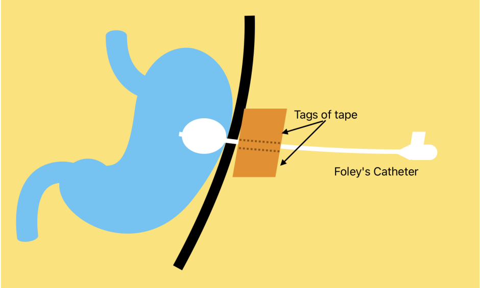
Click on the image to enlarge.
Children's Surgery Network, Western Cape Government, Red Cross War Memorial Children’s Hospital (RCWMCH), Clinical Guideline_Version 1_2025
Gastronomy