Guidelines Reviewed
Blunt and penetrating bladder injury Intraperitoneal injury Extraperitoneal injury Post-surgical bladder drainage Spontaneous (idiopathic) rupture of the urinary bladder Paediatric bladder injury Iatrogenic bladder injury Guidelines reviewed in this article include: European Association of Urology (EAU) guidelines on urological trauma (2023) [7] , EAU guidelines on paediatric urology (2022) [3] ,
Blunt and penetrating bladder injury
Post-surgical bladder drainage
Spontaneous (idiopathic) rupture of the urinary bladder
Guidelines reviewed in this article include: European Association of Urology (EAU) guidelines on urological trauma (2023) [7], EAU guidelines on paediatric urology (2022) [3], Urotrauma: AUA (2020) [2], Kidney and Uro-trauma: World Society of Emergency Surgery and the American Association for the Surgery of Trauma (WSES-AAST) guidelines (2019) [1], Management of blunt force bladder injuries: A practice management guideline from the Eastern Association for the Surgery of Trauma (EAST) (2019) [8], and EAU guidelines on iatrogenic trauma (2012) [27]. The EAU published updated guidelines in March 2023 but there are no changes related to bladder trauma from the 2022 edition.
In addition to blunt and penetrating bladder trauma, this review also includes aspects of guidelines pertaining to other forms of bladder injury including iatrogenic injuries, paediatric trauma, and spontaneous bladder rupture. Relevant recommendations reported in the included guidelines are summarised in Table 2.
Table 2: A summary of current guidelines relating to bladder trauma.
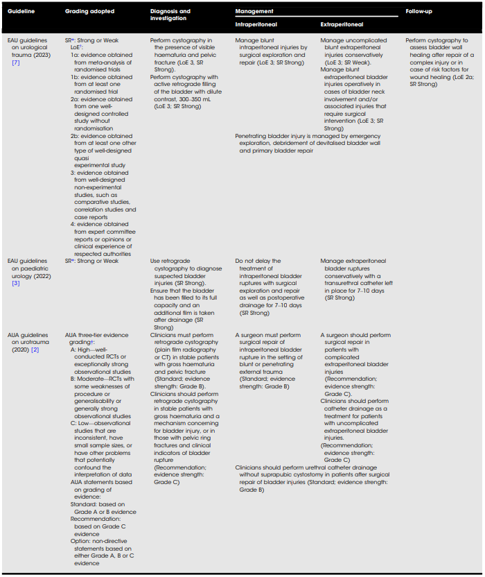
Click on image to enlarge.
Table 2 (continued)
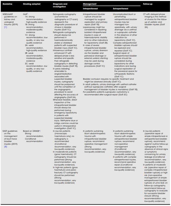
Click on image to enlarge.
Table 2 (continued)
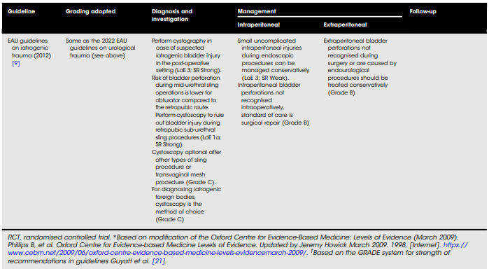
Click on image to enlarge.
Blunt and penetrating bladder injury
Diagnosis
There is consensus among the EAU (level of evidence [LoE] 3, strength of recommendation [SR] Strong) [7], AUA (Grade B) [2] and WSES-AAST (grade of recommendation [GoR] 1C) [1] guidelines that retrograde cystography (either plain film radiography or CT) should be performed to diagnose bladder injury in the setting of visible or gross haematuria and/or fracture of the pelvis, provided the patient is haemodynamically stable. Compared to plain film radiography, CT has the added advantage of diagnosing and defining other concomitant injuries [9]. However, delayed, or excretory phased CT is inferior to CT with cystography in delineating bladder injury (GoR 1B, WSES-AAST) [1]. The EAST guidelines base its imaging recommendations on the injury severity, with no investigation being recommended in low-risk patients (microscopic haematuria only) (Conditional recommendation) and retrograde CT cystography recommended in moderate (gross haematuria) and high-risk (gross haematuria and pelvic fracture) patients (Strong recommendation) [8].
Guideline: in cases of suspected bladder injury, CT with retrograde cystography is the investigation of choice. CT is superior at delineating injury and has the advantage of diagnosing concomitant injuries.
Intraperitoneal injury
There is consensus among all guidelines that, in general, intraperitoneal bladder injury, whether due to blunt or penetrating trauma, should be repaired via open surgical exploration (EUA: LoE 3, SR Strong; AUA: Grade B; WSESAAST: GoR 1B; EAST: Strong recommendation) [1,2,7,8]. Conservative management is only recommended in cases of minor uncomplicated intraperitoneal bladder injuries that are predominantly encountered during endoscopic bladder procedures (EAU: LoE 3, SR Weak) [7]. WSES-AAST is the only guideline to recommend laparoscopic management of intraperitoneal bladder injury, which may be considered in haemodynamically stable patients with isolated intraperitoneal injury and no other indications for exploratory laparotomy (GoR 2B) [1].
The role of laparoscopic nephrectomy in the setting of renal trauma has previously been explored [10]. Several case reports/series have also reported on the usefulness of laparoscopy in haemodynamically stable patients with intraperitoneal bladder injuries [11–16]. Minimally-invasive surgery has distinct advantages in terms of postoperative wound sepsis, hospital length of stay and pain reduction [11]. Important considerations prior to using this modality include operator skill and experience, haemodynamic stability of the patient, and the presence of other injuries [17].
Robot-assisted laparoscopic bladder repair has also been described in several cases; however, the majority of these cases were due to iatrogenic bladder injury [18]. The method of repairing extraperitoneal penetrating bladder injury has previously been described [19]; however, none of the guidelines provide any recommendation on the use of minimally invasive surgical repair of non-iatrogenic bladder injuries.
Guideline: in general, intraperitoneal bladder injury should be primarily repaired via open surgical approach. Where expertise is available, minimally invasive surgery may be considered in haemodynamically stable patients with isolated bladder injury.
Extraperitoneal injury
Again, there is general consensus among guidelines that uncomplicated extraperitoneal bladder injury may be managed conservatively with catheter drainage (EAU: LoE 3 SR Weak; AUA: Grade C; WSES-AAST: GoR 1C; EAST: Conditional recommendation) and that complex extraperitoneal injury must be managed surgically (EAU: LoE 3 SR Strong; AUA: Grade C; WSES-AAST: GoR 1C; EAST: Conditional recommendation) [1,2,7,8]. Complex extraperitoneal injury is defined as injuries involving the bladder neck, injuries associated with pelvic fractures, or injuries involving other neighbouring organs [1,7]. Although there is no explicit guidance on bladder injury management in patients requiring pelvic fixation, the assumption is that these patients should undergo surgical exploration.
Guideline: uncomplicated extraperitoneal bladder injuries may be managed conservatively. Complicated extraperitoneal bladder injuries should undergo surgical exploration.
Post-surgical bladder drainage
Both, the EAU [7] and EAST [8] guidelines do not make any recommendation regarding post-surgical bladder drainage. However, there is consensus among the AUA (Standard; Grade B) [2] and WSES-AAST (GoR 1B) [1] guidelines that bladder drainage post-surgical repair should preferentially be managed with transurethral catheterisation alone. Although the efficacy of both transurethral and suprapubic catheter drainage is comparable, the former is associated with fewer complications [7].
With regards to the duration of indwelling catheterisation, the only guidance is from the AUA who recommend catheter drainage for a period of 2–3 weeks (AUA: Grade C) for cases of extraperitoneal injuries being managed conservatively [2]; EAU guidelines suggest urinary catheter removal within 5– 10 days in healthy patients undergoing operative bladder repair and removal at Day 5 and 7 respectively for uncomplicated extraperitoneal and intraperitoneal bladder injuries being managed conservatively. It must be noted that these are not formal recommendations [7].
In cases where the repair is complex or concurrent trauma is significant, indwelling catheter time may need to be extended. The use of a drain and tailored antibiotics are also factors that will need consideration but there is currently no guideline that makes any strong recommendations for this.
Guideline: bladder drainage post-surgical repair should preferentially be managed with transurethral catheterisation for a duration of about 5–7 days. In patients being managed conservatively, transurethral catheterisation should be maintained for at least 7 days, with the exact duration to be tailored to the patient and the extent of injuries sustained.
Follow-Up
The AUA guidelines provide no guidance regarding the follow-up of patients with bladder injury. However, the EAU guidelines recommend that patients with complex injuries (involvement of the bladder trigone or ureteric reimplantation) or those with risk factors for impaired wound healing should undergo cystography to assess bladder wall healing or in patients with risk factors for impaired wound healing. (LoE 2a, SR Strong) [7]. Furthermore, the EUA guidelines suggest that bladder injuries being managed conservatively should be followed up with cystography at ~10 days after injury. If a leak persists, the patient should undergo cystoscopy to exclude the presence of bony fragments in the bladder with a repeat cystogram to be performed 7 days thereafter [7]. The WSES-AAST guidelines recommend the performance of a contrast enhanced CT with delayed phase when following-up patients with bladder injuries (GoR 2a) [1]. Besides these, there is a paucity of formal guidance pertaining to follow-up, specifically with regards to the investigation of choice and timing [7].
The EAST guidelines base their recommendations on the risk of urinary leakage with low risk described as operative repair of simple intraperitoneal or extraperitoneal bladder rupture, moderate risk described as operative repair of complex intraperitoneal bladder rupture and high risk described as nonoperative management of simple extraperitoneal bladder rupture. The guidelines recommend against routine follow-up cystography in low-risk patients (Conditional recommendation) and recommend routine follow-up cystography in moderate and high-risk patients (strong recommendation) [8]. See Fig. 2 for a summary of the suggested approach to bladder trauma.
Guideline: patients with simple injuries and no suspected leak being managed either conservatively or operatively do not routinely require cystography. Patients with complicated injuries, simple injuries with suspected persistent leak, complex repairs, or patients at risk of poor wound healing should have routine followup cystography performed at 7–10 days after repair.
Fig. 2 Suggested approach to management of suspected bladder trauma
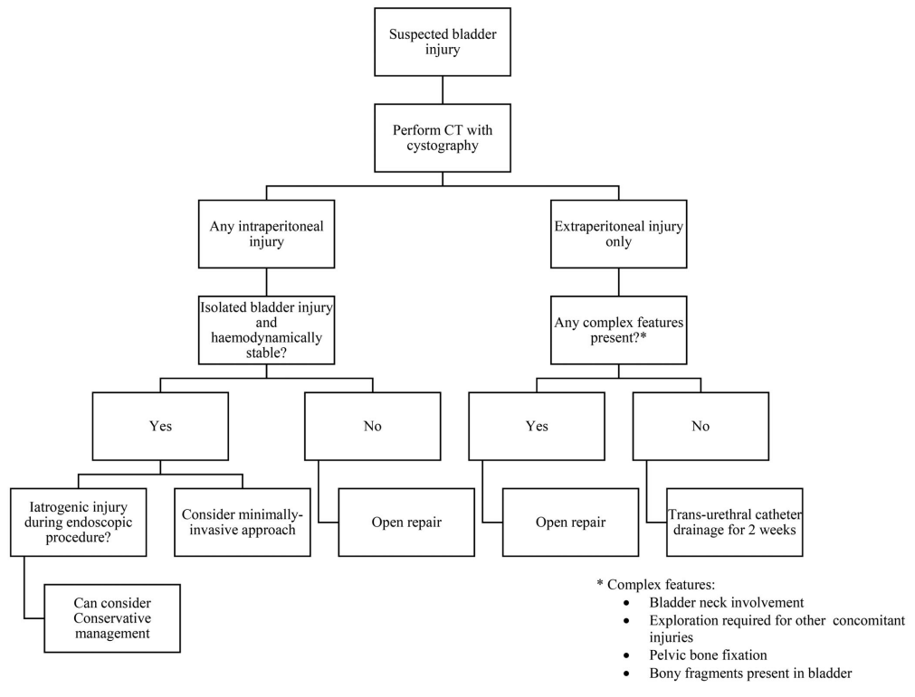
Click on image to enlarge.
Spontaneous (idiopathic) rupture of the urinary bladder
None of the current guidelines address the management of spontaneous or idiopathic bladder rupture. Although this condition is a distinct entity, the diagnosis and management should be based on the current standard of care as recommended by trauma guidelines until such time when there is more evidence to inform guideline recommendations. Although this entity is managed on an individual case by case basis, we have recently outlined various associations and risk factors that should alert the attending emergency team in this scenario [20].
Paediatric bladder injury
The bladder is the second most common urological organ that may be injured in paediatric trauma. It is widely recognised that the paediatric bladder is less protected than the adult bladder with injury commonly manifesting as a result of significant blunt abdominopelvic trauma [7]. In some estimates, paediatric bladder rupture may be associated with concomitant pelvic ring fractures in 57% of cases; however, the incidence of bladder injury in the setting of pelvic fractures may vary from 0.5% to 16.8% [7].
The EAU is the only body to have published standalone guidelines that pertain to paediatric trauma. This guideline was recently updated in March 2022, with no new changes or recommendations from the 2020 update [3]. The guideline includes management recommendations; however, due to the paucity of evidence there is no LoE provided, only grades of recommendation based on the Grading of Recommendations, Assessment, Development, and Evaluation (GRADE) system [21].
Diagnosis
Children with a history of blunt trauma, suprapubic pain and tenderness, an inability to void, haematuria and/or concomitant pelvic ring fractures must be investigated for bladder injury. Patients should undergo retrograde cystography (either plain film radiography or CT) with the bladder integrity to be interrogated at both full distention and when empty (EUA: SR Strong) [3].
Management
Management recommendations are similar to those of adult guidelines. Uncomplicated extraperitoneal injuries should be managed with catheter drainage for 7–10 days, while for complicated extraperitoneal injuries or intraperitoneal rupture surgical exploration and repair is recommended (EUA: SR Strong). Compared to the adult recommendation, the major difference is that drainage after surgical repair should be facilitated by placement of a mandatory suprapubic catheter [3]. This recommendation is also supported by the WSESAAST guidelines (GoR 2C) [1]. However, the guidelines do acknowledge that there is limited data to indicate that transurethral drainage may be as effective and result in a shorter period of diversion [22–25].
Follow-Up
Follow-up should be carried out at 7–10 days after the procedure or drainage and should include a repeat cystogram [3].
Iatrogenic bladder injury
The bladder is the most frequently injured urological organ during surgical procedures [26]. Iatrogenic trauma is classified as either external or internal iatrogenic bladder trauma (IBT). External IBT occurs most commonly during obstetric and gynaecological procedures, while internal IBT occurs most commonly during transurethral resection of bladder tumour (TURBT) [27].
The EAU is the only body to have published standalone guidelines pertaining to iatrogenic urological injury [27]. These guidelines were first published in 2012, with some recommendations being included in the March 2022 limited update of comprehensive urological trauma and no new changes in the latest 2023 update [7].
Diagnosis
Suspicion of external IBT should be confirmed by direct inspection. Signs include the presence of urinary extravasation, visualisation of blood and or gas in the urinary catheter or bag during the procedure [28]. Perioperative internal IBT should be excluded during cystoscopy. Signs include the presence of a dark space between detrusor muscle fibres, fatty tissue, or visualisation of bowel through a bladder wall defect [28]. Bladder injury not recognised intraoperatively presents similarly to traumatic bladder injury with abdominal pain, distention, and urinary symptoms. Cystography is the investigation of choice to exclude IBT (EAU: Grade B) [27].
Management
Due to a lack of evidence that two-layer vesicorraphy is superior to single-layer watertight closure, the former technique is no longer recommended (EAU: upgraded by panel consensus to Grade A) [7]. The management of IBT is still in keeping with the standard of care described in trauma guidelines. Injury recognised during the procedure should be primarily closed. The management of missed IBT depends on the site of perforation and the complexity of the injury [27]. Extraperitoneal injuries are generally managed conservatively, except in cases that are complicated with symptomatic extravesical collections. These patients require drainage with or without closure of the defect [27]. In cases of internal IBT due to TURBT, the bowel should be thoroughly examined to rule out other concomitant injuries [27].
Urologists should be vigilant of endoscopic bladder trauma and be sensitive to any sudden change in the irrigation, lighting, abdominal distention encountered during TURBT. A high index of suspicion should be present if the cystoscopic appearance is ‘out of the norm’. A standard whole bladder ‘cystoscopy check’ is thus recommended after any TURBT procedure.
Bladder Trauma: A Guideline of the Guidelines; Reddy D, Laher AE, Moeng M, Adam A_6; BJU International published by John Wiley & Sons Ltd on behalf of BJU International. 2024; 133(3):365–374. doi:10.1111/bju.16236._2024