Hip Dislocation
Traumatic hip dislocation is a specific term used to describe complete loss of contact between the articular surface of the femoral head and that of the acetabulum.
Clinical
Management
Reduction Techniques
Disposition
Traumatic hip dislocation is a specific term used to describe complete loss of contact between the articular surface of the femoral head and that of the acetabulum.
Hip dislocation is usually unilateral; very rarely it may be bilateral. After dislocation, the femoral head can either lie anterior (10%) to the acetabulum or posterior (90%) to it.
Clinical
The hip is commonly held in adduction and either internal rotation (in posterior dislocations) or external rotation (in anterior dislocations). Anterior dislocations may be associated with a palpable lump in the groin. In either case, the leg is usually shortened. The patient experiences extreme pain when any attempt is made to move the affected leg.
Examination PEARL
Traumatic hip dislocation is a high-energy injury. Check for other injuries!
Artificial hips dislocate with less force - Relatively minor forces, such as flexing the hip to pick an item up from the floor, can result in post-operative hip dislocation.
Management
- Traumatic hip dislocation is a true orthopaedic emergency.
- It is widely held that the sooner hip dislocation is reduced, the better the prognosis.
- There is a fourfold increase in the rate of osteonecrosis if reduction is delayed beyond 12 hours.
- Manage the patient as a whole using current resuscitation guidelines.
- For interest’s sake: the Stewart and Milford classification of hip dislocations is commonly used and is graded from I to IV.
Reduction Technique
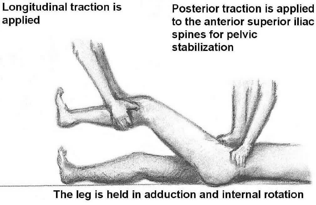
- Have an assistant stabilise the pelvis by grasping the bilateral anterior superior iliac spines and applying gentle posterior force.
- Apply longitudinal distraction of the injury by grasping the patient’s distal femur, bringing the femoral head from behind the acetabular rim.
- The leg should be held in a position of relative adduction and internal rotation.
- Several moments of persistent traction may be needed to relax the large musculature of the hip; this joint laxity helps to facilitate the subsequent manoeuvres.
- Once the leg has been brought out to length, bring the hip to 90° of flexion while allowing the ipsilateral knee to flex passively.
- Gently abduct, externally rotate, and extend the hip while distracting the femoral head anteriorly. Large amounts of rotational force should be avoided, since this has been associated with iatrogenic femoral neck fractures.
- When the femoral head returns to the acetabulum, an audible or palpable "clunk" can be appreciated.
- While the procedural sedation is still in effect, gently take the reduced hip through a full passive range of motion (ROM), being mindful of any additional fractures or injuries the patient may have sustained.
- The stable reduced hip should remain in anatomic position through these manoeuvres.
- Repeat hip dislocation during this passive ROM exercise is diagnostic of clinical joint instability. Longitudinal skeletal traction is required to stabilise such an injury.
- Place the affected extremity in a pillow brace to ensure stability during transport.
Disposition
- Once reduction has been achieved, repeat radiography is required to ensure concentric anatomic placement of the femoral head.
- Patient should be placed in an adduction pillow and admitted to Orthopaedics.
References
- Rockwood CA Jr, Green DP, Bucholz RW, eds. Rockwood and Green's Fractures in Adults. 4th ed. Philadelphia, Pa: Lippincott Williams & Wilkins; 1996:971-85.
- Mitchell JC, Giannoudis PV, Millner PA, Smith RM. A rare fracture-dislocation of the hip in a gymnast and review of the literature. Br J Sports Med. Aug 1999;33(4):283-4.
- Rockwood CA, Green DP, Bucholz PW, Heckman JD, eds. Fractures and dislocations of the hip. Fractures in Adults. Vol 2. 4th ed. Philadelphia, Pa: Lippincott-Raven; 1996:1756-1803.
- Yates C, Bandy WD, Blasier RD. Traumatic dislocation of the hip in a high school football player. Phys Ther. Jun 2008;88(6):780-8.