Hyperkalaemia
Hyperkalaemia
Introduction
Identification
Severity assessment
Management based on severity
Relevant history, examination
Investigations
Referral criteria
Discharge criteria
Treatment Algorithm
Introduction/ Description
Potentially life-threatening increase in extracellular potassium (K+) concentration
Identification/recognition/diagnosis of problem
HyperK+ is often only retrospectively diagnosed. Where prospectively diagnosed the cues were taken from the history and examination that pointed to the underlying cause. An early venous blood gas can provide an early diagnosis (see below). Causes of HyperK+ include the following:
- Impaired elimination:
- Acute and chronic renal failure
- Drugs interfering with K+ excretion: Spironolactone, ACE-inhibitors, Amiloride
- Hypoaldosteronism: Addison’s disease (primary), Chronic renal failure (secondary)
- Excessive intake
- Drugs (especially in overdose): K+ containing liquids (MistPotChlor),
- Excessive intake: Food sources (Dried fruit, Nuts, avocado, bananas, orange juice, white mushrooms, broccoli, spinach, French fries)
- Increased K+ shift from intra- to extracellular space
- Metabolic acidosis
- Diabetes Mellitus
- Acute cell tissue breakdown: Rhabdomyolysis, massive transfusion, DIC, haemolysis
- Drugs: Digoxin, Beta-blockers, Scoline.
Differential Diagnosis
- Acute Tubular Necrosis
- Congenital Adrenal Hyperplasia
- Digitalis Toxicity
- Electrical Burn Injuries
- Head Trauma
- Hypocalcaemia
- Metabolic Acidosis
- Rhabdomyolysis
- Thermal Burns
- Tumour Lysis Syndrome
Severity assessment (Triage)
- Mild: K+ 5.5-6.5 mmol/l
- Moderate: K+ 6.5-7.5 mmol/l
- Severe: K+ >7.5mmol/l
Management based on severity
Uncomplicated HyperK+ and HyperK+ due to reversible causes should be managed where presented. Alternatively stabilisation and initial correction of HyperK+ is required whilst awaiting transfer to secondary or tertiary referral centre. Referral is required if:
- Persistent HyperK+ despite optimal management
- Severe HyperK+ with cardiac toxicity
- Conditions causing the HyperK+ than can’t be managed at current facility
- If unsure whether to refer or not contact the Physician at your closest referral facility
Immediate action:
Non-drug treatment
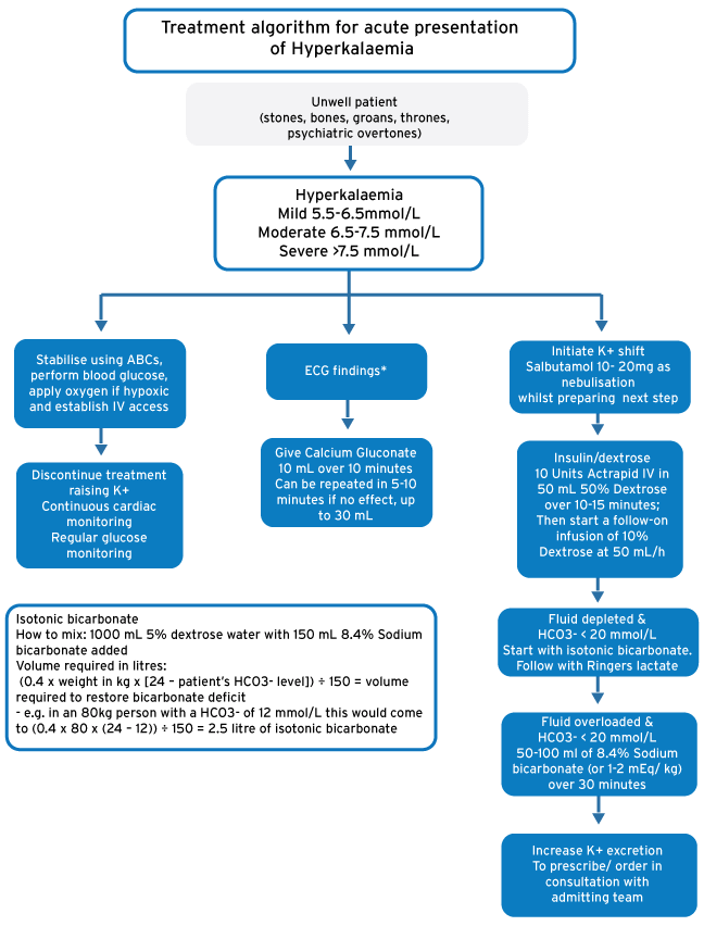
- Stabilise using ABCs, perform blood glucose, apply oxygen if hypoxic and establish IV access
- Discontinue all drugs that may cause or worsen HyperK+. Continuous cardiac and glucose monitoring is required whilst correcting HyperK+
Drug treatment
- If ECG changes present give Calcium. Some sources argue that Calcium should be given even in the absence of ECG changes. If the diagnosis is correct this is unlikely to cause harm. Caution is advised in digoxin toxicity and hyperCa2+
- Start with ß2-adrenergic agonists whilst preparing an insulin/ dextrose infusion. Patients on ß-blockers or in renal failure may not respond.
- 10 Units Actrapid IV in 50 mL 50% Dextrose over 10-15 minutes. Then start a follow-on infusion of 10% Dextrose at 50 mL/h whilst monitoring blood glucose
- Sodium Bicarbonate should be considered if a metabolic acidosis is present (HCO3- < 20 mmol/L). It is important not to infuse Sodium Bicarbonate in the same line as Ringers Lactate or Ca-Gluconate. The mode of administration differs depending on the patient’s volume status. See 4.1.3 and 4.2 below.
- Increase K+ excretion
- Ion-exchange resins: Kayexalate 30g PO or 60g PR (a Foleys catheter should be used to keep the resin in rectum for 2 hours). There is controversy in emergency medicine literature over the use of ion-exchange resins specifically with regards to their risk/ benefit ratio. Initiating ion-exchange resins should be done in consultation with a specialist
- Loop diuretics should be considered if fluid overload present or in consultation with a specialist: Furosemide 1-2 mg/kg slowly
- Renal replacement therapy (refer to tertiary referral guidance): Haemodialysis
Volume depletion and acute kidney injury (or non-anion gap acidosis)
Aggressively treat any cause of volume loss as the resulting reduced perfusion lead to recurrent ischaemic injury. Correct volume deficit with IV Isotonic fluids as follow
- Isotonic 1.26% bicarbonate (1000 mL 5% dextrose water with 150 mL 8.4% Sodium bicarbonate added): preferred starting resuscitation fluid for patients with hyperkalaemia and acidosis (HCO3- < 20 mmol/L) in renal failure. Calculate the bicarbonate deficit (0.4 x weight in kg x [24 – patient’s HCO3- level]) and divide by 150 mEq/L which will give you the volume of isotonic bicarbonate required in litres. Round this DOWN to the nearest full litre (example: a result of 1.7 L would mean you’ll use 1 L, likewise a calculation requirement of 0.6 L would mean that 1.26% bicarbonate is not used). Continuing to use isotonic bicarbonate after giving this volume would cause a metabolic alkalosis. Isotonic bicarbonate should be avoided in an anion gap acidosis (e.g. lactic or keto-acidosis)
- Ringers lactate (or similar): preferred follow on fluid for patients with hyperkalaemia and acidosis, to restore further volume deficit as clinically indicated. Ringers lactate does not cause lactic acidosis.
- Saline 0.9%: Should be avoided in hyperkalaemic, acidotic renal failure
| Calcium | Alternative consideration |
| Ca-Gluconate 10%, 10 mL over 10 minutes (can be repeated in 5-10 minutes if no effect, up to 30 mL). | Ca-Chloride 10% 5- 10 mL over 10 minutes (Ca-Gluconate is preferred as Ca-Chloride can cause damage on extravasation). |
| Salbutamol | |
| Salbutamol 10- 20mg as nebulisation will lower K+ levels by up to 1 mmol/L. | -- |
| Insulin/ dextrose | Alternative consideration |
| 10 Units Actrapid IV in 50 mL 50% Dextrose over 10-15 minutes; then start a follow-on infusion of 10% Dextrose at 50 mL/h | 10 Units Actrapid IV in 200 mL 10% Dextrose/ 500ml 5% Dextrose. The lower dextrose content reduces the risk of tissue damage in extravasation. |
| Sodium bicarbonate | Alternative consideration |
| If the patient is fluid overloaded and HCO3- < 20 mmol/L: 50-100 ml of 8.4% solution (or 1-2 mEq/ kg) over 30 minutes | If the patient is volume depleted and HCO3- < 20 mmol/L: start with isotonic bicarbonate followed by ringers lactate. See above |
Relevant history, Examination
History
A drug history is essential. HyperK+ is mostly associated with asymptomatic or non-specific symptoms. History may include muscle pain or weakness, that rarely progress to paralysis
Examination
An examination is usually unremarkable but this may depend on the underlying cause. General and systemic examination should be done to look for causes or precipitants
Investigations
- Early venous blood gas: acidosis and HyperK+. Beware though of a haemolysed sample which may result in a spuriously high K+ (e.g. blood gas taken after other samples obtained, prolonged tourniquet use before obtaining sample, struggled to obtain sample).
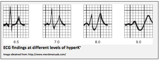
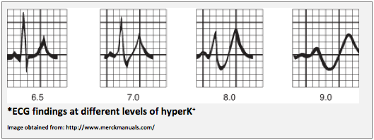
- ECG: Cardiac conduction abnormalities and cardiac arrhythmias may occur and is quite specific once present. A normal ECG does however not rule out HyperK+.
- Non-specific repolarisation abnormalities
- Peaked/tented T waves
- Widening of QRS-complex
- Loss of P-wave/ flattening P-waves
- Sine –Wave pattern
- Bradycardia
- AV-nodal blocks
- Cardiac arrest: PEA/ Asystole/ VF
- Renal function: Look for hyperkalaemia and renal failure
- Blood glucose: Look for hyperglycaemia
- FBC: Look for sepsis, DIC, haemolysis
- Other investigations to be guided by the suspected underlying cause
Referral criteria
- Referral for renal replacement is usually restricted to patients suffering with hyperK+ refractory to optimal medical therapy
Discharge criteria/checklist
- K+ Corrected
- Underlying cause found and treated/ corrected
- All offending drugs removed
- All drugs that may cause or aggravate HyperK+ adjusted
- Advised on decrease/ avoidance of high K+ containing foods
- Appropriate outpatient follow-up arranged
Treatment algorithm for acute presentation of Hyperkalaemia
References
Lehnhardt A, Kemper MJ. Pathogenesis, diagnosis and management of hyperkalemia. Pediatr Nephrol 2011; 26:377-384
Lawrence S, Weisberg MD. Management of severe hyperkalemia. Crit Care Med 2008(36)12
Rossiter D, Blockman M (eds.). South African Medicines Formulary. 11th Ed. Cape Town: Health and Medical Publishing Group; 2014
Cameron P, Jelinek G, Kelly A, et al. Textbook of Adult Emergency Medicine. 4th Ed. Elsevier; 2015