Hypocalcaemia
Hypocalcaemia
Introduction
Identification
Management based on severity
Relevant history, examination
Investigations
Referral criteria
Discharge criteria
Treatment Algorithm
Introduction/ Description
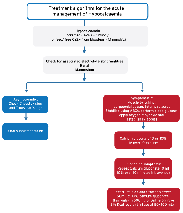
Corrected Ca2+ < 2.1 mmol/L is defined as HypoCa2+ (normal value 2.1–2.6 mmol/L). Corrected Ca2+: measured calcium (mmol/L) + [0.02 x (40 – measured albumin g/L)] (NHLS will calculate provided an albumin was ordered)
Identification/recognition/diagnosis of problem
HypoCa2+ is often only retrospectively diagnosed. Invariably some peripheral paraesthesia can be present. In more advanced cases muscle twitching, carpopedal spasm, tetany and seizures are seen. Where prospectively diagnosed the cues were taken from the history and examination that pointed to the underlying cause. An early venous blood gas can provide an early diagnosis in the form of an ionised Ca2+ level (see below). Causes of HypoCa2+ include the following:
- Vitamin D deficiency: malabsorption, limited exposure to ultraviolet light or low dietary intake, pregnancy and drugs: Anti-epileptic, diuretics, bisphosphonates, cisplatin, aminoglycosides
- Decreased/ Impaired Parathyroid Hormone (PTH): after thyroid surgery, hungry bones syndrome after parathyroidectomy, post radiation of parathyroid glands, metastatic disease to parathyroid, idiopathic, HIV infection
- Loss of Calcium: acute pancreatitis, acute respiratory alkalosis
- Other: chronic liver failure, chronic renal failure, Fanconi Syndrome
Differential Diagnosis
- Acute Renal Failure
- Hydrofluoric Acid Burns
- Hyperparathyroidism
- Hyperphosphataemia
- Hypoalbuminaemia
- Hypomagnesaemia
- Hypoparathyroidism
- Metabolic Alkalosis
- Pancreatitis, Acute
Management based on severity
Look for and treat the underlying cause. Correct associated electrolyte abnormalities, especially Mg2+. Continuous cardiac monitoring is required whilst correcting HypoCa2+.
Where metabolic acidosis is present, Calcium should be corrected first prior to correcting acidosis with Sodium Bicarbonate as this may cause a rapid fall in ionized Calcium.
Immediate action:
Non-drug treatment
Stabilise using ABCs, perform blood glucose, apply oxygen if hypoxic and establish IV access. This may include optimising airway for reduced level of consciousness.
Drug treatment
- Calcium gluconate 10 ml 10% over 10 minutes Intravenous. (Not indicated for asymptomatic patients with chronic renal failure.)
- May be repeated until neurological signs has improved/ cleared.
- Then start a Calcium infusion as follow: dilute 50mL of 10% calcium gluconate (ten vials) in 500mL of Saline 0.9% or 5% Dextrose and infuse at 50- 100 mL/hr. Titrate the rate of infusion to achieve normocalcaemia and continue until treatment effect
- Calcium chloride can be used as an alternative to calcium gluconate, but it is more irritant to veins and should ideally only be given via a central line
- For asymptomatic patients:
- Start on oral Calcium supplementation (consult your pharmacist for available preparations)
- Other treatment options: Will depend on the underlying cause and include Vit D supplementation (oral and intramuscular preparations available)
- Ensure other electrolytes within normal limits
Relevant history, Examination
History
Specific enquiry regarding a family history of HypoCa2+ or previous head and neck surgery/radiation
Examination
Usually unremarkable, except if neurological features present and will depend on the underlying cause. General, systemic and neurological examination should be done to look for signs and causes. Check for presence of Chvostek and Trousseau’s sign
| Neurological signs |
| Muscle twitching, carpopedal spasm, tetany, seizures |
| Chvostek sign |
| Tapping on the cheek 2cm anterior to earlobe, below the zygomatic arch over the facial nerve. Twitching of the lip to spasm of the facial muscles seen |
| Trousseau’s sign |
| Inflation of Blood pressure cuff (above the patient’s systolic BP) for 3 minutes Causes carpopedal spasm |
Investigations
- Blood gas: a blood gas can give an early indication of abnormal Ca2+. It should be noted that ionized (or free) Ca2+ is reported in a blood gas and not total Calcium. Ionised Ca2+ has different reference ranges: 1.1-1.3 mmol/L (about half the reference range of total Ca2+). A total, corrected Ca2+ should always be ordered.
- ECG – Prolonged QT-interval
- Renal function, Mg2+, PO43-– Look for renal/ electrolyte abnormalities
- Albumin – used to calculate corrected Ca2+
- Calcium – confirm hypocalcaemia
- Other tests to be guided by suspected underlying causes including parathyroid hormone, amylase
Referral criteria
Work-up of underlying cause of HypoCa2+ that can’t be done at presenting facility should be referred onwards. If unsure whether to refer or not contact the Physician at your closest referral facility
Discharge criteria/checklist
- Ca2+ Corrected
- Underlying cause found and treated/ corrected where possible
- Drugs causing/ aggravating HypoCa2+ discontinued/ decreased
- Appropriate outpatient follow-up arranged
Treatment algorithm for the acute management of Hypocalcaemia
References
Cooper MS, Gittoes NJL. Diagnosis and management of hypocalcaemia. BMJ 2008;336:1298-302
Rossiter D, Blockman M (eds.). South African Medicines Formulary. 11th Ed. Cape Town: Health and Medical Publishing Group; 2014
Cameron P, Jelinek G, Kelly A, et al. Textbook of Adult Emergency Medicine. 4th Ed. Elsevier; 2015