Internal Oral Signs/Symptoms: Teeth and Gingival and Mucosal Signs and Symptoms
Internal oral signs/symptoms: Teeth and gingival and mucosal signs and symptoms
Intra-oral trauma
Management of soft tissue dental injuries
Management of hard tissue dental injuries
Common intra-oral traumatic lesions
Gingival conditions
Muscosal Conditions
Intra-oral trauma
Soft tissue injuries
- Check all soft tissues of the mouth and throat
- If bleeding or blood clots interfere with proper vision, areas should be cleaned with a wet gauze
- Where is the crown fragment? (aspiration?/embedded in soft tissue?). If a soft tissue wound (lip) is present and a piece of tooth is unaccounted for, the wound should be cleaned, palpated and verified with radiographs to ensure that a foreign body is not present.
- Check for tooth avulsions/ missing teeth
- Check for displacement injuries-- Is the tooth displaced? Can it be re-positioned? Is it necessary to splint the teeth?
- Check for mobility in a lateral as well as axial direction-- Is the tooth mobile? Note: Mobility is normal in erupting teeth.
Management of soft tissue dental injuries
Type of injury | Description | Management | |
Primary dentition | Permanent dentition | ||
Concussion | Tooth is sensitive but is not loose or displaced out of the arch. May be tender to the touch. |
| |
Subluxation | Tooth is loose but not displaced. Bleeding from the gingival crevice may be noted. |
| No displacement No treatment required Soft diet Slight displacement Re-position and place flexible splint for 2 weeks Refer to dentist |
Luxation | Tooth is loose and displaced |
| |
Lateral luxation | Displacement of the tooth in a lateral direction. Usually associated with bone fracture (more than one tooth moves at the same time) |
| |
Intrusion | Tooth knocked into the socket |
|
|
Extrusion | Tooth partially displaced out of the socket |
| |
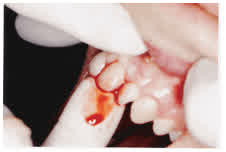
- Re-positioning of tooth back into the arch by placing one finger in front and one at the back
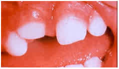
- Intrusion injury: more than half of the crown visible— allow to re-erupt
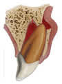
- Lateral luxation—associated with fracture of buccal bone
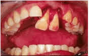
- Extrusive luxation
Management of soft tissue dental injuries continued
Type of injury | Description | Management | |
Primary dentition | Permanent dentition | ||
Avulsion | Tooth completely knocked out of the socket. Tetanus might be indicated in cases where the tooth is contaminated. |
|
|
Transport medium for avulsed teeth:
Saliva is a septic medium and avulsed teeth should ideally not be transported in saliva as it would affect the prognosis. In the absence of the most optimal transport medium i.e. Hanks Balanced Salt Solution (HBSS), milk has been proven to be the best transport medium. In the absence of milk, keeping the tooth in the cheek could be considered but this is not ideal as besides it being a septic medium, the tooth could be aspirated or swallowed.
Hard tissue injuries
- Check for fractures of teeth-- Is the tooth fractured?
- Check occlusion. Are the teeth biting together properly? If not, this could indicate a jaw fracture
- Assess if a bony fracture has occurred i.e. more than one tooth moves at the same time
- Sublingual haematomas are associated with fractures of the mandibule
Management of hard tissue dental injuries
Type of injury | Management: Primary and permanent dentitions |
Small fracture of tooth crown (pulp not exposed) | No immediate attention required Routine dental appointment required for restoration |
Large fracture (pulp is exposed—red/ dark red pin-point/ bleeding visible) | Requires immediate referral to dentist for pulp treatment and restoration |
Complicated crown fractures | Requires immediate referral to dentist for possible extraction/ pulp treatment |
Alveolar bone fracture (more than one tooth moves at the same time) | Rigid splint for 4 weeks Refer to maxillo-facial surgeon Refer to dentist for follow-up |
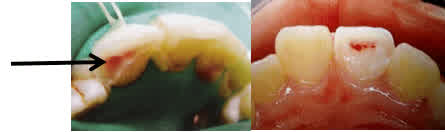
Pulp exposures
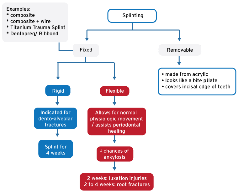
- Splinting
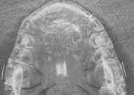
Removable splint
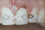
Titanium Trauma Splint
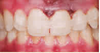
Dentapreg/Ribbond
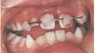
Composite and wire
Common intra-oral traumatic lesions:
Cheek ulceration as a result of biting a cheek accidentally during function. Treat with pain medication and a topical anaesthetic spray if required.
‘Pizza burn’ – Really hot food may result in burn trauma with the formation of a palatal ulcer. Healing time is about 10 days with pain medication and topical anaesthetic spray if required.
Lip ulceration in children as a result of ‘chewing’ on the lip after a dental appointment while the lip is still anaesthetised. The result may be an ulceration of alarming proportions. Treatment involves putting the patient and parents at ease and following the pain medication / topical anaesthetic regimen together with a chlorhexidine rinse.
Orthodontic wires causing soft tissue ulceration – Cover the area with a piece of wax if possible. Otherwise isolate the offending sharp wire with gauze or cotton wool and let them contact the orthodontist who is on call for that practice.
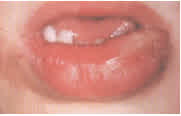
Lip biting after local anaesthetic had been administered
Gingival conditions
1. ANUG (Bacterial – Source intraoral)
ANUG is an acronym for Acute Necrotising Ulcerative Gingivitis. This was also known as ‘Trench Mouth’ during World War I. The main symptoms will be severe gingival discomfort. The patients are usually young adults who smoke, have poor oral hygiene and who are under stress. Clinical presentation will reveal halitosis and the breakdown (necrosis) of the triangle-shaped gum areas between the teeth.
Treatment for adults comprises Amoxicillin (500mg three times a day for 5 days) with Metronidazole (200mg or 400mg three times a day for 7 days). Pain medication and an antibacterial mouth rinse containing chlorhexidine should also be prescribed. Remember, no intake of alcohol along with the Metronidazole. They should report to their dentist after one week for further evaluation and treatment.
2. Periodontal abscess (Bacterial – Source intraoral)
Periodontitis is a chronic bacterial infection which causes bone breakdown around teeth. This results in deep pocketing between the gingival and the teeth. These pockets may become blocked and an acute swelling might occur. If the swelling can be visualised then it should be lanced and massaged to drain and the same treatment regimen instituted as in ANUG.
3. Pericoronitis (Bacterial – Source intraoral)
‘Peri-‘means ‘around’, ‘corona’ means crown (of tooth in this case), and ‘itis’ refers to inflammation’. This is most commonly seen in young people who have partially erupted wisdom teeth where the partial gingival covering has become infected. Emergency treatment involves rinsing under the flap of tissue over the tooth with a large syringe and chlorhexidine (or just water) and prescribing a broad spectrum antibiotic.
4. Dry socket (Osteitis)
The first step of healing after tooth removal is the formation of a stable blood clot in the socket. Sometimes this blood clot does not form optimally or it gets dislodged through eating or excessive rinsing. The bony surface of the socket then becomes infected by oral bacteria. This is accompanied by severe pain. The patient will give a history of a tooth being extracted in the preceding 10 days. Theoretically it will heal without further intervention but can take up to 6 weeks and there will be accompanying severe pain for an extended period. Optimal treatment involves local anaesthesia followed by cleaning out the socket with a curette and establishing bleeding so that a clot can form. A broad spectrum antibiotic should also be prescribed. If you are not able to clean the socket manually then at least rinse the socket with a syringe and water and prescribe the antibiotic.
Mucosal conditions – Vesiculo-Bullous diseases (Vesicles and bullae)
Erythema Multiforme (Auto-immune):This primarily affects the lips with an ulcerative, bloody, crusty appearance. The patient may give a history of previous episodes. Treat with 1mg/kg of Prednisone as a single dose in the mornings for 5 days. With this regimen no tapering of steroids needs to be done and can be repeated if more than 6 weeks between acute flare ups.
Oral Lichen Planus (Auto-immune):This autoimmune disease affects 1-2% of the population and may cause extensive bilateral ulcerations of the oral mucosa involving the buccal mucosa (cheeks), tongue, lips and gingiva. If they present with severe ulcerations then they may give a history of previous treatment for the disease. Treat with 1mg/kg of Prednisone as a single dose in the mornings for 5 days.
Bullous pemphigoid (Auto-immune): Occurs less than Oral Lichen Planus and patient will generally give a history of diagnosis and treatment. If severe flare up treat with 1mg/kg of Prednisone as a single dose in the mornings for 5 days.
Infectious mononucleosis (Also known as glandular fever or kissing disease) :Caused by Epstein Barr virus and can occur in children or young adults. Multiple small intraoral vesicles along with other symptoms such as fever, fatigue, sore throat and swollen lymph nodes. Most common in 15-17 year old age group. Treatment is palliative. Rule out Strep throat.
Acute herpetic gingivostomatitis (Herpes Simplex 1 / 2 virus) – Children or young adults:The majority of the population gets exposed to the Herpes virus early in their lives without developing any symptoms. A small percentage, however, develop an acute presentation which is extremely painful. The whole mouth is covered in vesicles making talking and eating difficult. The treatment is palliative involving pain medication along with a topical anaesthetic spray or an antibacterial rinse.
In children, primary herpetic gingivostomatitis is usually accompanied by fever. It is a self-limiting condition which resolves without treatment within 10 to 14 days. Citrus juices and spicy or salty food should be avoided. The child should increase their fluid intake to prevent dehydration. Nutritional supplements may also be indicated. Topical oral anaesthetic gels may reduce pain and discomfort. Paracetamol may be prescribed for pain and fever. Chlorhexidine mouth rinses are also beneficial to prevent secondary infection. In children younger than 7 years, it may be gently dabbed on with a piece of gauze.
References:
Douglass AB & Douglass JM (2003). Am Fam Physician. Common dental emergencies. Feb 1; 67(3):511-517.
Parthiscar A & Har-El G (2001). The annals of Otology, Rhinology and Laryngology. Deep neck abscess: a retrospective review of 210 cases. 110(11):1051-1054.
Fundamentals of Pediatric Dentistry (3rd Edition). RJ Mathewson, RE Primosch. Pg 287 + 297+ 301.
Dentistry for the child and adolescent (6th Edition). RE McDonald, DR Avery. Pg 503—563.
The management of traumatised anterior teeth of children. JA Hargreaves, JW Craig, HL Needleman.
Flores MT, Andersson L, Andreasen JO et al. (2007). Guidelines for the management of traumatic dental injuries II (b). Avulsion of permanent teeth. Dental Traumatology 23: 130-- 136.
Flores MT, Malmgren B, Andersson L, et al. (2007). Guidelines for the management of traumatic dental injuries III (c). Primary teeth. Dental Traumatology 23: 196-- 202.
McIntyre JD, Lee JY, Trope M, Vann WF (2007). Management of avulsed permanent incisors: a comprehensive update. Paediatric Dentistry 29 (1): 56—63.