Intestinal Obstruction
Surgical Emergencies - Intestinal Obstruction
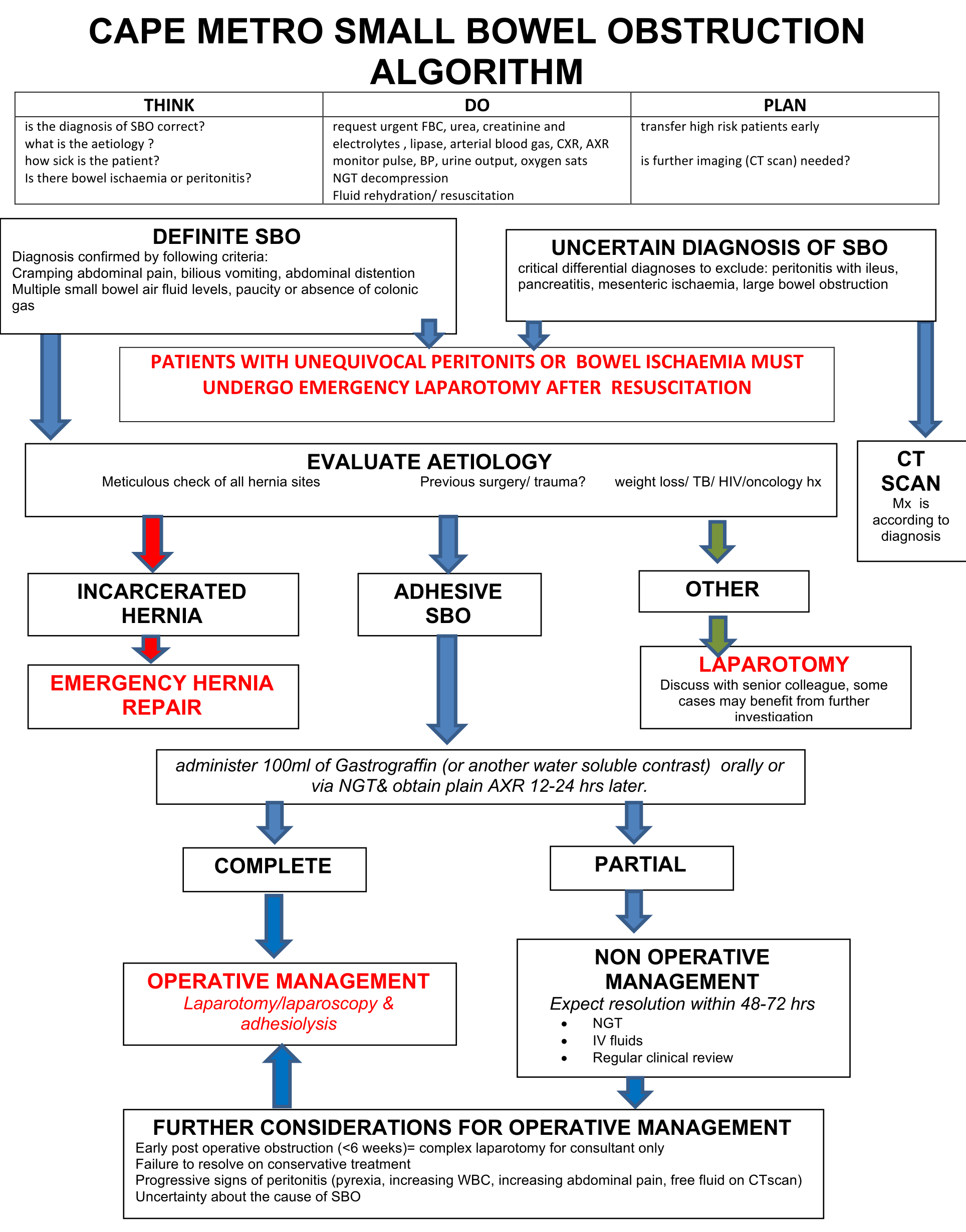
Cape Metro small bowel obstruction algorithm
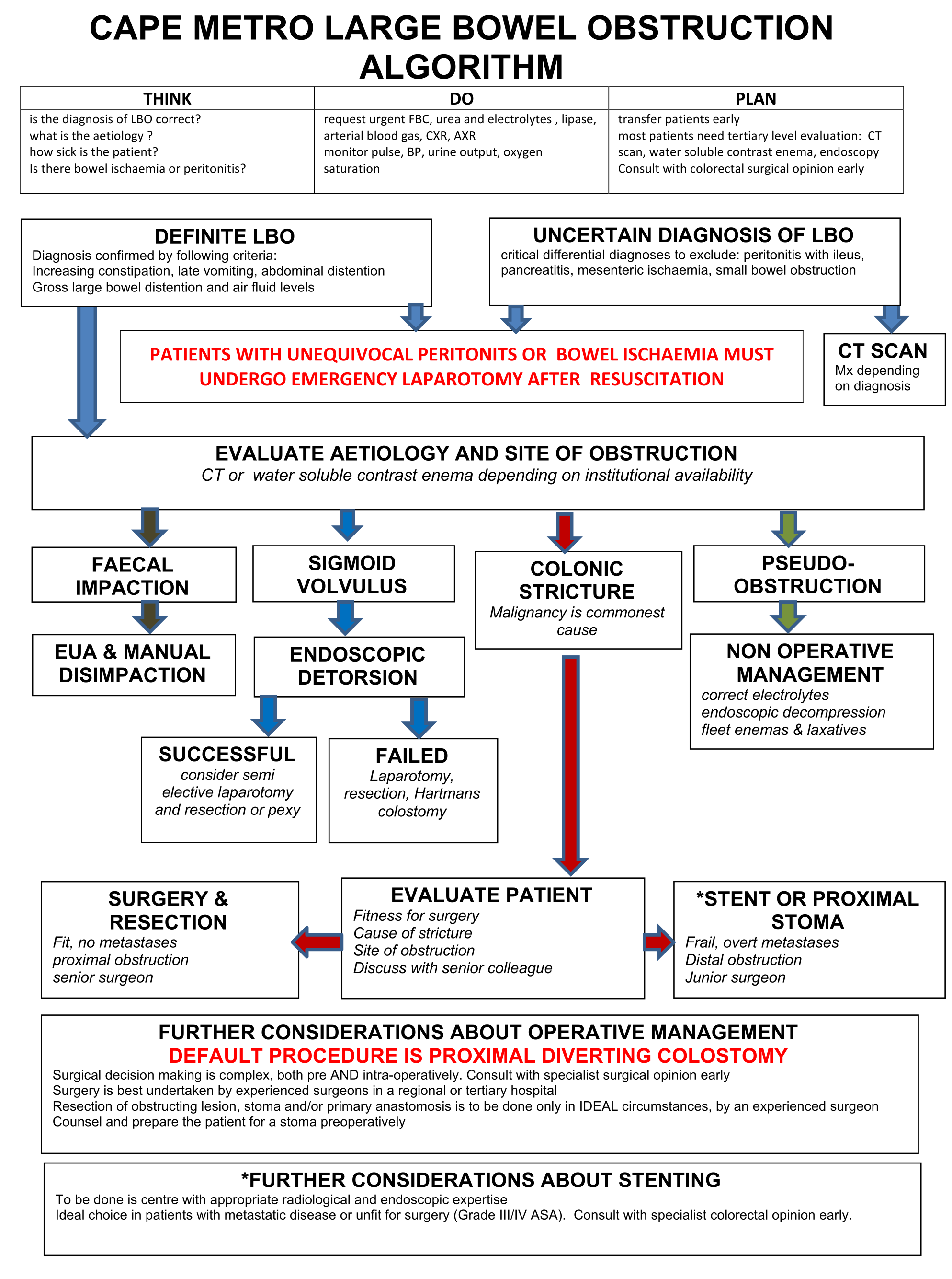
Cape Metro large bowel obstruction algorithm
The immediate goal in the management of patients with any bowel obstruction is the correction of fluid deficit and restoration of haemodynamics with intravenous fluids. Important investigations are FBC, urea and electrolytes with serum amylase, abdominal and chest X-rays, ECG in older patients or those with a history of ischemic heart disease, arterial blood gases in patients with respiratory co-morbidity and DIC screen if sepsis or peritonitis is suspected. Small bowel obstruction usually requires an operation unless a decision was made to carefully manage the patient non-operatively with intravenous fluids and nasogastric tube drainage in cases of adhesive bowel obstruction. If there is abdominal tenderness and any signs of peritonism, immediate surgery is usually required. A full thorough examination of all hernia sites is mandatory. Always consider a diaphragmatic hernia in a patient with bowel obstruction and a pleural effusion.
Diagnosis
- All patients being evaluated for small bowel obstruction (SBO) should have at least a supine AXR.
- All patients with inconclusive plain films for complete or high grade SBO should have a CT as it gives incremental information over plain films in regard to differentiating grade of obstruction and etiology, and may lead to changes in planned management. Multiple signs on CT suggesting strangulation should suggest operative intervention. If a CT scan is performed no oral contrast should be given as presence of oral contrast precludes the assessment of IV contrast in the bowel wall. In simple terms if you give oral contrast you cannot asses the presence of bowel ischaemia.
- The oral administration of 100ml of a water soluble contrast (Gastrograffin), followed by a plain AXR taken 12 hours later allows confident stratification for surgical intervention vs conservative treatment in cases of adhesive SBO. If the contrast passes into the colon within 12 hrs patients will almost always settle.
Management
- Patients with plain film finding of small bowel obstruction and clinical markers (fever, leukocytosis, tachycardia, metabolic acidosis and continuous pain) or peritonitis on physical exam warrant surgery.
- Patients without the above mentioned clinical picture, and a partial SBO or a complete SBO can undergo non-operative management safely; although, complete obstruction has a higher level of failure.
- Patients without resolution of their SBO by day 3-5 of non-operative management should undergo surgery.
- If a contrast test has been done, and the contrast has reached the caecum on the follow up AXR, the obstruction is expected to settle on conservative treatment, on the other hand if the contrast is still in the SB, the obstruction can be deemed complete and surgery can be undertaken without further delay.
- In a highly selected group of patients the laparoscopic treatment of small bowel obstruction should be considered
Large bowel obstruction
- The majority can be temporised until a CT scan is done to elucidate the cause and site of obstruction. This allows for planning of the operative or endoscopic management. An instant limited water soluble enema can also be of assistance in the evaluation of these cases.
- Large bowel obstruction needs immediate surgical intervention if there is tenderness over the caecum with caecal dilatation. This often denotes impending caecal rupture
- Stenting should be considered for left sided bowel obstruction either as a bridge to surgery of definitive palliative treatment. This can be performed at any hospital with the requisite skills.
Cape Metro Small Bowel Obstruction Algorithm
Cape Metro Large Bowel Obstruction Algorithm
Bowel Obstruction Guidelines, 2023