Intra-Osseous Infusions
Intra-Osseous Infusions
Fluids and drugs suitable for intraosseous use
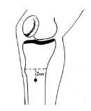
Insertion of infusion in the proximal tibia
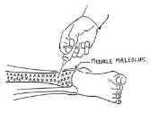
Insertion of infusion in the distal tibia
Anatomy
The medullary cavities of the long bones are comprised of a network of spongy venous sinusoids, which drain to a central venous canal. The system is fed and drained by vessels to and from the systemic circulation.
Areas
(Broad flat bones with minimal overlying soft tissue)
- Proximal tibia.
- Distal tibia.
Requirements
- Needles:
- Preterm- or term neonates: No 23G butterfly or LP needle (with stylet no 18 or 20)
- Children <18/12: LP needle (with stylet) No 18 or 20
- Older children: Bone marrow needle No 13 - 16
- Iodine en alcohol
- Sterile swabs
- 1% Lignocaine
- Intravenous fluids and infusion set
- Assistant
Indications
- Use only when ability to place traditional venous line has been exhausted.
- Acute life threatening circumstances such as cardiac arrest, severe dehydration or shock.
Fluids and drugs suitable for intraosseous use
The average achievable flow rate is 10 ml/min with gravitational assistance.
An infusion control pump should be used.
- FLUID :
- Crystalloid (Neonatalyte; 0.2% NaCl + 5% D/W; 0.9% NaCl), blood and stabilised human serum.
- DRUGS :
- Sodium bicarbonate
- Atropine
- Dopamine
- Antibiotics
- Anti-convulsants
- Sodium bicarbonate
Complications
Osteitis and cellulitis. Prevent these by using sterile technique and correct method of needle insertion. Avoid infected areas or fractured areas.
Extravasation of the infusion-fluid (usually as a result of the pressure pump or prolonged infusion). Stop the infusion and apply a pressure dressing over the area.
Us the alternate limb if a repeat infusion is required.
Epiphyseal plate injury can occur if the needle is inserted more proximally or distally than the recommended insertion point.
Sketch 1
Sketch 2
Insertion of infusion in the proximal tibia (sketch 1 above)
- Use sterile gloves.
- Clean the upper 1/3rd of the tibia with iodine and alcohol.
- Infiltrate the skin and peritoneum with local anaesthetic (1% lignocaine) after determining the site for needle insertion.
- Steps 1 & 3 can be omitted if the neonate is in cardiac arrest or comatose.
- Palpate the tibial tuberosity with the index finger and the medial aspect of the tibia with the thumb. A line is drawn between the two palpated points. The point of insertion is midway between the two palpated points and 2 cm below (distally). (See sketch 1).
- The needle is inserted perpendicular to the bone cortex with a twisting action (be careful as the needle can be pushed right through the bone in small neonates).
- The medullary space is ½ - 1 cm deep and has less resistance to needle insertion than initially felt advancing through the cortex.
- Confirm that the needle is correctly placed by removing the stylet (spinal or bone marrow needle) and aspirating blood or rinsing through with 1 - 2 ml of sterile 0.9% NaCl. Observe for fluid extravasation and ease of flow. If resistance to flow is experienced, the needle is more than likely too deep.
Insertion of infusion in the distal tibia (sketch 2 above)
The optimal site for needle insertion is the medial surface of the tibia, proximal to the medial malleolus.
Sterile technique and needles should be used in the same manner as for proximal tibia infusion.