Intrauterine Growth Restriction
The guidelines recommend management of common obstetric conditions at primary and referral level, based on local experience and evidence-based practice. They are not intended as strict protocols nor as standard operating procedures, but rather as a guide to safe effective management of common obstetric problems. The guidelines should be most useful to interns, medical officers and registrars.
Intrauterine growth restriction (IUGR) refers to the failure of a fetus to achieve its growth potential. IUGR can be classified into 2 main groups:
- Symmetric – both the head & body show growth failure. This may result from genetic or chromosomal defects, intrauterine infection, or exposure to teratogens. Ultrasound may reveal structural abnormalities. Some fetuses may appear symmetrically growth impaired, but are normal small babies, or may be suspected to be small because of wrong pregnancy dates.
- Asymmetric – the head continues to grow, but the body shows growth deceleration. This may result from pre-eclampsia, vascular disease (as in diabetes or lupus), or from isolated placental insufficiency. Ultrasound findings are those of placental insufficiency, such as oligohydramnios & abnormal Doppler wave-forms (umbilical vessels, middle cerebral artery, ductus venosus)
In a nutshell
- When both the anomaly scan and umbilical artery waveforms are normal, the small fetus should be classified as a ‘normal small fetus’
- When the anomaly scan is abnormal and the waveforms are normal, the fetus is an ‘abnormal small fetus’– symmetric IUGR
- When the anomaly scan is normal & Doppler waveforms are abnormal, there is asymmetric IUGR
Factors causing growth restriction
| FETAL FACTORS Chromosomal Congenital infection Genetic syndromes Multiple gestation | MATERNAL FACTORS Constitutional Nutrition Genetic (e.g. phenylketonuria) Hypertension, current or previous Cardiac disease Autoimmune disease Diabetes mellitus Kidney disease Medication Smoking, alcohol & recreational drugs Exposure to toxins Residence at high altitude |
| PLACENTAL FACTORS Abnormal trophoblast invasion (e.g. pre-eclampsia) Abnormal cord insertion Placental lakes Placental location (e.g. praevia) Tum |
Pathophysiology of impaired placentation in asymmetric IUGR
- There is minimal or no trophoblastic invasion into maternal uterine spiral arteries beyond the decidual-myometrial junction. Cellular mechanisms may involve reduced trophoblast motility, increased trophoblast apoptosis, altered natural killer cell function, & endothelial cell interaction
- This results in failure of conversion of the small muscular spiral arteries into large vascular channels: the uteroplacental circulation cannot then develop into a healthy low-resistance-to-flow system
- The consequence is placental insufficiency &/or pre-eclampsia, with fetal growth restriction
SCREENING FOR IUGR
Methods to identify women at risk for IUGR include the following:
- Antenatal history-taking & assessment for factors listed in the above
- First-trimester ultrasound screening at 11-14 weeks for fetal abnormalities & for uterine artery Doppler indices as for pre-eclampsia screening
- Third-trimester symphysis-fundal height measurement & uterine palpation to identify the small-for-dates uterus
Diagnosis and follow-up
Ultrasound scanning is done to confirm IUGR based on biometry & umbilical artery Doppler indices.
- If the umbilical artery resistance index is abnormally increased, consider delivery if estimated fetal weight ≥1.5 kg or gestation ≥34 weeks, otherwise follow up with weekly middle cerebral artery pulsatility indices (MCA PI) as long as these remain normal. Give betamethasone to accelerate lung maturity
- If middle cerebral artery indices are abnormal, admit to hospital for twice-weekly ductus venosus Doppler & daily NST
- Deliver if the ductus venosus Doppler is abnormal or the NST is pathological
- Individualise management with estimated fetal weight <900 g
ALGORITHM
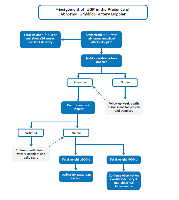
Timing of delivery with IUGR in the presence of pre-eclampsia
- If fetus ≥1.5 kg (≥34 weeks) give betamethasone & consider delivery
- From 32-34 weeks factors such as availability of neonatal services are important points when making a decision
- From 26-32 weeks, follow with multivessel Doppler studies & NSTs
- In cases of severe pre-eclampsia, the maternal condition precludes fetal observation & may necessitate delivery in the woman’s interest
Mode of Delivery
- Maternal well-being must be balanced against the likely neonatal outcome
- Caesarean section in the baby’s interest can rarely be justified as a means of delivery at ≤24 weeks’ gestation because of the poor neonatal prognosis.
- If the parents insist on caesarean section, seek a second senior opinion
- In the presence of a reassuring NST, vaginal delivery is generally preferable with continuous CTG in labour, but at gestation <32 weeks, caesarean section may be preferred as the success rate of induction is reduced.