Patient-Controlled Analgesia and Neuraxial Analgesia Techniques
Clinical guidelines on the use of patient-controlled analgesia Chart 1: Proposed prescription chart Neuraxial techniques PCA and neuraxial analgesia techniques offer alternative nonconventional methods for postoperative pain management. Clinical guidelines on the use of patient-controlled analgesia Rationale for use PCA refers to a method of analgesia through which a patient self-administers smal
Clinical guidelines on the use of patient-controlled analgesia
Chart 1: Proposed prescription chart
PCA and neuraxial analgesia techniques offer alternative nonconventional methods for postoperative pain management.
Clinical guidelines on the use of patient-controlled analgesia
Rationale for use
PCA refers to a method of analgesia through which a patient self-administers small doses of an analgesic, usually an opioid, delivered with a programmable infusion pump.
Efficacy
The proposed benefits of PCA, compared to conventional parenteral opioid regimens, include the following:
- Improved pain control
- Higher patient satisfaction
- Decreased risk of overdose
- Less labour-intensive for the nursing staff
However, IV PCA with opioids may result in higher opioid consumption and pruritus compared to intermittent opioid administration. There may also be no difference in efficacy in settings with a high nurse-to-patient ratio.
The decision to provide PCA depends on availability and resources (financially and staffing), and is taken after discussion with the patient. Ongoing communication regarding, for example, the adjustment of demand dosing, may influence the success of PCA management.
PCA is only effective after the initial rapid pain control under supervision of the prescribing physician, for example, in the postoperative care unit. Initial doses should be individualised, taking into account factors such as previous opioid use and age.
The concept of PCA continues to develop in children. Patient-controlled epidural analgesia (PCEA), SC PCA and intranasal (IN) PCA are extensions of this method. PCA, when used with adequate monitoring, is a well-tolerated technique with high patient and staff acceptance. It can now be regarded as a standard for the delivery of postoperative analgesia in children above the age of 5 years.
Cost
Although equipment cost is higher, the possible benefits of reduced adverse effects and nursing time must also be considered.
Standards of care
The safety of PCA can be improved upon by adopting standardised protocols regarding methods and documentation (see Chart 1). Operator errors may be reduced with the use of ‘smart pump’ technology. Although technical device errors may be more common than operator errors, very few technical errors seem to be associated with patient harm.
Chart 1: Proposed prescription chart
Prescription chart
Instructions
- Nursing staff are not allowed to change settings on the pump
- PCA boluses must only be administered by the patient, not by the nursing staff
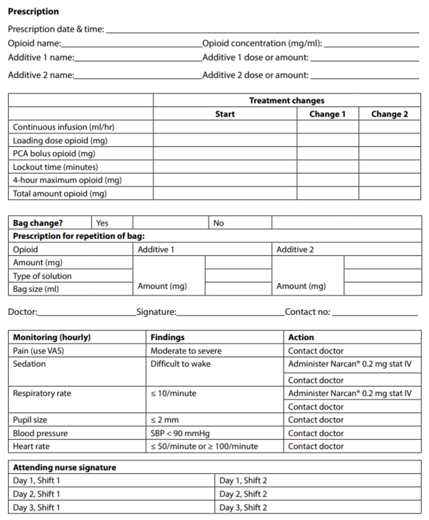
Click on the image to enlarge
Additional instructions
Clinical practice points
- The changing pain score and the presence of side effects should be detected through appropriate routine monitoring of patients, in order to prevent complications such as an inadvertent overdose or inappropriate use (e.g. nurse or doctor administration of the intended PCA bolus). The masking of pain from new problems, such as compartment syndrome of the limbs, urinary retention, pulmonary embolism (PE) or myocardial infarction, should also be uncovered.
- Monitoring addresses the prevention of complications, and assures that repeated assessment is conducted with regard to the adequacy of pain management.
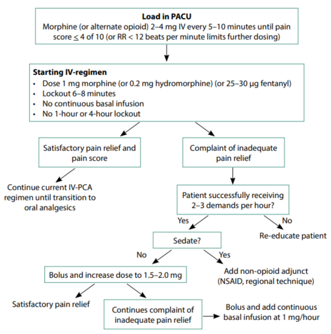
Figure 1: Proposed treatment algorithm for patient-controlled analgesic management (adapted, not revised; used with permission)
3. Intensive and frequent monitoring is essential in patients at risk of developing respiratory depression and other side effects. If peripheral oxygen saturation cannot be continuously monitored, administering oxygen by nasal cannula or face mask for the duration of IV opioid administration may be indicated.
4. Standardised prescriptions within institutions also prevent complications arising from the use of supplemental medication, such as opioids by other routes, or sedatives (see Table I).
5. A standardised treatment algorithm may improve pain management by integrating pain assessment and side effects with a clear reaction pathway. A treatment algorithm for PCA management is detailed in Figure 1.
Medication
Opioid analgesics
Generally, there are no major differences in the efficacy of various opioid drugs for PCA. On an individual basis, one opioid may be better tolerated than another, and it may be beneficial to change to an alternative agent if a patient is experiencing intolerable side effects.
The following opioid analgesics are available for PCA use in South Africa:
- IV morphine: Included in the adult hospital level Essential Medicines List. A background infusion increases the risk of respiratory depression with no additional benefit.
- IV tramadol: Recommended for inclusion in the adult hospital level Essential Medicines List, for regional hospitals only. It provides effective analgesia comparable to morphine.
- IV pethidine: Pethidine should not be prescribed. It may cause neurotoxicity owing to the accumulation of norpethidine.
- IV remifentanil: Offers analgesia equivalent to morphine and may be associated with less nausea and vomiting.
- IV fentanyl: There is limited evidence of a difference between IV fentanyl and morphine.
- IV oxycodone: It has similar effects to morphine.
Adjuvant medicines in PCA
The opioid dose-sparing effect of concurrent NSAIDs, ketamine, pregabalin, lignocaine, clonidine and dexmedetomidine, may contribute to a reduction of opioid adverse effects. The following adjuvant medicines may be added to the opioid solution.
Antiemetic drugs
- The routine addition of antiemetic drugs to a PCA infusion is not advocated as there is no additional benefit over selective administration when indicated.
- Droperidol is an effective antiemetic, but may cause unacceptable sedation at the dose necessary to prevent nausea and vomiting.
- The benefit of adding 5-hydroxytryptamine-3 antagonists (e.g. ondansetron) to PCA, is unclear. 5-HT-3 receptor antagonists should not be used with tramadol.
Ketamine
- The addition of ketamine to PCA may benefit patients after major surgery with regard to analgesia as well as the adverse effects of opioids.
Dexmedetomidine
- There is some evidence that the use of dexmedetomidine, when added to IV morphine PCA, may improve analgesia and reduce morphine-related side effects without increasing sedation or the haemodynamic side effects.
Hydroxyzine
- There is no evidence to support the addition of hydroxyzine to PCA.
Equipment and programme variables/settings
Several PCA products are available in South Africa. Systems can be broadly categorised into the following two groups:
- Durable (often bulky) pumps with disposable cartridges, which usually offer multiple programmable options.
- Pumps composed of disposable components with built-in mechanisms for bolus administration. These do not allow background infusion administration or provide programmable options.
The effectiveness of the latter systems may be compromised by the fact that dose adjustments cannot be made, although these may provide a cost benefit.
All PCA infusion systems must incorporate anti-syphon valves, as well as anti-reflux valves in nondedicated lines
Pump settings
Bolus dose
- The optimal bolus dose should provide adequate pain relief with minimal adverse effects. Age and a history of previous opioid exposure can influence the response to the bolus dose. The usual PCA bolus dose for morphine is 1 mg (15–20 µg/kg).
Lockout time
- The lockout time interval should allow the analgesic to reach its peak effect. The optimal lockout time for morphine is 7–11 minutes.
Dose limits
- An overdose with PCA is usually due to the effect of large doses accumulating after hours or days. Limiting the maximum dose over several hours may be the most effective way of preventing an overdose. However, sound evidence of a benefit that can be attributed to these limits, does not exist. The impact of decreased clearance should be kept in mind (e.g. liver impairment, kidney failure, cardiac failure and any cause of a decreased cardiac output).
Background infusions
- The risk of respiratory depression is higher when a background infusion is used, and it does not improve pain control or sleep, or reduce the number of PCA demands. It may be useful in opioid-tolerant patients, but its routine use is not advised.
Loading dose
- No good evidence of any benefit exists when using this option on programmable pumps. Pain should be controlled first by titrated LDs before PCA is started.
Total amount and concentration
- Drug concentrations should be standardised within institutions to reduce the occurrence of programming errors.
Examples of PCA management regimens are provided in Table I.
Table I: Examples of IV patient-controlled analgesia management regimens
| Morphine with Opioid dilution of 1 mg/ml | ||
| Additive | Additive dilution | Pump settings for opioid |
| Dexmedetomidine or ketamine droperidol or ondansetron |
2–5 μg/ml 2 mg/ml 15–100 μg/1 mg morphine, or 5 mg/100 ml 16 mg/100 ml |
- Bolus of 1 mg (1 ml) - Lockout time of 7–11 minutes - Maximum 30 mg in 4 hours |
| Tramadol with Opioid dilution of 5 mg/ml | ||
| Additive | Additive dilution | Pump settings for opioid |
| Dexmedetomidine | 1–2 μg/ml | - Bolus of 10–20 mg - Lockout time of 5–10 minutes |
Table I continued
| Opioid | Opioid dilution | Pump settings for opioid |
| Remifentanil | 50 μg/ml | - Bolus of 50 μg over 5 minutes (background 0.075– 0.15 μg/kg/ minute) - Lockout time of 5 minutes |
| Sufentanil | 1 μg/ml | - Bolus of 4–6 μg (background of 1.15 μg/hour) - Lockout time of 1 minute |
| Fentanyl | 1 μg/ml | - Bolus of 30–40 μg - Lockout time of 5–8 minutes - Maximum 300 μg in 4 hours |
Neuraxial techniques
Epidural analgesia
Epidural analgesia (i.e. the provision of pain relief by the continuous administration of agents into the epidural space via an indwelling catheter) has become a widely-used technique for the management of acute pain in adults and children. Epidural analgesia at the correct level for an appropriate duration may decrease pulmonary complications, ventilator requirements or MI, and may improve bowel motility. However, data on improved outcomes are controversial, and the focus should be on pain relief and patient satisfaction.
Clinical practice points regarding epidural analgesia
- The decision to perform an epidural, and the technique selected, should be appropriate for the intensity of pain anticipated and congruent to the level of tissue damage (i.e. head and neck, torso or limbs).
- All techniques of epidural analgesia, except when using a lipophilic opioid only, for all types of surgery, provide better postoperative pain relief than parenteral opioids (including PCA).
- The complete absence of pain is seldom achievable, and never realistic, even with neuraxial techniques. The objective should therefore be the balance between analgesia, patient satisfaction, safety and available resources. A VAS count of < 4/10 (40/100) is usually a satisfactory goal.
- Combinations of low concentrations of LAs and opioids provide better analgesia than either component alone, and reduce the dose requirements of both drugs.
- Permanent neurological damage with epidural techniques is a rare, but devastating, occurrence and all efforts should be made to prevent, diagnose and treat it immediately. Immediate decompression of a haematoma or an abscess increases the likelihood of recovery. Therefore, the risks and benefits should be discussed with the patient and informed consent obtained.
- The insertion of an epidural catheter at spinal level matching the dermatome of the surgery (i.e. catheter incision-congruent analgesia) results in optimal postoperative epidural analgesia by infusing analgesic agents to the appropriate incisional level, providing superior analgesia and minimising side effects.
- Infusions of epidural LA plus opioid combinations in a general ward have been advocated to be safe, but the precondition is supervision by an anaesthesia-based pain or similar service, with 24-hour medical staff cover, and monitoring by well-trained nursing staff. In many hospital wards, this may not be available or possible.
- Epidural catheters are inserted under sterile conditions for obvious reasons. A sterile theatre gown, mask, cap and gloves must be worn. It is prudent to use an alcoholic chlorhexidine solution to disinfect the skin, adhere to antiseptic technique at insertion, maintain sterility at the puncture and infusion ports, and to remove the catheter before or on day five. Chlorhexidine-impregnated dressings of epidural catheters reduce the incidence of catheter bacterial colonisation. Whenever the epidural filter gets disconnected from the catheter, both the filter and the catheter must be regarded as contaminated and the epidural catheter must be removed.
- The level of the epidural is determined by the level of trauma. The benefits of postoperative epidural analgesia are optimised when the epidural catheter is inserted in a location which corresponds to the dermatomes covered by the surgical incision (i.e. catheter incision-congruent analgesia) resulting in a lower dose of the drug being administered and a decreased incidence of drug-induced side effects, such as pruritus, nausea, vomiting, urinary retention, motor block and hypotension. Although an incision-congruent level of block supplies somatic analgesia, visceral analgesia requires a block that includes a few segments above and below the incision level (Table II). According to NYSORA, a generally accepted guideline for dosing epidural anaesthesia in adults is 1–2 ml per segment to be blocked.
Table II: Level of the epidural
| Surgery | Level of the epidural |
| Thoracotomy | T4–T8 |
| Upper abdominal | T6–T8 |
| Middle abdominal | T8–T10 |
| Lower abdominal | T8–T12 |
| Lower extremity | L1–L4 |
Epidural local anaesthetics
LAs available for epidural use in South Africa are outlined in Table III.
Table III: Local anaesthetics for epidural use available in South Africa
| Duration of action | Drug | Formulation |
| Short-acting | Lignocaine | Various |
| Bupivacaine | Macaine® 0.5% (5 mg/ml), with or without adrenaline Macaine® 0.1% (1 mg/ml) polybag |
|
| Long-acting | Levobupivacaine | Chirocaine® 5.0–7.5 mg/ml |
| Ropivacaine | Naropin® 5.0, 7.5 and 10 mg/ml, and 2 mg/ml as the 100/200 ml polybag |
There are no consistent differences between ropivacaine, levobupivacaine and bupivacaine when given in low doses for regional analgesia in terms of quality of analgesia and motor blockade.
Epidural opioids
The behaviour of epidural opioids is governed largely by their lipid solubility. Morphine is the least lipid soluble of opioids administered epidurally. As it has a prolonged analgesic effect, it can be given either by intermittent bolus dose or by infusion. With bolus dose regimens, the risk of respiratory depression is higher and analgesia is less effective. Lipophilic opioids (e.g. fentanyl, sufentanil and alfentanil) have a faster onset, but shorter duration of action, compared with hydrophilic drugs. Contrary to epidural morphine, epidural lipophilic opioids reach analgesic systemic concentrations, which contributes substantially to their analgesic effects.
The levels of the required level of the epidural blocks are outlined in Table II.
Epidural test doses
This procedure is standard when the epidural commences, but should be repeated before top-up doses for analgesic purposes. Although migration of the catheter to the intravascular or intrathecal space from the epidural space is uncommon, aspiration and the administration of a test dose of adrenaline and LA before bolus dosing for pain may prevent complications (e.g. high or total spinal anaesthesia and neurotoxicity) associated with the accidental administration of LAs into this space. After negative aspiration, 45 mg of lignocaine plus 15 µg of adrenaline, can be administered as a test dose. This is prepared as follows:
- Adrenaline 2 mg is diluted to 10 ml = 20 µg/ml.
- Add 1 ml of this adrenaline dilution (20 µg) to 3 ml of lignocaine 2% (20 mg/ml) in a 5 ml syringe. This solution contains 60 mg of lignocaine plus 20 µg of adrenaline in 4 ml. Therefore, 3 ml will contain 45 mg of lignocaine and 15 µg of adrenaline.
- Since the dead space of the epidural catheter and filter is approximately 1 ml, 4 ml can be administered in a bolus. If the filter and catheter has been primed with the solution, 3 ml is administered.
According to NYSORA: “The intrathecal injection of 45 mg of lidocaine should produce a significant motor nerve block if the catheter is in the subarachnoid space, although recent evidence suggests that this is not always reliable. A change in heart rate of 20% or greater (or, alternatively, an increase in heart rate of 10 to 25 beats per minute) within 1 minute suggests that the catheter has been placed in (or has migrated into) a vessel and should be replaced. If the heart rate does not increase by 20% or greater or if a significant motor nerve block does not develop within 5 minutes, the test dose is considered negative. Exceptions to this rule have been observed in labouring patients, anaesthetised patients, and patients receiving β-adrenergic blocking agents.”
Patient-controlled epidural analgesia
The use of patient-controlled epidural analgesia (PCEA) is based on the individualisation of therapy, similar to other patient-controlled techniques. The optimal settings of PCEA will vary, based on the epidural vertebral level, type of surgery, as well as patient factors.
Systemic analgesia in combination with epidural analgesia
The use of epidural analgesia does not preclude the use of systemic analgesia. Patients with epidural analgesia should have concomitant paracetamol prescribed to treat other aches and pains, and facilitate the withdrawal of epidural analgesia.
Table IV: Complications of epidural analgesia treatment
| Complications | Incidence | Treatment | Remarks |
| Nausea and vomiting | Common | Ondansetron or class equivalent, droperidol or prochlorperazine | Dose-dependent |
| Pruritus | Common | Antihistamine, naloxone or droperidol, HT3 receptor antiemetics (ondansetron, granisetron) | Dose-dependent |
| Urinary retention | Common | Catheterise | |
| Postdural puncture headache | Uncommon, unless dura is perforated | Bed rest, intravenous fluids, caffeine, analgesics, neostigmine plus atropine If no improvement of symptoms within 48 hours, consider an epidural blood patch | A blood patch is controversial |
| Hypotension | Uncommon | IV volume Decrease the dose and rate of the epidural infusion |
Prevent hypovolaemia |
| Motor block | Common with higher concentrations of LA | Decrease the dose of local anaesthetic | It may be more frequent with bupivacaine |
| Respiratory depression | Uncommon | If due to opioid: naloxone Supportive measures (see text below for specific interventions) |
Sedation is an early warning sign Sedation can be caused by hypoventilation due to opioids, hypotension, high block, etc. |
| Local anaesthetic systemic toxicity (LAST) | Uncommon | LAST: intralipid CPR |
Bupivacaine > levobupivacaine ≥ ropivacaine |
| Epidural haematoma or abscess | Rare | Surgical decompression within 8 hours | Urgent neurosurgical consultation and imaging to confirm the diagnosis |
| Permanent neurological damage | Rare | Prevent | Warn patient of risk before performing the procedure |
LA – local anaesthetic, LAST – local anaesthetic systemic toxicity, CPR – cardiopulmonary resuscitation
Respiratory depression by epidural solutions
A strategy to detect and treat this complication should be in place. The incidence of respiratory depression is between 1% (decreased RR) and 15% (desaturation) depending on the criteria used, but is clinically significant in less than 1% of patients. High-risk patients (e.g. sleep apnoea and obesity) should be identified preoperatively.
Patients receiving neuraxial opioids should be monitored clinically for adequacy of ventilation, oxygenation and level of consciousness 1–2-hourly for the first 24 hours after injection with morphine, or continuously during an infusion. The absence of a decreased RR is not a reliable warning sign of respiratory depression, but it is almost always preceded by sedation. (Exclude motor block, high block, sympathetic nervous system block, migration of catheter, etc.)
Supplemental oxygen should be available to patients receiving neuraxial opioids, and administered to those with an altered level of consciousness, respiratory depression or hypoxaemia. In the infrequent event of life-threatening apnoea, the airway should be maintained, and the patient bag-mask ventilated pending naloxone administration. The naloxone dose should be titrated to effect (40–80 μg IV increments), followed by an infusion at a dose sufficient to maintain an adequate RR until the effect of the opioid has worn off (e.g. 1–2 μg/kg/min).
The reason for the respiratory depression should be assessed and the dose of the neuraxial infusion of opioids decreased if this is the cause. If an epidural infusion is stopped, alternative pain treatment should be prescribed to prevent rebound pain.
The patient should be transferred to a higher level of nursing for care and monitoring.
Duration of epidural analgesia
The epidural catheter is commonly left in place for 2–4 days, but it is impossible to scientifically determine what the maximum safe time would be to persist with a percutaneous catheter owing to the rarity of epidural infections.
Drugs and doses used in epidural analgesia are outlined in Table V.
Table V: Drugs and doses used in epidural analgesia
Lipophilic opioid infusion
| Fentanyl | |||
| Dose | Onset | Duration | Remarks |
| Dilute the single dose in 10 ml normal saline: - Loading dose = 50–100 μg - Infusion dose = 25–100 μg/hour |
5–10 minutes | 2–4 hours | - Limited spread in CSF - Early respiratory depression is most likely - Opioids alone via the epidural route seem to be of limited benefit |
| Sufentanil | |||
| Dose | Onset | Duration | Remarks |
| Dilute the single dose in 10 ml normal saline: - Loading dose = 10–50 μg - Infusion dose = 10–20 μg/hour |
5–10 minutes | 2–4 hours | - Limited spread in CSF - Early respiratory depression is most likely - Opioids alone via the epidural route seem to be of limited benefit |
Hydrophilic opioid infusion
| Morphine | |||
| Dose | Onset | Duration | Remarks |
| Loading dose = 1–5 mg Infusion dose = 0.1–1 mg/hour |
30–60 minutes | 6–24 hours | - Extensive spread in CSF - Early and delayed respiratory depression is possible |
Local anaesthetics
| Bupivacaine* | |||
| Dose | Onset | Duration | Remarks |
| 5–8 ml/hour of 1.25–2.5 mg/ml solution or incremental doses of 3–5 ml of 1.25–2.5 mg/ml solution | 10–20 minutes 30 minutes for optimal effect |
3–4 hours | - Establish the block with a 0.5% bolus of 15–30 ml - Recommendation: Limit to 2 mg/kg in 4 hours and 400 mg/24 hours |
| Levobupivacaine | |||
| Dose | Onset | Duration | Remarks |
| 10–15 ml/hour of 1.25 mg/ml, or 5–7.5 ml/hour of 2.5 mg/ml | 150–240 minutes | - Minimal to moderate motor block - Dilution stable for up to 7 days at 20°C - Maximum dose over 24 hours of 400 mg |
|
| Ropivacaine | |||
| Dose | Onset | Duration | Remarks |
| 2 mg/ml Bolus:10–20 ml 6–14 ml/hour |
15–20 minutes | 140–200 minutes | Establish the block for surgery with 15–25 ml of 7.5 mg/ml for a lumbar epidural, or 5–15 ml for a thoracic epidural |
* Not commonly used alone in an analgesic infusion, CSF – cerebrospinal fluid
Local anaesthetic-opioid infusions
| Drug | Dose | Remarks |
| Ropivacaine 2 mg/ml + fentanyl 4 μg/ml | 6–14 ml/hour | This combination is marketed as a polybag in some countries (not in South Africa) |
| Bupivacaine 1 mg/ml + fentanyl 4 μg/ml | 5–10 ml/hour | Prepare by adding 20 ml of 0.5% bupivacaine to 40 μg fentanyl (4 x 100 μg ampoules) and 72 ml normal saline (total volume = 100 ml) |
| Bupivacaine 0.625 mg/ml +fentanyl 2–5 μg/ml | 8–16 ml/hour |
Local anaesthetic-opioid epidural PCA (infusion + demand)
| Levobupivacaine 1.25 mg/ml + fentanyl 4 μg/ml | |||
| Continuous infusion (ml/hour) | Demand dose (ml) | Lockout (minutes) | Remarks |
| Initial rate of 4 ml/hour | 2 | 10 | Stability proven for up to 40 hours at 20°C |
| Bupivacaine 1 mg/ml + fentanyl 5 μg/ml | |||
| Continuous infusion (ml/hour) | Demand dose (ml) | Lockout (minutes) | Remarks |
| 6 ml/hour (3–4 ml/hour for thoracic block) | 2 | 10–15 | |
| Ropivacaine 1–2 mg/ml + fentanyl 2 –5 μg/ml | |||
| Continuous infusion (ml/hour) | Demand dose (ml) | Lockout (minutes) | Remarks |
| 3–5 ml/hour | 2 | 10–20 | |
Spinal (intrathecal) analgesia
A single injection of intrathecal LA plus an opioid is an acknowledged part of a postoperative analgesia strategy. It is as effective as, or even better than, other established techniques, although the duration of the relief is limited to the first 24 hours and side effects are common.
Drugs used for intrathecal analgesia
LAs are often combined with opioids to provide a smooth transition from the anaesthetic technique to the analgesic plan.
Drugs for intrathecal analgesia are detailed in Table VI.
Table VI: Drugs for intrathecal analgesia
| Drug (preservative free) | Intrathecal single dose | Onset | Duration |
| Fentanyl | 5–25 μg | 5–10 minutes | 1–4 hours |
| Sufentanil | 2–10 μg | 5–10 minutes | 2–6 hours |
| Morphine | 50–300 μg | 45–75 minutes | 18–24 hours |
The longer duration of morphine has established it as the drug of choice, but this advantage is gained at the increased risk of respiratory depression. At doses of 100–800 µg, intrathecal morphine for pain relief, following a range of surgical procedures, produces a high degree of patient satisfaction and effective analgesia during the first 24 hours. In particular, the lower dose of 100–200 µg offers effective analgesia, with a low risk of adverse effects (e.g. for hip replacement in the elderly). However, higher doses are required for thoracotomy and abdominal surgery. In general, there is a ceiling analgesic effect for doses above 200 μg. Significant side effects associated with intrathecal morphine are as follows:
- Respiratory depression in 3% of patients (partial pressure of carbon dioxide [PaCO2 ] ≥ 50 mmHg, and/or an RR ≤ 8/minutes)
- Pruritus (itching) in up to 30% of patients
- Nausea and vomiting in 25% of patients
- Urinary retention in 35% of patients
When intrathecal morphine is administered, the patient should be nursed in an area where there is a high level of awareness of the risk of respiratory depression. The monitoring and treatment of these complications is similar to that employed with epidural opioids. The analgesic efficacy rivals that of PCA, but does not last for longer than a day. A multimodal plan of alternatives should be in place to prevent (preferably) or treat rebound pain. Lipophilic opioids may be suitable for outpatient surgery, but morphine is not.
Indwelling spinal catheters are not established as a routine technique for the treatment of short-term pain.
Clinical practice points
- Intrathecal morphine offers improved analgesia and is opioid-sparing for up to 24 hours.
- Intrathecal morphine doses of ≥ 300 µg increase the risk of respiratory depression.
- The incidence of respiratory depression and pruritus after major surgery is higher with intrathecal morphine than with IV PCA opioids, but there is no difference in the incidence of nausea and vomiting.
- The lowest effective dose should be used in all circumstances.
Neuraxial techniques and concurrent anticoagulant medication
Neurological compromise due to neuraxial haemorrhagic complications is rare, but devastating. It can be difficult to decide whether it is worth the risk to perform spinal or epidural procedures. The risk of haematoma is almost impossible to determine, but has been calculated at about 1:150 000 for epidural anaesthesia and 1:220 000 for spinal anaesthesia. Owing to the paucity of scientific evidence, the clinician has to rely on clinical judgement, consensus expert opinion and knowledge of pharmacology, to decide whether it is worth the risk to perform the spinal or epidural for anaesthesia per se, or as an analgesic technique. This risk is increased by the following factors:
- Anticoagulation (the most important risk factor)
- Any other coagulopathy
- Advanced age (older than 65 years)
- Female gender
- Difficult needle placement
- Abnormalities of the vertebral canal or spinal cord
- Indwelling catheter techniques (especially during sustained anticoagulation)
Absolute recommendations cannot be made in many clinical situations and the opinions of experts in the field should be considered. For example, the Consensus Conference of the American Society of Regional Anesthesia and Pain Medicine can be found at http://www.asra. com.
Medications, indications and information continue to evolve. Knowledge should be current.
Every case should be individualised according to risk versus benefit in each particular situation. What may be feasible to the expert in a specialised environment may not be a worthwhile risk to the regular anaesthetist in routine practice. Err on the side of safety. Take care to prevent a combination of anticoagulation effects. Neuraxial techniques should not be performed in patients who receive more than one anticoagulant simultaneously (especially when early postoperative use of other anticoagulant drugs is anticipated).
It is prudent to raise the level of vigilance as the haemostatic compromise increases, and to evaluate the patient every two hours if the risk is deemed to be high. The most common presenting symptoms of a neurologically significant haematoma are a progressive sensory and motor block (68%) or bowel/bladder dysfunction (8%); however, severe radicular back pain is uncommon. Spinal cord ischaemia tends to be reversible in patients who undergo surgical decompression within 8 hours of onset of neurological dysfunction. The implication of this is that the anaesthetist cannot perform the neuraxial technique and consider that to be the end of their commitment to the patient’s care.
The novel oral anticoagulants have a long duration of action, and care must be taken when considering the timing of neuraxial anaesthetic. Generally, the decision whether or not to continue the anticoagulant up to the time of the surgical procedure, is made by weighing the risk of thrombosis if the anticoagulant is stopped versus the bleeding risk of the procedure. This should be a multidisciplinary discussion.
Recommendations on timing of neuraxial techniques after stopping the anticoagulant are usually pharmacologically based. For example, recommended time intervals between discontinuation of a drug during therapeutic anticoagulation and subsequent neuraxial block are 5 half-lives (and is dependent on renal function). This will allow for resolution of 97% of anticoagulant effect. With lower levels of anticoagulation associated with prophylaxis, only a 2-half-life interval is required. Subsequent dosing of anticoagulation therapy is based on 8 hours (the time it takes for a platelet plug to become stable) minus the time it takes the anticoagulant to reach peak effect.
It can be foreseen that inadvertent or unavoidable anticoagulation would be instituted in a patient with a neuraxial catheter in situ. In this case, the treating physicians should consider the safest compromise and optimal timing to remove the catheter.
A reasonable approach to the practical management of neuraxial anaesthesia and analgesia in the patient on anticoagulation medication is summarised in Table VII. The recommendations are not absolute and the risk versus benefit must be considered in every clinical scenario.
Table VII: Clinical approach to neuraxial analgesia in the patient on medication with anticoagulation effects
| Herbal (ginkgo, garlic and ginseng) | |
| Is epidural or spinal reasonable? | Yes |
| Time from last dose to neuraxial block insertion | Anytime |
| Administration of drug while catheter in situ | Can continue |
| Time from dose (while catheter in situ) to catheter removal | Anytime |
| Time from block/removal of catheter to next dose | Anytime |
| Remarks | Combination with others may be unsafe |
| NSAIDs (COX-2 inhibitors preferred) | |
| Is epidural or spinal reasonable? | Yes |
| Time from last dose to neuraxial block insertion | Anytime |
| Administration of drug while catheter in situ | Can continue |
| Time from dose (while catheter in situ) to catheter removal | Anytime |
| Time from block/removal of catheter to next dose | Anytime |
| Remarks | Combination with others may be unsafe |
| Aspirin | |
| Is epidural or spinal reasonable? | Yes |
| Time from last dose to neuraxial block insertion | Anytime |
| Administration of drug while catheter in situ | Can continue |
| Time from dose (while catheter in situ) to catheter removal | Anytime |
| Time from block/removal of catheter to next dose | Anytime |
| Remarks | Combination with others may be unsafe |
| Dipyridamole | |
| Is epidural or spinal reasonable? | Yes |
| Time from last dose to neuraxial block insertion | Anytime |
| Administration of drug while catheter in situ | Can continue |
| Time from dose (while catheter in situ) to catheter removal | Anytime |
| Time from block/removal of catheter to next dose | ≥ 6 hours |
| Remarks | Combination with others may be unsafe |
| Dipyridamole/ aspirin combination | |
| Is epidural or spinal reasonable? | Yes |
| Time from last dose to neuraxial block insertion | 24 hours |
| Administration of drug while catheter in situ | Stop combination drug, aspirin may be continued |
| Time from dose (while catheter in situ) to catheter removal | N/A |
| Time from block/removal of catheter to next dose | ≥ 6 hours |
| Remarks | Increased risk of bleeding with combination therapy |
| SC UFH prophylaxis (low-dose) ≤ 15 000 U/day | |
| Is epidural or spinal reasonable? | Yes |
| Time from last dose to neuraxial block insertion | ≥ 4–6 hours or confirm normal aPTT |
| Administration of drug while catheter in situ | Can continue |
| Time from dose (while catheter in situ) to catheter removal | ≥ 4–6 hours or confirm normal aPTT |
| Time from block/removal of catheter to next dose | ≥ 1 hour |
| Remarks | Consider HITT if ≥ 4 days on heparin, and perform a platelet count |
| SC UFH prophylaxis (high-dose) ≤ 20 000 U/day | |
| Is epidural or spinal reasonable? | Caution advised |
| Time from last dose to neuraxial block insertion | ≥ 12 hours and confirm normal aPTT |
| Administration of drug while catheter in situ | Safety not established |
| Time from dose (while catheter in situ) to catheter removal | N/A |
| Time from block/removal of catheter to next dose | ≥ 1 hour |
| Remarks | Consider HITT if ≥ 4 days on heparin, and perform a platelet count |
| SC UFH therapeutic ≥ 20 000 U/day | |
| Is epidural or spinal reasonable? | Caution advised |
| Time from last dose to neuraxial block insertion | ≥ 24 hours and confirm normal aPTT |
| Administration of drug while catheter in situ | Safety not established |
| Time from dose (while catheter in situ) to catheter removal | N/A |
| Time from block/removal of catheter to next dose | ≥ 1 hour |
| Remarks | Consider HITT if ≥ 4 days on heparin, and perform a platelet count |
| IV UFH therapeutic | |
| Is epidural or spinal reasonable? | Caution advised |
| Time from last dose to neuraxial block insertion | ≥ 4–6 hours and confirm normal aPTT |
| Administration of drug while catheter in situ | Caution advised ≥ 1 hour after block |
| Time from dose (while catheter in situ) to catheter removal | ≥ 4–6 hours and confirm normal aPTT |
| Time from block/removal of catheter to next dose | ≥ 1 hour |
| Remarks | Bloody or difficult needle placement may increase bleeding risk with subsequent IV heparin |
| IV heparin during surgery (5 000–10 000 U for vascular surgery) | |
| Is epidural or spinal reasonable? | Yes |
| Time from last dose to neuraxial block insertion | Conduct ≥ 1 hour before heparinisation |
| Administration of drug while catheter in situ | |
| Time from dose (while catheter in situ) to catheter removal | ≥ 2–4 hours and confirm normal aPTT |
| Time from block/removal of catheter to next dose | |
| Remarks | “Bloody tap” is not an absolute indication to cancel surgery |
| IV heparin (cardiopulmonary bypass) | |
| Is epidural or spinal reasonable? | Not known |
| Time from last dose to neuraxial block insertion | Conduct ≥ 1 hour before heparinisation |
| Administration of drug while catheter in situ | |
| Time from dose (while catheter in situ) to catheter removal | ≥ 4-6 hours and confirm normal aPTT |
| Time from block/removal of catheter to next dose | |
| Remarks | Certainly not routine practice; some place the epidural 12 hours preoperatively |
| SC LMWH prophylaxis once daily dose | |
| Is epidural or spinal reasonable? | Yes |
| Time from last dose to neuraxial block insertion | ≥ 12 hours |
| Administration of drug while catheter in situ | Can continue: 1st postop dose ≥ 12 hours after block; 2nd dose given ≥ 24 hours after 1st dose |
| Time from dose (while catheter in situ) to catheter removal | ≥ 12 hours |
| Time from block/removal of catheter to next dose | ≥ 4 hours |
| Remarks | Traumatic placement: no need to postpone surgery, but delay LMWH 24 hours |
| SC LMWH prophylaxis bd dose | |
| Is epidural or spinal reasonable? | Caution advised, bd dose associated with increased risk of spinal haematoma |
| Time from last dose to neuraxial block insertion | ≥ 12 hours |
| Administration of drug while catheter in situ | No |
| Time from dose (while catheter in situ) to catheter removal | N/A |
| Time from block/removal of catheter to next dose | ≥ 4 hours |
| Remarks | Traumatic placement: no need to postpone surgery, but delay LMWH 24 hours |
| SC LMWH therapeutic | |
| Is epidural or spinal reasonable? | No |
| Time from last dose to neuraxial block insertion | If feasible, stop and wait ≥ 24 hours; consider checking antifactor Xa |
| Administration of drug while catheter in situ | No |
| Time from dose (while catheter in situ) to catheter removal | N/A |
| Time from block/removal of catheter to next dose | ≥ 4 hours |
| Remarks | Traumatic placement: no need to postpone surgery, but delay LMWH 24 hours |
| Warfarin prophylaxis | |
| Is epidural or spinal reasonable? | Yes, if ≤ 24 hours after first dose |
| Time from last dose to neuraxial block insertion | Can do if ≤ 24 hours after first dose |
| Administration of drug while catheter in situ | Yes, monitor INR daily |
| Time from dose (while catheter in situ) to catheter removal | INR ≤ 1.5 and ≤ 48 hours after first dose If ≥ 3, cut warfarin |
| Time from block/removal of catheter to next dose | N/A |
| Remarks | Warfarin is usually started the evening before surgery |
| Warfarin established, therapeutic | |
| Is epidural or spinal reasonable? | No |
| Time from last dose to neuraxial block insertion | N/A |
| Administration of drug while catheter in situ | N/A |
| Time from dose (while catheter in situ) to catheter removal | N/A |
| Time from block/removal of catheter to next dose | N/A |
| Remarks | Can conduct if feasible to stop for 5 days; verify normal INR |
| Direct thrombin inhibitors (parenteral) | |
| Is epidural or spinal reasonable? | No |
| Time from last dose to neuraxial block insertion | Not recommended |
| Administration of drug while catheter in situ | N/A |
| Time from dose (while catheter in situ) to catheter removal | N/A |
| Time from block/removal of catheter to next dose | Not known |
| Remarks | Given IV, with an effect for up to 3 hours; no antagonist |
| Direct thrombin inhibitors (oral) dabigatran | |
| Is epidural or spinal reasonable? | Carefully and individually considered; advised against in pts with CrC of ≤ 30 ml/min |
| Time from last dose to neuraxial block insertion | ≥ 5 days |
| Administration of drug while catheter in situ | No |
| Time from dose (while catheter in situ) to catheter removal | N/A Unanticipated administration, wait 34–36 hours |
| Time from block/removal of catheter to next dose | ≥ 6 hours |
| Remarks | Highly dependent on renal excretion |
| Anti-factor Xa inhibitors: fondaparinux | |
| Is epidural or spinal reasonable? | Not known, limited clinical experience |
| Time from last dose to neuraxial block insertion | Not known |
| Administration of drug while catheter in situ | N/A |
| Time from dose (while catheter in situ) to catheter removal | N/A |
| Time from block/removal of catheter to next dose | Avoid catheter |
| Remarks | AntiFxa effect for days |
| Direct oral anti-factor Xa agents (rivaroxaban#, apixaban##) | |
| Is epidural or spinal reasonable? | Not known |
| Time from last dose to neuraxial block insertion | ≥ 72 hours |
| Administration of drug while catheter in situ | No |
| Time from dose (while catheter in situ) to catheter removal | N/A Unanticipated administration, wait 22–26# hours or 26–30## hours |
| Time from block/removal of catheter to next dose | ≥ 6 hours |
| Remarks | Newer drugs in this class: edoxaban and betrixaban |
| Antiplatelets thienopyridines (clopidogrel* and prasugrel**) | |
| Is epidural or spinal reasonable? | Most likely no |
| Time from last dose to neuraxial block insertion | If feasible, wait ≥ 5–7* days or ≥ 7–10** days |
| Administration of drug while catheter in situ | No |
| Time from dose (while catheter in situ) to catheter removal | N/A |
| Time from block/removal of catheter to next dose | Stat if no LD given; if LD, ≥ 6 hours |
| Remarks | Given p.o.; inhibits ADP platelet aggregation |
| Antiplatelets ticagrelor | |
| Is epidural or spinal reasonable? | Not known |
| Time from last dose to neuraxial block insertion | ≥ 5–7 days |
| Administration of drug while catheter in situ | No |
| Time from dose (while catheter in situ) to catheter removal | N/A |
| Time from block/removal of catheter to next dose | Stat if no LD given; if LD, ≥ 6 hours |
| Remarks | Antiplatelet effect within 30 minutes with maximum effect within 2 hours |
| Antiplatelets (GPIIb/ IIIa antagonists) abciximab+ eptifibatide++ tirofiban++ | |
| Is epidural or spinal reasonable? | No |
| Time from last dose to neuraxial block insertion | Waiting time of 4–8++ hours or 24–48+ hours |
| Administration of drug while catheter in situ | No |
| Time from dose (while catheter in situ) to catheter removal | N/A |
| Time from block/removal of catheter to next dose | N/A |
| Remarks | Contraindicated for 4 weeks after surgery; usually also on others (i.e. aspirin and LMWH) |
| Fibrinolysis and thrombolysis | |
| Is epidural or spinal reasonable? | No |
| Time from last dose to neuraxial block insertion | ≥ 48 hours and normal clotting studies including fibrinogen |
| Administration of drug while catheter in situ | No |
| Time from dose (while catheter in situ) to catheter removal | N/A |
| Time from block/removal of catheter to next dose | Remove and wait 10 days before starting Rx |
| Remarks | Effect may last 27 hours; also has an effect on platelets |
ADP – adenosine diphosphate, aPTT – activated partial thromboplastin time, bd – twice daily, CrC – creatinine clearance, COX – cyclo-oxygenase, HITT – heparin-induced thrombocytopenia and thrombosis, INR – international normalised ratio, IV – intravenous, LD – loading dose, LMWH – low-molecular-weight heparin, N/A – not applicable, NSAIDs – nonsteroidal anti-inflammatory drugs, p.o – per os, Rx – treatment, SC – subcutaneous, Stat – immediately, UFH – unfractionated heparin
Bibliography
- Assouline B, Tramèr MR, Kreienbühl L, Elia N. Benefit and harm of adding ketamine to an opioid in a patient-controlled analgesia device for the control of postoperative pain: systematic review and meta-analyses of randomized controlled trials with trial sequential analyses. Pain. 2016;157(12):2854-64. https://doi.org/10.1097/j.pain.0000000000000705.
- Collyer T. Regional anaesthesia and patients with abnormalities of coagulation. Anaesthesia. 2013;68:966-72. https://doi.org/10.1111/anae.12508.
- Dinges H, Otto S, Stay DK, et al. Side effect rates of opioids in equianalgesic doses via intravenous patient-controlled analgesia: a systematic review and network meta-analysis. Anesth Analg. 2019;129(4):1153-62. https://doi.org/10.1213/ANE.0000000000003887.
- Eikelboom JW, Hirsh J, Spencer FA, Baglin TP, Wietz JI. Antiplatelet drugs: antithrombotic therapy and prevention of thrombosis, 9th ed. American College of Chest Physicians Evidence-Based Clinical Practice Guidelines. Chest. 2012;141:e89S-e119S. https://doi.org/10.1378/chest.11-2293.
- Gogarten W, Vandermeulen E, Van Aken H, et al. Regional anaesthesia and antithrombotic agents: recommendations of the European Society of Anaesthesiology. Eur J Anaesthesiol. 2010;27:999-1015. https://doi.org/10.1097/EJA.0b013e32833f6f6f.
- Grass JA. Patient-controlled analgesia. Anesth Analg. 2005;101(5S):S44-S61. https://doi.org/10.1213/01.ANE.0000177102.11682.20.
- Horlocker TT, Vandermeuelen E, Kopp SL, et al. Regional anesthesia in the patient receiving antithrombotic or thrombolytic therapy: American Society of Regional Anesthesia and Pain Medicine Evidence-Based Guidelines. 4th ed. Reg Anesth Pain Med. 2018;43:263-309. https://doi.org/10.1097/AAP.0000000000000763.
- Ituk U, Wong AD. Epidural and combined spinal-epidural anesthesia: Techniques. UpToDate; 2020. Available from: https://www.uptodate.com/contents/epidural-and-combined-spinal-epidural-anesthesia-techniques. Accessed 7 Jun 2020.
- Ituk U, Wong AD. Overview of neuraxial anaesthesia. UpToDate; 2020. Available from: https://www.uptodate.com/contents/overview-of-neuraxial-anesthesia. Accessed 5 May 2020.
- Leffert L, Butwick A, Carvalho B, et al. The Society for Obstetric Anesthesia and Perinatology consensus statement on the anesthetic management of pregnant and postpartum women receiving thromboprophylaxis or higher dose anticoagulants. Anesth Analg. 2018;126:928. https://doi.org/10.1213/ANE.0000000000002530.
- Mariano ER. Management of acute perioperative pain. UpToDate; 2020. Available from: https://www.uptodate.com/contents/management-of-acute-perioperative-pain. Accessed 24 Mar 2020.
- Mariano ER. Patient-controlled analgesia regimens for opioid-naïve patients. UpToDate; 2020. Available from: https://www.uptodate.com/contents/image. Accessed 15 Jun 2020.
- Mariano ER. Patient-controlled analgesia. UpToDate; 2020. Available from: https://www.uptodate.com/contents/management-of-acute-perioperative-pain. Accessed 24 Mar 2020.
- McNicol ED, Ferguson MC, Hudcova J. Patient-controlled opioid analgesia versus non‐patient-controlled opioid analgesia for postoperative pain. Cochrane Database Syst Rev. 2015;6:CD003348. https://doi.org/10.1002/14651858.CD003348.pub3.
- Narouze S, Benzon HT, Provenzano D, et al. Interventional spine and pain procedures in patients on antiplatelet and anticoagulant medications (second edition): guidelines from the American Society of Regional Anesthesia and Pain Medicine, the European Society of Regional Anaesthesia and Pain Therapy, the American Academy of Pain Medicine, the International Neuromodulation Society, the North
- American Neuromodulation Society, and the World Institute of Pain. Reg Anesth Pain Med. 2018;43:225-62. https://doi.org/10.1097/AAP.0000000000000700.
- Quan C. Morphine spinal and evidence of safety. South Afr J Anaesth Analg. 2018;24(3 Suppl 1):79-80.
- Rosenquist R. Neuraxial anaesthesia/analgesia techiques in the patient receiving anticoagulant or antiplatelet medication. UpToDate; 2020. Available from: https://www.uptodate.com/contents/neuraxial-anesthesia-analgesia-techniques-in-the-patient-receiving-anticoagulant-or-antiplatelet-medication. Accessed 27 Apr 2020.
- SASRA Guidelines for Regional Anaesthesia; 2016. Available from: https://journals.co.za/doi/abs/10.10520/EJC-6c938f80b.
- Schug SA, Palmer GM, Scott DA, Halliwell R, Trinca J; on behalf of the APM:SE Working Group of the Australian and New Zealand College of Anaesthetists and Faculty of Pain Medicine. Acute pain management: scientific evidence. 4th ed. Melbourne: ANZCA & FPM; 2015.
- Toledano RD, Van de Velde M. Epidural anesthesia and analgesia. NYSORA; 2020. Available from: https://www.nysora.com/regional-anesthesia-for-specific-surgical-procedures/abdomen/epidural-anesthesia-analgesia/. Accessed 7 Jun 2020.
- Van Dyk D. Anticoagulation and bridging in Obstetric Anaesthesia. UCT Part II Anaesthesia refresher course; 2019.
- Wilson G. Anaesthesia in patients with platelet and inherited coagulation disorders. UCT Part II Anaesthesia refresher course; 2019.
- Wilson G. New oral anticoagulants and anaesthesia. UCT Part II Anaesthesia refresher course; 2015.
- Wu CL, Cohen SR, Richman JM, et al. Efficacy of postoperative patient-controlled and continuous infusion epidural analgesia versus intravenous patient-controlled analgesia with opioids: a meta-analysis. Anesthesiology. 2005;103(5):1079-88. https://doi.org/10.1097/00000542-200511000-00023.
South African Society of Anaesthesiologists (SASA) Guidelines for the Management of Acute Pain in Specific Scenarios 2022
Drugs mentioned
- Morphine25
- Fentanyl13
- Bupivacaine9
- Adrenaline7
- Lignocaine6
- Ropivacaine6
- Dexmedetomidine5
- Ketamine5
- Levobupivacaine5
- Droperidol4
- Naloxone4
- Ondansetron4
- Sufentanil4
- Warfarin4
- Aspirin3
- Chlorhexidine2
- Hydroxyzine2
- Pethidine2
- Remifentanil2
- Adenosine
- Alfentanil
- Apixaban
- Atropine
- Clonidine
- Clopidogrel
- Dipyridamole
- Dipyridamole/ Aspirin
- Eptifibatide
- Granisetron
- Neostigmine
- Paracetamol
- Prasugrel
- Pregabalin
- Prochlorperazine
- Rivaroxaban
- Ticagrelor
- Tirofiban