Perfusing Ventricular Tachycardia
Perfusing ventricular tachycardia
Introduction
Identification
Management of a perfusing wide complex tachycardia
Referral criteria
Treatment Algorithm
Introduction/ Description
Ventricular Tachycardia (VT) is a broad complex tachycardia originating in the ventricles. There are several different varieties of VT — the most common being Monomorphic VT.
Ventricular tachycardia may impair cardiac output with consequent hypotension, collapse, and acute cardiac failure. This is due to extreme heart rates and lack of coordinated atrial contraction (loss of atrial kick). The presence of pre-existing poor ventricular function is strongly associated with cardiovascular compromise. Decreased cardiac output may result in decreased myocardial perfusion with degeneration into VF. Prompt recognition and initiation of treatment (e.g. electrical cardioversion) is required in all cases of VT.
Identification/recognition/diagnosis of problem
Ventricular tachycardia diagnosis is made using a twelve lead ECG. It can be classified based on:
- Morphology
- Monomorphic
- Polymorphic VT
- Torsades De Pointes (Polymorphic with QT prolongation)
- Right Ventricular Outflow Tract Tachycardia
- Fascicular Tachycardia
- Bidirectional VT
- Duration
- Sustained: duration > 30 seconds or requiring intervention due to hemodynamic compromise.
- Non-sustained: three or more consecutive ventricular complexes terminating spontaneously within < 30 seconds.
- Clinical Presentation
- Haemodynamically stable.
- Haemodynamically unstable — e.g hypotension, chest pain, cardiac failure, decreased conscious level.
Causes of VT include
- Myocardial ischaemia / infarction
- Torsade de pointes (Polymorphic VT – QT prolongation)
- Pacemaker-mediated tachycardia (PMT)
- Drug overdose (digitalis, TCAs, lithium, cocaine, diphenhydramine)
- Sodium channel blocking agents
- Hyperkalaemia
Common Electrocardiographic Features of Ventricular Tachycardia
Ventricular tachycardia can be difficult to differentiate from other causes of broad complex tachycardia.
The following characteristics aid in the identification of VT:
- Rapid heart rate (> 120 BPM)
- Broad QRS complexes (> 120 ms/ 3 small blocks)
- Regular Rhythm
Differential Diagnosis
- Ventricular fibrillation (VF)
- Supraventricular tachycardia (SVT) or atrial tachycardia (AT) with aberrant conduction
- Electrocardiographic (ECG) lead motion artifact
- Inappropriate rate-responsive pacing
- Dual-chamber pacemaker tracking of AT
- Pacemaker failure
- Pacemaker syndrome
- Premature ventricular contraction
- Multifocal AT
- Wolff-Parkinson-White syndrome
- Accelerated idioventricular rhythm
Management of a perfusing wide complex tachycardia
Immediate action
Key treatment points
- 80% of fast regular wide complex rhythms will be ventricular tachycardia.
- Rather than spending valuable time trying to decide if the rhythm is SVT with aberrancy or VT, cardiovert the patient and scrutinise the ECG later when the patient is out of a potentially lethal arrhythmia.
- Synchronised cardioversion is the treatment of choice for WCT.
- Cardioverting a supraventricular tachycardia (SVT) with aberrancy is a safe and effective treatment option.
- The use of adenosine for a diagnostic aid in WCT should only be used by experienced clinicians and for the sake of this guideline it is not included.
Non-drug treatment
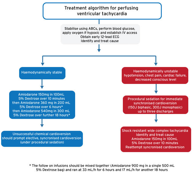
- Stabilise using ABCs, perform blood glucose, apply oxygen if hypoxic and establish IV access
- Obtain early 12-lead ECG
- Identify and treat cause
- For unstable wide complex tachycardia: sedation for immediate synchronised cardioversion (150J biphasic, 300J monophasic) up to three discharges
Drug treatment
- For stable wide complex tachycardia: give Amiodarone as follow:
- Initial rapid 150 mg Amiodarone infusion: add 150 mg to 100 mL 5% Dextrose and infuse over 10 minutes
- Followed by giving an Amiodarone 900 mg maintenance infusion as follow
- Add Amiodarone 900 mg to 500 mL 5% Dextrose
- Infuse Amiodarone 360 mg (200 mL of mixed solution) over 6 hours (or 33 mL/h for 6 hours)
- Infuse the remaining Amiodarone 540 mg (300 mL of mixed solution) over 18 hours (or 17 mL/h for 18 hours)
- Elective synchronised cardioversion can also be useful in stable wide complex tachycardia and use will depend on the situation as well as resource availability.
Shock resistant wide complex tachycardia
- Identify and treat the cause of the wide complex tachycardia
- Infuse the initial rapid 150mg Amiodarone (as above) over 10 minutes and then reattempt synchronised cardioversion
Referral criteria
- Unlike most SVTs patients that presented with possible VT should not be discharged. Refer the patient to the appropriate inpatient team for management. Continuous cardiac monitoring is required for onward admission and a transfer may be required if not locally available.
Treatment algorithm for perfusing ventricular tachycardia
Key treatment points
- 80% of fast regular wide complex rhythms will be ventricular tachycardia.
- Rather than spending valuable time trying to decide if the rhythm is SVT with aberrancy or VT, cardiovert the patient and scrutinise the ECG later when the patient is out of a potentially lethal arrhythmia.
- Synchronised cardioversion is the treatment of choice for WCT.
- Cardioverting a supraventricular tachycardia (SVT) with aberrancy is a safe and effective treatment option.
- The use of adenosine for a diagnostic aid in WCT should only be used by experienced clinicians and for the sake of this guideline it is not included.
References
Rossiter D, Blockman M (eds.). South African Medicines Formulary. 11th Ed. Cape Town: Health and Medical Publishing Group; 2014
Cameron P, Jelinek G, Kelly A, et al. Textbook of Adult Emergency Medicine. 4th Ed. Elsevier; 2015