Procedural Protocols
SADA COVID-19 Guidelines, May 2020
Non-Aerosol-Generating Procedures
Responsibilities of the Dental Assistant Pre- and Intra-operatively
Recommendations for the Use of Specialised Equipment
Other Risk Reduction Considerations
Classification of Contact
- Non-Aerosol-Generating Procedures
- Aerosol-Generating Procedures
We recognise that many procedures do not fall strictly into the above categories and would be a combination of the two. The strictest classification should then apply.
Non-Aerosol-Generating Procedures
Definition
For the purpose of this document, this would be defined as procedures with minimal droplets and no AGP (more than expected but in line with non-facemask social contact). All patients should be treated as though they are potentially infected, and no aerosols should be produced during clinical interventions. It would include all procedures that are done without any rotary/vibrating instruments,suction (https://www.ncbi.nlm.nih.gov/pmc/articles/PMC3322898/) or pressurised air.
Procedures that could fall into this category include clinical extra- and intra-oral examinations without the aid of a 3-in-1 syringe, intra-oral x-rays and dental impressions (be aware of an increased risk of coughing or gagging), as well as denture repairs and changing elastics for orthodontic patients.
Persons at Risk
- It must be accepted that all members of the OHC team are at risk.
- All staff entering the red zone should wear appropriate PPE in line with their function in this area. Administrative staff are less at risk and should be instructed to avoid the red zone as much as possible. Everybody should adhere to the PPE protocol set out above.
Standard Precautions
All staff members should wear masks at all times. Social distancing should be practised during tea breaks/meals.
- It is expected that hand disinfection protocols will be performed much more frequently and diligently than those proposed as public guidelines.
- Appropriate protective clothing to be worn by staff in line with their job description. For clinical staff, scrubs or similar attire are preferred. Additional protective clothing for routine and emergency changes during a working day is advocated, with changing to personal street clothing upon leaving the premises.
- Personal cough etiquette and respiratory hygiene to be adhered to as per government proposals. It is advocated that all staff are formally informed and educated and that they police each other.
Pre-Procedural
- The assistant should stand behind the patient when placing the bib. Refrain from using bib chains or clips, as these may increase the potential cross-infection risk disease from patient to patient or from patient to health care worker (Molepe et al., 2015).
- Cleanse the peri-oral area with either a water face wash at the basin or, as an untested opinion, using swab with 0.2% povidone-iodine (povidone-iodine is known to be virucidal).
Patient Hand Hygiene on Entry and Exit
- Patients sanitise/disinfect their hands again with the use of an alcohol-based hand sanitiser, dispensed into their hands by the dental assistant. Requesting patients to refrain—as far as practically possible—from touching any surfaces, such as working surfaces.
- Add a visual display of hand disinfection protocol for patient education and reassurance.
Appropriate PPE
- Head:
a. Cover head with a disposable surgical cap
b. Face to be covered by a visor/face mask
c. Eyes—protective eyewear with side shields or goggles
d. Nose and mouth to be covered, preferably with an N95(or equivalent) mask but at minimum a 3-layered surgical mask - Cover exposed skin at neck area with polar/buff neck clothing, preferably a fluid-resistant material or part of the gown design.
- Cover body with a fluid-resistant gown or at least an apron with at least full-length arm coverage clothing and legs to the below the knee.
- Hands—Gloves—latex gloves offer the best protection against bacteria and viruses, whilst synthetic gloves provide better chemical protection. Of the different types of latex-free gloves, nitrile gloves offer a higher degree of protection against viruses, besides the chemical protection they provide (https://www.cadth.ca/sites/default/files/pdf/htis/oct- 2013/RC0486%20Disposable%20gloves%20final.pdf).
Hand Hygiene
General Dental Operating Procedure
- Wet hands and apply sufficient liquid soap to create a proper lather.
- Temperature of water ideally between 35°C and 45°C
- Rub palms together in a circular motion. Rotate clockwise and anti-clockwise
- Rub back of hands with fingers linked through the other hand and then alternate
- Interlink facing fingers into clasped hands followed by rubbing palms and fingers together
- Cup fingers together, with right hand over and left hand under. With fingers interlocked,scrub the backs of them against palms and then alternate
- Clean thumbs by enclosing your right hand around your left thumb as you rotate and thenalternate.
- Rub palms with fingers in a circular motion, then alternate
- Dry hands with disposable paper hand towels
- Apply alcohol-based hand sanitiser
- Don gloves
- Apply alcohol-based hand sanitiser on donned gloves. Avoid touching any surfaces prior to touching the patient (https://www.cdc.gov/handhygiene/)
Sterile Surgical Protocol Pertaining to Surgical Interventions
Refer to routine sterile protocol procedures as prescribed in hospital/sterile environments, plus additional measures during crisis management. Donning procedure: as prescribed above by illustrations and video.
Aerosol-Generating Procedures
Definition
Aerosols are produced when a current of air moves across the surface of a film of a liquid. The greater the force, the smaller the droplets formed. OHC AGPs are defined as any patient and medical care procedure that results in the production of airborne particles (aerosols). AGPs can produce particles <5 μm in size, which can remain suspended in the air and travel over a distance, causing infection when inhaled. Therefore, AGP creates the potential for airborne transmission of infections that may otherwise be transmitted via droplets (Shiu E, Leung N, Cowling B). Controversy around airborne versus droplet transmission of respiratory viruses exists. This has implications: implication for infection prevention, and we need to accept the probability of airborne spread is high (Curr Opin Infect Dis. 2019;32(4):372–9) (NIPC Scotland: AGPs guidelines reviewed November 2019) (https://hpspubsrepo.blob.core.windows.net/hps-website/nss/2892/documents/1_2019-10-11-combined-nipcm-methodology-v1.pdf).
Aerosol-Generating Procedures in Dental Practice
The list of procedures is vast, but for the purpose of this definition, it is more inclusive to concentrate on aerosol-producing and air abrasion dental equipment used to perform OHC procedures. This would include the following:
- Use of 3-in-1 syringe in dental oral examination to facilitate better vision of the intra-oral working area
- Use of ultrasonic scalers to aid in oral hygiene and periodontal procedures
- Use of high and low speed hand pieces with or without water coolant. Surgical hand pieces, air or electric turbine hand pieces, also when used to adjust prosthesis intra-orally and peri-orally.
- Maxillofacial and oral surgery where high-speed devices are used
- Use of Lasers
Persons at Risk
Anybody in the area of expected higher viral load (red zone) is at risk of acquiring the virus upon entering this area. Special mention should, however, be made of persons directly in contact with the patients, such as OHC personnel and those present in the room to assist. In this time the following scenario can be imagined: In a time of need, a staff member, correctly donned for distant contact, may be asked to open certain items that were not considered when planning for a specific procedure. An example may be that a sterile package needs to be opened but is not easily reachable, the implication being that the assistant’s hands may contaminate multiple surfaces to reach this. The distant staff member correctly trained and donned could open this package whilst maintaining social distancing. This is an ideal opportunity for the reuse of an appropriate mask if treated as explained above.
Appropriate Clothing, Hygiene, and PPE
Head Protection
- Types: caps and covers (disposable or reusable)
- Additional hair cover is recommended when working in a sterile or high risk environment with long hair and beards.
Respiratory (Nose and Mouth) Protection
The purposes of respiratory protection are (i) to prevent or reduce transmission of air droplets and aerosols between health care workers and the patient and (ii) to prevent air droplets and aerosols into oral and nasal mucous membranes of OHC workers during procedures.
Type of PPE for Respiratory Protection
- Respirators (without valves) are indicated for high risk clinical procedures AGP
- N95 (or equivalent) respirators without valves c one shaped or duckbill shaped are indicated during any AGP procedure
- Respirators with valve s are recommended w here prolonged contact with the patient is expected 1 hour)
Eye Protection
- Type of eye protection: Goggles, face masks and face shields
- Goggles and face shields protect the eyes from splashes and spatter of contaminated (saliva) air droplets and contaminated aerosols
- Face masks an d face shields protect the eyes and other parts of the face against the spatter of contaminated air droplets and contaminated aerosols
- Face shields or face masks are indicated when doing AGP
- Protective safety glasses and face shields should undergo thorough disinfection with 70% isopropyl after each procedure
Body Protection
- Types: Gowns (disposable and reusable) and overalls
- Must meet the following criteria: sterile if reusable, long sleeves to cover arms and water resistant
- Cloth gowns are not recommended, as they are not water resistant
- Commercially available reusable gowns should be water resistant washable and sterilisable according to manufacturer's recommendations
- Plastic aprons are not recommended for dental and surgical purposes because they do not provide full body and arm cover
Shoe Protection
- Types: Overshoes and shoe covers (disposable).
- It is suggested that overshoes and shoe covers should be single patient use. Any airborne droplet will gravitate to ground level and will contaminate foot wear. Inadvertent contact may be a route for mucosal entry after touching these soiled areas
Hand Protection (Gloves)
Types
- Latex or latex free (nitrile) gloves-short cuff and non sterile. Indicated for all general procedures. Latex or latex free surgical gloves sterile, individually wrapped and long cuff. Indicated for all surgical procedures.
- Gloves are used to protect health care workers from direct contact with blood, saliva and body fluids and secretions.
- Gloves should be worn with every patient intervention/procedure; they are for single use purposes only and should be discarded after each procedure and patient use.
- Double gloving in dentistry is impractical, as dexterity is lost, which is crucial in our profession. The donning of a new set of non sterile gloves before starting doffing is advised.
Non-Clinical Staff Selection and Utilisation of PPE
- Reception and non clinical staff: Surgical mask or cloth mask, N 95 (or equivalent) if in the Red zone and at least 30 min thereafter.
- Cleaning staff: Surgical mask and gloves
Non-AGP and AGP on High-Risk Patients
Health care workers in contact with a suspected or confirmed COVID-19 case should wear a surgical mask or, if available, an FFP2 respirator—tested for fit, eye protection (goggles) and face shields, a cover all and gloves (EDCD Technical Report, 2020). It is recommended that these procedures be treated in a specialised environment, such as Airborne Infection Isolation Room (AIIR) or theatre environments that meet these standards as set out under practice ventilation requirements. Laminar flow, appropriate HEPA protected.
Guidance for the Selection and Use of Personal Protective Equipment in Healthcare Settings
CDC has developed this slide set for use by staff development, infection control and occupational health personnel for training healthcare personnel on how to select and use PPE to protect themselves from exposure to microbiological hazards in the healthcare setting.
Available Formats for download:
Special Precautions
General:
Isolation Devices
Fine aerosols generated by AGP (e.g. use of air turbines, ultrasonic and sonic devices and 3-in-1 syringes) produce fine aerosols (<5 μm) or less in diameter that can suspend in the air. Spatter droplets are much larger than aerosol particles (<50 μm). Both aerosol particles and spatter droplets can contain infectious agents (bacteria or viruses). Respiratory tract viruses are a recognised hazard for health care workers, including dentists. SARS-CoV-2 has predominantly respiratory transmission through aerosols <10 μm (Shiu E, Leung N, Cowling B). Controversy around airborne versus droplet transmission of respiratory viruses: implication for infection prevention (Curr Opin Infect Dis. 2019;32(4):372–9) and droplets (<50 μm) (Izetti, Nisi, Gabriele, 2020). It is therefore reasonable to assume that any method for reducing the viable bacterial or viral load in saliva and/or oral environment and/or limiting the effects of viral diffusion could lower the risk of cross-contamination and are therefore critically important for infection control (Izetti, Nisi, Gabriele, 2020).
It is of utmost importance to consider all methods that can minimise the risk of transmission of potentially infectious agents, such as SARS-CoV-2, to dentists, dental auxiliary staff, dental assistants and patients.
Pre-procedural Mouth Rinse
To this end, the most important method for reducing the viable bacterial and viral load is through pre-procedural rinsing with povidone-iodine (0.23% to 1%) or 1% hydrogen peroxide rinse.
Rubber Dam
Contamination from aerosols and spatter (air droplets) dissemination remains a significant hazard to dental personnel when AGP are done.
Studies have shown that during conservative dental procedures (15–30 minutes) without the use of a rubber dam, the airborne bacterial load increased from 8.8 to 25.1 CFU. This means that patients, dentists and assistants, as well as surfaces and objects in the operative area, are at a risk of exposure to airborne contamination 2.5 to 3X greater than the norm (Legnani et al., 1994).
Various studies have shown that the use of rubber dam isolation during AGP resulted in a 98.8% bacterial reduction (El-Din, Ghoname, 1997; Cochrane, Muller, 1989; Samaranayake, Reid, Evans, 1989; Al-Aman, Awad, Edher et al., 2017). This reduction increased with antiseptic mouth rinse used before the rubber dam application.
When resources are limited or under strain, the presence of physical constraints (e.g. wearing eyeglasses), or due to personal preferences, dentists and their assistants have the choice to use rubber dam isolation for eye protection instead of using a face shield and N95 respirator when performing AGP.
Several studies have confirmed that the viral load in human saliva is very high and that preoperative mouth rinses can reduce but not eliminate it (https://journals.sagepub.com/doi/10.1177/0022034520914246; Peng et al. 2020 (https://www.nature.com/articles/s41368-020-0075-9.pdf?origin=ppub). In terms of the coronavirus, Wang et al. (2004) (https://doi.org/10.3201/eid1007.031113) examined the oral cavity of SARS patients and found a large amount of SARS-CoV RNA in their saliva ((7.08×10³) to (6.38×10⁸) copies/mL). This suggests a strong possibility of coronavirus transmission through oral droplets. According to Chowell et al. (2015) (https://doi.org/10.1186/s12916-015-0450-0), evidence shows that the majority of SARS-CoV and MERS-CoV cases are associated with nosocomial transmission in hospitals, resulting, partly, from the use of AGPs performed on patients with respiratory disease. Based on the current epidemiological data, 2019-nCoV has higher transmissibility than SARS-CoV and MERS-CoV (Chen, 2020) (https://doi.org/10.1016/j.micinf.2020.01.004). Therefore, modification of standard precaution and infection control regimen targeted toward 2019- nCoV is essential during this outbreak (Ge et al. 2020 (https://www.ncbi.nlm.nih.gov/pmc/articles/PMC7089481/pdf/11585_2020_Article_417.pdf).
Viral transmission can occur when viral particles are aerosolised by a cough, sneeze, or during dental care. According to Froum and Strange (2020), particles can potentially travel up to a distance of 6 m from an infected person and have the potential to incite secondary infections (https://www.perioimplantadvisory.com/periodontics/oral-medicine-anesthetics-and-oral-systemic-connection/article/14173521/covid19-and-the-problem-with-dental-aerosols).
Dentists and auxiliary staff who treat patients during aerosolisation procedures are at very high risk of infecting themselves or to re-infect other patients. The highest risk occurs from splatter and droplet transmission to the midface of the dentist and/or dental assistant, as well as the nasal area of the patient (Nejatidanesh et al. 2013) (http://europepmc.org/article/med/23930175#free-full-text).
The water coolant from a high-speed hand piece could generate aerosols during restorative, prophylaxis and surgical procedures (Farah, 2019) (https://www.ncbi.nlm.nih.gov/pubmed/30834225). When combined with bodily fluids in the oral cavity, such as blood and saliva, bioaerosols are created (Ge et al. 2020) (https://www.ncbi.nlm.nih.gov/pmc/articles/PMC7089481/pdf/11585_2020_Article_417.pdf).
These bioaerosols are commonly contaminated with bacteria, fungi and viruses, and have the potential to float in the air for a considerable amount of time and be inhaled by the dentists or patients (Grenier, 1995) (https://aem.asm.org/content/aem/61/8/3165.full.pdf); (Jones and Brosseau, 2015) (https://www.ncbi.nlm.nih.gov/pubmed/25816216). Ultrasonic and sonic transmission during nonsurgical procedures had the highest incidence of particle transmission, followed by air polishing, air/water syringe and high-speed hand piece aerosolisation (Harrel and Molinari, 2004) (https://www.ncbi.nlm.nih.gov/pubmed/15127864).
Therefore, taking additional measures, such as the use of dental rubber dams to reduce exposure to contaminated aerosols during treatment, is very important (Spagnuolo, De Vito, Rengo, and Tatullo, 2020). Several published guidelines, not only for endodontics procedures, but for almost all aerosol-generating dental procedures recommend the use of rubber dam when possible (American Association of Endodontics, 2020) (https://mddsdentist.com/wp-content/uploads/2020/03/COVID-JOE-Preprint-3.18.20-v2.pdf); American Dental Association, 2020a (https://www.ada.org/en/publications/ada-news/2020-archive/march/ada-adds-frequently-asked-questions-from-dentists-to-coronavirus-resources); Indian Endodontic Society et al., 2020 (https://www.google.com/url?sa=t&rct=j&q=&esrc=s&source=web&cd=2&ved=2ahUKEwij6bm-iPfoAhVkoXEKHS7JC-4QFjABegQIARAB&url=https%3A%2F%2Fida.org.in%2F&usg=AOvVaw1qx0nI8HBbOfQYZS_Ge04v).
The rubber dam provides a barrier of protection from the primary source and can virtually eliminate all pathogens emerging from respiratory secretions during dental procedures that generate aerosols (Ge et al. 2020) (https://www.ncbi.nlm.nih.gov/pmc/articles/PMC7089481/pdf/11585_2020_Article_417.pdf). According to Harrel and Molinari (2004) (https://www.ncbi.nlm.nih.gov/pubmed/15127864), if the rubber dam is placed correctly, the only source of contamination would be the tooth that is undergoing treatment. The application of a rubber dam during cavity preparation showed a reduction in the spread of microorganisms by 90% (Cochran et al. 1989) (https://doi.org/10.14219/jada.archive.1989.0131). Samaranayake, Reid and Evans (1989) (https://www.ncbi.nlm.nih.gov/pubmed/2681303) reported that the use of a rubber dam could significantly reduce airborne particles in a 3-foot diameter of the operational field by 70%. The efficacy of a rubber dam isolation as an infection control procedure in paediatric dentistry has also been reported. They showed a 98.8% bacterial reduction up to 1 m from the patient (El-Din and Ghoname (1997) (http://www.emro.who.int/emhj-volume-3-1997/volume-3-issue-3/article16.html).
In contrast to previous studies, Al-Amad et al. 2017 (https://core.ac.uk/download/pdf/82393007.pdf) demonstrated that despite the clinical value of the rubber dam, it can result in significantly higher aerosol levels on various areas of the dentist’s head. They advised that dentists should cover their heads with suitable protective wear during AGPs.
Another device that can be used for isolation of the oral cavity during restorative procedures is Isolite (Isolite Systems, 2017) (https://optident.co.uk/app/uploads/2018/03/Isolite-Systems-Brochure.pdf). Isolite delivers quick, easy, reliable isolation with uninterrupted retraction and continuous evacuation of fluids and oral debris with a reduction of aerosols. By inserting the soft Isolite mouthpiece, immediate tongue protection, cheek retraction and throat protection are accomplished whilst the built-in bite block allows the patient to comfortably rest his or her jaw and keep the mouth open at all times. The system also provides unique shadowless illumination in the patient's mouth, which reduces eye strain for the practitioner.
Dahlke et al. (2012) (https://www.ncbi.nlm.nih.gov/pubmed/23115148) evaluated the spatter-reduction effectiveness of a non-isolated control consisting of HVE with a dental rubber dam in conjunction with HVE and the Isolite system. The results of this in vitro study showed that the rubber dam with HVE and the Isolite system reduced spatter significantly compared to HVE alone. However, a more recent in vivo study compared aerosol and spatter reduction of a saliva ejector and the Isolite system during and after ultrasonic scaling (Holloman et al., 2015) (https://jada.ada.org/article/S0002-8177(14)00002-6/pdf).
The results of the study showed no significant difference between the two groups in aerosol and spatter reduction. The authors concluded that there was a significant amount of contamination during ultrasonic scaling in both groups and that additional measures should be taken with these devices to reduce the likelihood of disease transmission.
In conclusion, the rubber dam with an appropriate preceding mouth rinse appears to be the best option for working field isolation devices in order to eliminate AGP. It is also important to note that when the rubber dam is used, extra high-volume suction for aerosol and spatter should be used during procedures in conjunction with regular suction (Samaranayake & Peiris, 2004) (https://www.ncbi.nlm.nih.gov/pubmed/2681303)
Suction
During clinical procedures known to cause aerosol and splatter formation, it is mandatory to incorporate the use of an HVE. Studies have shown that effective use of an HVE can remove more than 90% of the aerosol. The most efficient way to remove aerosols is with four-handed dentistry (https://pubmed.ncbi.nlm.nih.gov/9766105/)(https://www.ncbi.nlm.nih.gov/pubmed/25581979).
When evaluating suction systems in an OHC environment, consideration should be given to the following:
- Efficiency
- Maintenance/disinfection
- Installation
Efficiency: The ideal power of an HVE should be a minimum flow rate of 300 l/min
Maintenance/disinfection: After procedures involving blood or aerosol, a glass of cold water should be passed through the system. It is essential to clean filters and disinfect with the correct solutions at the end of every day (see later: sterility recommendations).
Installation: All suction systems have an exhaust that removes toxic waste. Ideally, these exhausts should be outside and if potentially close to a public place should have a filter of an appropriately strong UV-C system to sterilise the air (removing particles of 0.01 μm or less, efficiency of 99.99%). The filter should provide particle removal to 0.01 μm with a Dispersed Oil Particle (aerosol) efficiency of not less than 99.9%. Some practices will not be able to install an external exhaust; here filters are mandatory. Also refer to ventilation in this document: http://www.bcga.co.uk/assets/HTM_2022_S1.pdf
In conclusion, modified suction devices that are fitted to the HVE do not remove aerosol/splatter at the source, so we must accept there will be a compromise in reduction of exposure to operators. External suction devices must be considered as an additional layer of protection and not a replacement of the use of HVE. The ideal control of aerosol would be with isolation procedures, such as a rubber dam in combination with an HVE used by an assistant.
The Use of Dental Auxiliaries
Two-Handed Dentistry
Two-handed dentistry is defined as dentistry where the OHC worker is the only person working on the patient. There is no dental auxiliary available to assist with any of the normal procedures, such as suctioning, instrument control and management and, most importantly, surgery decontamination following patient treatment. The clinician is the sole operator and manages all functions when treating, aftercare, admin, billing and seeing the patient out. Once this is complete, surgery disinfection and sterilisation need to take place.
There are many disadvantages to two-handed dentistry:
- Time: A single operator needs to carry out all the functions themselves.
- Bringing patient into the surgery along with patient preparation prior to entering the surgery
- Patient and surgery preparation preoperatively and intra-operatively
- Patient post-treatment management including billing and clinical notes
- Surgery decontamination before the next patients are seen
- Cross-contamination
- Two handed dentistry requires that all items necessary for treatment a re prepared so that the operator does not need to open drawers or touch any item that can be contaminated with aerosol. This means having to glove up and de-glove not just between patients but during the procedure to open a drawer or open items needed for normal practice. All this may lead to cross infection and contamination of various items in the surgery. All of which need to be decontaminated after the treatment has been completed.
- Trying to save money by reducing staff may not only put your patients at risk but also yourself and your staff. Reducing aerosol is essential to prevent the spread of virus droplets. Without having a second set of hands to hold the HVE, the risk of droplet spread and aerosol production is extremely high. Although one can hold the HVE and work at the same time, the efficacy of this is not tested. At this point, there is no current data to suggest that two handed dentistry leads to increased aerosols; however, it is not recommended by this working group.
Several options are available to operators working without an assistant. They include using the operating instrument in one hand and the HVE in the other hand, HVE devices that attach to the operating instrument and various ‘dry field’ devices that attach to an HVE. For air polishing and air abrasion, devices are available that combine a barrier device to help contain the abrasive material and a vacuum to remove the abrasive material and the airborne particles created by the procedures. All of these instruments are available commercially from multiple sources.
Four-Handed Dentistry
Four-handed dentistry had been advised by the CDC. Glene Robinson (1968) summarised the concept of four-handed dentistry: Four-handed dentistry involves the coordinated work of both the dentist and the assistant, working as a team to perform operations in a manner that has been carefully and deliberately planned.
It involves the use of a trained, competent, chairside assistant to work constantly with the dentist in performing the technical procedure during any dental procedure in the dental setup. The CDC has stated categorically that any person who will be producing aerosols needs to employ four-handed dentistry (https://www.cdc.gov/coronavirus/2019-ncov/hcp/dental-settings.html). This is due to the fact that a second person or dental assistant is needed to adequately reduce the aerosols produced by the instrumentation used in dentistry, thereby reducing the risk of COVID spread. Meng et al. (2020) in the Journal of Dental Research has also advised the use of four-handed dentistry to help reduce aerosols.
There are, however, no references for this (https://journals.sagepub.com/doi/full/10.1177/0022034520914246). In a letter, the president of the FDI referred to this publication and endorsed the use of four-handed dentistry (https://www.fdiworlddental.org/news/20200317/journal-of-dental-research-publishes-covid-19-guidelines-from-researchers-based-in).
Auxiliary Responsibilities
- Help with patient management before bringing them into the surgery
- Help with patient management before, during and after treatment
- Reduce aerosol to a minimum level possible
- Reduce the operators’ need to touch, and thus contaminate, other areas with contaminated gloves
- Reduce aerosol spread and cross-contamination from the operator and patient
- Reduce treatment times
- Improve disinfection protocols of the surgery and instrumentation following treatment
Responsibilities of the Dental Assistant Pre- and Intra-operatively
The job of the dental assistant is to ensure that the surgery is ready for the acceptance of the patient into the surgery. Following disinfection of the surgery as described in this document. It is important to ensure that all items and instrumentation that are going to be needed during the procedure or surgery are packed out and anything that is in drawers is laid out prior to any procedure being done. All instrumentation that is unpacked and left on the surface should be in sterile bags and opened only once the patient is seated so that the patient can see that all instrumentation is sterile. Making sure that all instrumentation is available prior to treatment ensures that there is no need to unnecessarily open any drawers or cabinets during treatment. All water bottles used for coolant should be disinfected between patients, and the filling of the bottles should be carried out, preferably in a green or appropriate zone where there is no risk of aerosol entering the bottles whilst they are being filled. Ensure that the bottles have caps so that the water is not exposed to aerosols whilst being carried.
Due to the ability of the aerosol to remain in the air for a long period following treatment, opening and closing drawers could lead to contaminated aerosols resting on instruments, burs or any other items that are in the drawers and cabinets. There are also many nooks and crannies where the aerosol may sit, resulting in contaminated areas that are not easily cleaned or disinfected. Once the patient is seated and the appropriate pre-procedural mouth rinse has been provided, all instrumentation can be opened. It is important that your assistant ensures that during AGP suctioning with the HVE is utilised at all times, no matter the procedure type. The HVE should be as close as possible to the area of aerosol formation to limit the aerosol spread from the mouth.
Once your assistant has seen the patient out of the surgery, the decontamination and disinfection of the surgery can begin again, making it ready for the next patient.
It is important for your assistant to understand and be trained in all aspects of disinfection and cross-contamination control so that the correct protocols are followed. It is also critical that our assistants understand that we are now dealing with not just blood-borne contaminants but also aerosol-borne contaminants that spread far more easily. It is essential that they understand how the spread of airborne and aerosol infections takes place. We need to change the thinking of our auxiliaries so that they understand the risks and the severity of cross-contamination and the implications they have for our practices.
It is advised that in order to reduce the amount of aerosol production that we utilise a dental assistant for four-handed dentistry in order to reduce the risk of SARS-CoV-2 spread.
Recommendations for the Use of Specialised Equipment
Loupes and Microscope Disinfection
Magnifying loupes and the DOM allow both dentists and oral hygienists to perform tasks not possible without improved visual acuity. However, loupes cannot be compared to the comfort, versatility, illumination and visual acuity offered by the DOM (Van Faunhofer & Johnston, 1992).
Loupes and the DOM can collect debris and aerosol spray from most dental procedures. Ideally, all areas of the loupe and microscope should be disinfected with a high level of disinfectant after each patient. Zwicker et al. (2019) showed that even visually clean eyewear used by the dental profession can be a source of contamination, and thorough disinfection is recommended between patients.
Follow your manufacturer’s standards of care guidelines for your surgical microscope to ensure that you are cleaning within the tolerances of your instrument.
Dental Loupes
Ideally, all areas of the loupe should be disinfected with a high-level disinfectant after each patient. However, facing the reality that most dentists using loupes have only one set of loupes and that some loupes will not tolerate constant use of disinfectants, the infection control challenge is obvious (Christensen, 2003) (https://endoexperience.com/documents/Magnification-Christensen.pdf).
Most of the time, ethyl alcohol is the recommended solution of choice to disinfect the loupe telescopes and frame surfaces. The lenses can be cleaned with alcohol or quaternary ammonium disinfectant.
Even though the optical lenses of most of the loupes are manufactured with protective coatings, they are still susceptible to damage. Without proper care, the lenses may become cloudy, scratched, and difficult to use.
If your dental loupe is water resistant, you can use a water lavage to wash off the debris. After such, wipe the lenses and the loupes with cleaning wipes followed by a microfibre cloth.
If it is not water resistant, you can use a can of compressed air to remove debris. Once done, wet a soft cotton swab or tissue with mild soap, 70% isopropyl alcohol or other lens cleaning solutions that the dental loupe manufacturer has recommended and then carefully wipe away loose particles and major debris. Finally, dry the loupes by successively wiping it with another set of microscope cleaning wipes or with the use of a microfibre cloth.
Dental Operating Microscope
All the system's mechanical surfaces can be wiped clean with a damp cloth. Do not use any aggressive or abrasive cleaning agents. Remove any residue using a mixture of 50% ethyl alcohol and 50% distilled water plus a dash of household dishwashing liquid.
For disinfection of mechanical surfaces, the maximum application concentrations for disinfection solutions are either 60% alcohol, 2% glutaraldehyde or 0.2% quaternary compounds. For regular cleaning of the surgical microscope’s objective lenses and eyepieces, it is recommended that special, manufacturer-produced cleaning kits be used.
Some manufacturers also supply sterilisable caps for handgrips and knobs and custom-made sterile drapes to cover the microscope head and body to protect the microscope from aerosol spray during procedures. Disinfected plastic bags can also be used to protect the DOM from aerosol and should be replaced after each patient.
Lasers
Lasers in dentistry have evolved dramatically over the years. Initially, lasers were limited to soft tissue treatment, such as gingivectomies and frenectomies. Newer lasers are now able to cut bone and tooth structure as well as soft tissue.
Lasers used in dental practice can be classified by various methods:
- According to the lasing medium used, such as gas laser and solid laser
- According to tissue applicability, hard tissue and soft-tissue lasers
- According to the range of wavelengths
- We will further classify them for this document as lasers that utilise water (wet lasers) for cooling and those that are dry (dry lasers) (i.e. those that produce aerosol and those that do not).
Wet Lasers: ErCr;YSGG 2780 nm/Er;YAG 2940 nm
These lasers are now able to cut bone, tooth and soft tissue but have a multitude of applications in all fields of dentistry. These types of lasers work on heating water molecules within the tissue, which differs from the dry lasers. The introduction of lasers that cut bone and teeth required the need for coolant to reduce lateral heat production during treatment, thereby reducing damage to the pulp and adjacent structures. It is this coolant combined with air spray that has the ability to produce aerosol. Therefore, it is recommended that all lasers that utilise water cooling be handled and managed in exactly the same way and with the same protocols as AGP.s. Wet lasers also have the ability to create plumes; however, there is no evidence to show that this plume has viral inclusions or that it is infective in any way.
Strict AGP reducing protocols should be in place when utilising a laser.
- During treatment, HVE is essential. This has two important functions: (1) reduce aerosol release and (2) help with tissue cooling through airflow.
- Keeping the HVE as close to the laser as possible is advised.
- Where possible, it is essential to reduce the amount of water as well as the airflow from the laser hand piece in order to reduce the amount of aerosol formation.
- When utilising lasers for either tooth preparation or endodontics, it is mandatory to use a rubber dam during these procedures to reduce the aerosol formation, as set out with normal use of rotary instruments in these fields.
- Typically, in soft-tissue procedures, the water and air ratios can be very low; this may cause the tissue temperature to rise more than is ideal, with the consequence of slower healing as a side effect. It is important to use high magnification to observe the tissue for any carbonisation or a noticeable band of denaturation. Should this become evident, then the air/water ratios should be adjusted to improve tissue cooling. Guidelines for the Waterlase Iplus laser (2780 nm) suggest a minimum of 20% water and 30% air.
- The pulse duration also has an influence on the thermal reaction in tissue. Typically, visible thermal reaction decreases as the pulse duration shortens. Iplus (2780 nm) has two options: S-mode has a pulse duration of 700 ms and H-mode has a pulse duration of 60 ms. It is better to operate at the shortest possible pulse duration available if air/water ratios are decreased. The specific procedure and suggested laser parameters should still be respected.
- Hard tissue (Bone / Enamel /Dentine/ Cementum/Caries): The water percentage plays a crucial role as a mediator to the ablation of hard tissue, and an over-compromise might have a detrimental effect on the living tissue. However, it is still possible to use the minimum amount of water required to perform procedures safely.
Guidelines for Waterlase iPlus
a. Enamel—80% water / 60 %air
b. Bone—60% water/ 50–40% air
c. Cementum—similar to bone
d. Caries—the possibility of decreasing water to a point lower than for enamel, but it is not advised due to the risk of a rise in pulpal temperatures above safe levels.
The use of lasers to treat periodontal disease can be carried out with either a dry or wet laser. When utilising a wet laser, it is better to reduce the air flow and water coolant and make sure your HVE is as close as possible to the area you are working on. This will collect as much of the aerosol as possible, and the airflow will help cool the area being treated by the laser.
No matter what procedure is being carried out with lasers, it is advised that the normal PPEs and ventilation are used to reduce the risk from AGP and plume.
Dry Lasers: Diodes (810 Nm, 940 nm, etc. and some CO2 Systems 10600 nm)
Dry lasers do, however, create a plume, and there is some data to show that there are viral particles present in the plume. No known transmission of illness of HIV and HBV due to laser use has, however, been recorded (CDC). MMWR 2003;52(No. RR-17; http://www.cdc.gov/oralhealth/infectioncontrol/guidelines/index.htm).
These lasers are limited to soft issue work, periodontal and endodontic decontamination. These lasers do not use water for cooling and therefore do not generate aerosol. Dry lasers heat blood and haemoglobin within the tissue. Lasers that do not utilise water (dry lasers), namely a diode laser or a carbon dioxide laser (or wet lasers used without water), do not create aerosol at all and are deemed safer to use, as no aerosol is produced. However, these lasers do create a plume (defined as the vapours, smoke and particulate debris produced during these surgical procedures), and there is some data to show that there are viral particles and some carcinogens present in the plume. Data from dermatology and otolaryngology both show the viability of virus DNA in the plume. No known transmission of illness of HIV and HBV due to laser use has, however, been recorded (CDC). MMWR 2003;52(No. RR-17; http://www.cdc.gov/oralhealth/infectioncontrol/guidelines/index.htm).
To date, nothing has been shown with regards to SARS-CoV-2. New data (May 2020), however, may come out later and may require further adjustment of our techniques and protocols. Although we do not have enough data at this point, it is strongly recommended that HVE be utilised at all times to remove the plume and that we take all the normal precautions for aerosol management. The close use of the HVE as close to the cutting area whilst using these lasers is also advised to help reduce the heat generation of the laser.
Alternative Therapies Using Laser
- Photo-biomodulation (PBM): The use of laser energy to promote healing and reduce inflammation and pain.
- Laser anaesthesia was used to anaesthetise teeth.
- Photo-Dynamic Therapy is the painting of tissue with a toluene blue or other dyes onto the infected tissue. The addition of lasers will lead to the formation of oxygen radicals that will kill bacteria.
None of these techniques use water or create a plume. Hence, they are deemed safe to use at any stage.
How Do I Disinfect my Laser Equipment?
- All laser hand pieces need to go through the normal sterilisation protocols with autoclaves.
- It is not enough to wipe off these hand pieces between patients.
All tips that are used either need to be sterilised, if possible, and if not, then cold sterilisation can be used, but proper protocols need to be followed. Combustible disinfectants must be avoided. - Certain tips need to be initiated (for gingivectomy and other soft-tissue procedures), which means that they are run along a piece of blue articulating paper to make them cut more effectively.
- Tips need to be replaced more often than tips that are utilised non-initiated (for periodontal and endodontic applications).
- Dry lasers that utilise a single cable of glass fibre need to have at least 1 mm of the tip cut off after each patient and the cable disinfected according to the manufacturer’s recommendations.
- There are certain hand pieces for biostimulation and bleaching that cannot be autoclaved, and these hand pieces need to be disinfected with adequate disinfection material, as described in this document. These hand pieces should also be covered with plastic covers to reduce contamination of the hand pieces.
- The main laser unit should have plastic covers to protect it from aerosol and should be wiped down between patients according to the protocols advised in this document.
- The water bottles used for the wet lasers should be refilled in a room that has not been contaminated by aerosol sealed and disinfected in that room and then returned to the laser units.
- Most wet laser hand pieces have positive pressure when changing tips and when removing the hand pieces, thus making them more easily autoclavable.
Other Risk Reduction Considerations
Disinfection of Dental Impressions (Laboratory Work)
According to the British Dental Association (2013), it is the responsibility of the dentist to disinfect impressions that are sent to dental laboratories to prevent cross-contamination and spread of disease. Communication between the dental practice and the dental laboratory is essential to ensure that appropriate disinfection protocols are implemented without overlap. Repeated exposure to disinfectants could compromise the quality of the impression and the restoration or appliance (https://www.ncbi.nlm.nih.gov/pmc/articles/PMC6511629/#CIT0002).
Dental impressions invariably are contaminated with patient saliva or blood and are considered potentially infectious https://www.researchgate.net/publication/257678834_The_effect_of_water_and_sodium_hypochlorite_disinfection_on_alginate_impressions. Such fluids can contain viral and bacterial pathogens, including HIV and the hepatitis A, B and C viruses. Although most infectious agents do not survive for extended periods outside the body, many pathogens, if present in high enough numbers, can survive several days in protein-containing body fluids. Microorganisms can be transferred from contaminated impressions to dental casts, and oral bacteria can remain viable in set gypsum materials for up to seven days.
Disinfection Methods
Optimally, impressions should be decontaminated at chairside immediately after removal from the patient's mouth. Impressions should be rinsed thoroughly under running tap water before disinfection to remove as much bioburden as possible.
PPE must be utilised, and adequate ventilation must be in place when hazardous materials, such as disinfectants, are used. The OHC provider must keep in mind that different impression materials would react differently to disinfectant procedures. In selecting an impression disinfectant, material compatibility is a requirement (see Table 1). 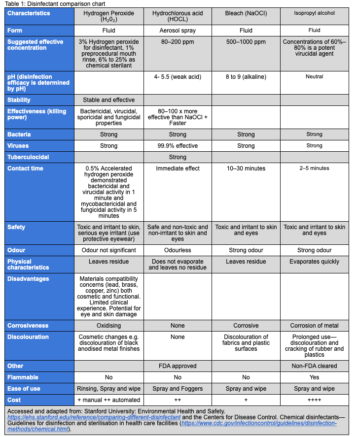
Click on image to enlarge
Both immersion and spraying have been recommended for disinfection of impressions. Spraying uses less solution, and often the same disinfectant can be used for general disinfection of operatory surfaces. Spraying does increase the chances for staff exposure to hazardous chemicals. Using stream spray can minimise aerosolisation.
When using impression materials that are predisposed to distortion from immersion into a disinfectant, spraying may provide an acceptable alternative. Impressions can be sprayed thoroughly and placed in airtight plastic bags. After the appropriate contact time, the impression can be removed, rinsed thoroughly with tap water, shaken gently to remove adherent water, and then poured.
For adequate inactivation of microbial contaminants, the contact time between impression and disinfectant should be at least equal to the time for tuberculocidal activity, as recommended by the manufacturer of the germicide.
Alginate Impressions
A number of studies have evaluated the effects of disinfection on irreversible hydrocolloid (alginate) impressions. The results varied greatly depending on the techniques and materials evaluated. Generally, however, distortion has been found to be minimal and not clinically significant. Based on these findings, disinfection via immersion in diluted sodium hypochlorite or iodophor is recommended. Impressions can be submerged or sprayed with disinfectants like diluted hypochlorite, iodophors and surface disinfectants. It should be done within the prescription of the manufacturer’s instructions (https://www.dentaleconomics.com/macro-op-ed/article/16393662/impression-disinfection).To emerge for 10 minutes in 0.5% sodium hypochlorite (https://www.ncbi.nlm.nih.gov/pmc/articles/PMC3410321/;) (https://pubmed.ncbi.nlm.nih.gov/29358546/).
Elastomeric Impressions
Rubber-base silicone impressions can be disinfected adequately by immersion in an iodophor, diluted sodium hypochlorite solution, chlorine dioxide, glutaraldehyde, or complex phenol for the time recommended for tuberculocidal activity. However, the method of disinfection should be verified with the material manufacturer to prevent distortion of the impression or loosening of the adhesive bond between the impression tray and the impression material. Phenols with a high alcohol content, for example, can desiccate some impression materials.
ZOE and Compound Impressions
Limited data are available on disinfection of zinc oxide eugenol (ZOE) and compound impressions. Current ADA recommendations for ZOE impressions suggest disinfection via immersion in a 1:213 iodophor or 2% glutaraldehyde solution prepared according to the manufacturer's instructions. Compound impressions reportedly can be disinfected via immersion in a 1:10 sodium hypochlorite solution or with an iodophor (https://www.sciencedirect.com/topics/medicine-and-dentistry/iodophor).For 10 minutes in a 0.5% sodium hypochlorite solution (https://pubmed.ncbi.nlm.nih.gov/22505972/).
Wax Bites and Wax Rims
Because of the delicate nature of wax, the wax is prevented from being safely immersed in disinfectant without distortion. Wax rims and wax bites can be disinfected using an iodophor, chlorine dioxide, or sodium hypochlorite spray and a spray-wipe-spray technique. Following the second spray, the wax bites can be enclosed in a sealed plastic bag for the proper contact time and should remain wet with disinfectant for the time recommended to halt tuberculocidal activity.
Wax bites can be thin and be deformed easily. It can be immersed but with care as above.
Impression Trays
Impression trays should be rinsed thoroughly under running water to remove residual blood and saliva. Consistent with dental infection-control guidelines for semi-critical instruments, chrome-plated and aluminium impression trays can be cleaned, packaged and heat-sterilised. Single-use, plastic impression trays provide a disposable alternative to heat sterilisation.
Conclusions
- Know your environment and the dangers and risks that you pose to your own OHC facility but also to the population at large.
- Identify high-risk patients and potential positive patients appropriately; all other patients should be treated as potentially positive.
- Positive COVID-19 patients should be treated in very specialised environments such as laminar flow, HEPA filtered (H13, H14) hospital theatres or specially designed AIIR. These could be constructed/adapted in an environment outside of the hospital, decreasing the load on our healthcare facilities, especially if AGP are unavoidable.
- The goal should always be to aim to better isolate your working space and thereby decrease the risk of spread, change from a possible risk situation to a relatively safe situation (rubber dam isolation and use of HVE).
- Implement additional safety and sterility measures until vaccination.
- Take all measures to avoid AGP in the initial relaxing stages.
- Health care workers at high risk: > 65 or with comorbidities as well as environmental high-risk practices (Health and Safety certified) should stay in self-quarantine/closed until such time as vaccination or Level 1 is reached. Professional insurance companies should be encouraged to see these above groups as actively not being able to function normally and should be compensated such as would