Pulmonary Embolism
Pulmonary Embolism
Introduction
Identification
Severity assessment
Management based on severity
Relevant history, examination
Investigations
Referral
Discharge criteria
Preventative measures
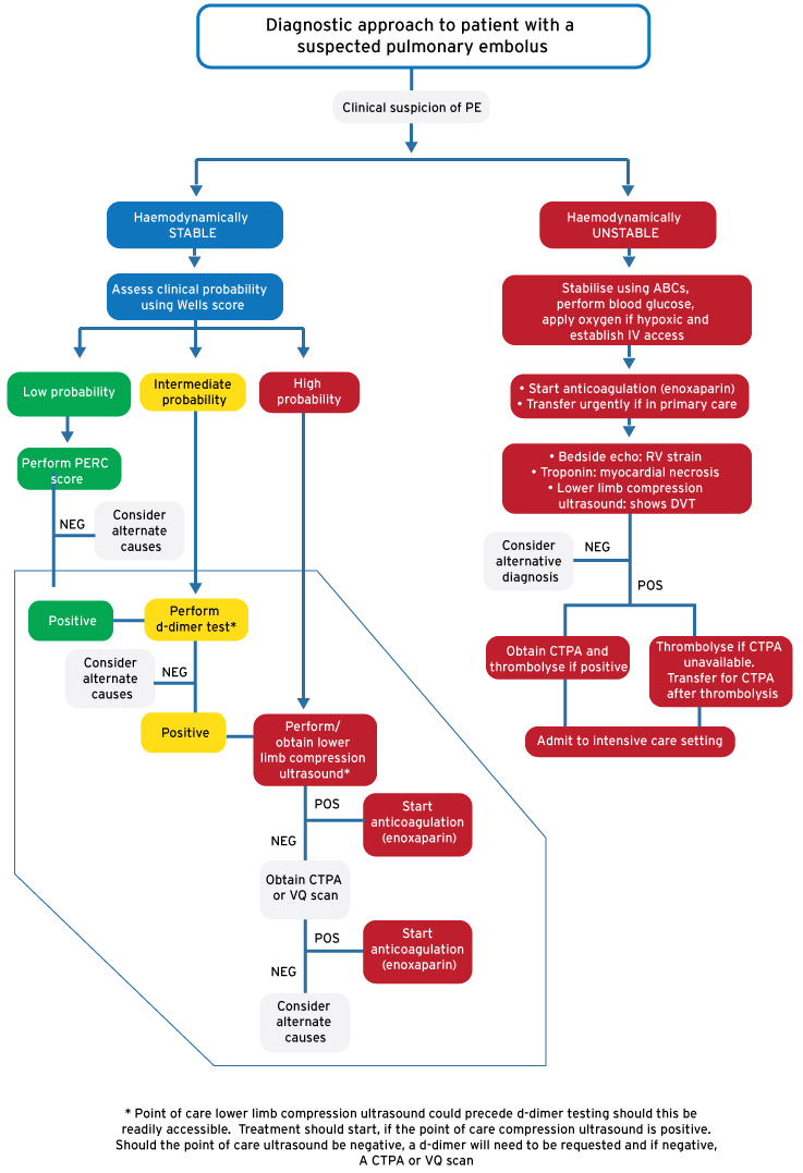
Diagnostic approach to patient with a suspected pulmonary embolus
Introduction/ Description
Pulmonary embolism (PE) occurs when a thrombus is deposited within the pulmonary circulation resulting in right ventricular outflow obstruction and/ or ventilation perfusion defects. PE has a mortality of 30-35% if untreated. It can be very elusive as it often presents with non-specific symptoms. It is particularly challenging in the resource limited setting.
Identification/ recognition/ diagnosis of problem
PE is often diagnosed after ruling out alternate diagnosis. Symptoms associated with PE include dyspnoea (common), pleuritic chest pain, substernal chest pain, cough, haemoptysis and syncope. PE is diagnosed by firstly determining the pre-test probability through using a clinical risk score (Wells and PERC) followed by performing definitive testing in those patients with higher probability as described below.
The PERC rule is performed on Wells low probability score. HIV and Tuberculosis are very prevalent in South Africa and both conditions promote coagulation and can contribute to venous thromboembolism. This needs to be considered during risk stratification.
| Wells probability score | |
| Clinical signs and symptoms of DVT (minimum of leg swelling and pain with palpation of the deep veins) | 3.0 |
| An alternate diagnosis is less likely than PE | 3.0 |
| Heart rate is >100 bpm | 1.5 |
| Immobilisation at least 3 days, or surgery in previous 4 weeks | 1.5 |
| Previous PE/DVT | 1.5 |
| Haemoptysis | 1.0 |
| Malignancy (on treatment or treated in the last 6 months, or palliative) | 1.0 |
| Interpretation | |
| Low probability | < 2.0 |
| Moderate probability | 2.0 – 6.0 |
| High probability | > 6.0 |
PERC (pulmonary embolism rule out criteria)
(Further investigation is required for any yes answers)
- Low risk of PE (decided either through clinical evaluation or low risk Wells)
- Is the patient older than 49 years of age?
- Is the pulse rate above 99 beats min?
- Is the pulse oximetry reading <95% while the patient breathes room air?
- Is there a present history of hemoptysis?
- Is the patient taking exogenous estrogen?
- Does the patient have a prior diagnosis of venous thromboembolism (VTE)?
- Has the patient had recent surgery or trauma? (Requiring endotracheal intubation or hospitalisation in the previous 4 weeks.).
- Does the patient have unilateral leg swelling?
Differential diagnosis
- Anxiety disorder
- Aortic dissection
- Asthma
- Costochondritis/ muscular
- Myocardial infarction
- Pericarditis
- Pneumonia
- Pneumothorax
- Rib fracture
Severity assessment
- Once diagnosed, severity is judged by assessing the haemodynamic status of the patient.
- Massive PE is recognised by right ventricular dysfunction and haemodynamic compromise not due to a cause other than PE
- Sub-massive PE is recognised by right ventricular strain or myocardial necrosis without haemodynamic compromise
- Non-massive PE is not associated with right ventricular strain, myocardial necrosis or haemodynamic compromise
Markers useful for risk stratification in acute PE
Clinical markers
- Shock
- Hypotension – Defined as a systolic blood pressure of <90 mmHg or a pressure drop of >40 mmHg for >15 min
Markers of right ventricular dysfunction
- RV dilatation, hypokinesis or pressure load on echocardiography
- RV dilation on spiral CT
- BNP or NT-proBNP elevation
Markers of myocardial injury
- Cardiac Trop T or I positive
The severity of PE should be understood as an individual estimate of PE-related early mortality risk rather than the anatomical shape and distribution of intra-pulmonary emboli. The latest guidelines suggest replacing terms such as ‘massive’, ‘sub-massive’ or ‘non-massive’ with the estimated level of the risk of PE-related early death, i.e. Low (<3%), Intermediate (3-15%) and High (>15%)
Management based on severity
Immediate action
Non drug treatment
Protect airway, supply oxygen, ensure adequate ventilation, IV access, fluid boluses
Drug treatment
- Massive PE: Inotropic support is often required. After checking for contraindications, thrombolytics are indicated.
- Sub-massive PE: Controversy exists over whether thrombolytics are of benefit in this group. Local guidance should be followed and specialist advice sought.
- Low risk PE: passive anti-coagulation
Added tables/ dose charts
| Trombolytic | Dose |
| tPA (alteplase)* | 100mg over 120 minutes followed by anti-coagulation |
| Streptokinase | 1 million units over 24 hours |
| Anticoagulants | Dose |
| Enoxaparin* | 1mg/kg SC 12 hourly |
| Heparin | 80 units/kg IV bolus, followed by 18 units/kg/hour IV infusion |
*Drugs of choice
Relevant history, Examination
Signs include tachypnoea, tachycardia, signs of DVT, fever (temp >38.5), hypoxia and/ or signs of RV strain or obstructive shock
Investigations
ECG often transient changes may be seen in a large pulmonary embolus.
- Sinus tachycardia
- T wave inversion in leads V1 - V3
- S1Q3T3 pattern
- a prominent S wave in lead I
- a Q wave and inverted T wave in lead III
- Complete or incomplete Right Bundle Branch Block
Point of care lower limb compression ultrasound should either follow or precede d-dimer testing should this be readily accessible. Treatment should start, if the point of care compression ultrasound is positive. Should the point of care ultrasound be negative, a d-dimer (if not already performed) will need to be requested and if negative, a CTPA or VQ scan ordered
Transthoracic ECHO and IVC assessment. PE is more likely with the following findings:
- Right ventricular dilatation
- Right ventricular size does not change from diastole to systole
- D shaped left ventricle
- Acute tricuspid regurgitation
- Inferior vena cava >2.5cm and < 50% collapse
- 40% of patients with a PE will have right ventricular abnormalities on echo
Imaging
- CTPA (gold standard for evaluating PE)
- The advantages of CTPA is that is fast (can be used with a single breath), accurate (sensitivity and specificity >90%) and it has the ability to diagnose other intrathoracic pathology
- The disadvantages are artefact (patient movement can cause between 5-10% of CT’s to be unreadable), contrast load (requires contrast and is subject to the contraindications applicable to its usage) and radiation exposure
- V/Q scan
- The result is given as a probability. Result should be used in conjunction with Pre-test probability score. Many studies are non-diagnostic. It is difficult to interpret in COPD and Asthma. V/Q scan is usually used when a patient has a contra-indication to intravenous contrast use. A good rule of thumb is to perform a chest x-ray first and to perform VQ scan only in those with a normal appearance.
- Pregnancy:
- Both CTPA and V/Q scans expose the foetus to low levels of radiation and are thus equivocal in this regard. CTPA exposes the breast tissue to higher levels of radiation when compared to V/Q scans. V/Q scans are the preferred imaging modality in centres where both modalities are available. If you suspect a PE in a pregnant patient then a shielded CXR and a lower limb ultrasound should be the initial investigations performed. Ultrasound is safe and non-invasive. If the patient has a DVT and clinical signs that suggest PE then treatment should be commenced and no further imaging is required. If the patient has asthma or COPD or if you have no access to a V/Q scan then a CTPA should be performed.
Referral/ admission criteria
- Admit all patients with PE for anti-coagulation and observation. Start warfarin on day 1 or 2 of LMWH or unfractionated heparin therapy and overlap until desired INR (2.0-3.0), THEN discontinue heparin.
- Clinically stable patients with a high suspicion for PE, no contraindications to coagulation, and a lack of imaging availability should be admitted, anticoagulated and studied when resources are available in the morning.
- Unstable patients need to be admitted to a HCU/ICU. Refer as needed.
Discharge criteria/ checklist
- Patient needs to be haemodynamically stable
- Any concomitant conditions under control
- INR must be therapeutic
- Social situation adequate
Preventative measures
- Identify patients at risk
- Mechanical – Compression stockings or pneumatic compression devices
- Chemical – Enoxaparin 40mg SC once daily or heparin 5000 IU SC 8 hourly. Renal adjusted doses should be considered in acute/ chronic kidney injury as per SAMF guidance.
Diagnostic approach to patient with a suspected pulmonary embolus
References
Jaff MR, et al. Management of massive and sub-massive pulmonary embolism, iliofemoral deep vein thrombosis, and chronic thromboembolic pulmonary hypertension: a scientific statement from the American Heart Association. Circulation. 2011;123(16):1788-830
Konstantinides S, et al. 2014 ESC Guidelines on the diagnosis and management of acute pulmonary embolism. European Heart Journal (2014) 35, 3033–3080, doi:10.1093/eurheartj/ehu283
Marx JA, et al. Rosen’s Emergency Medicine: Concepts and clinical practice, 8th edition
Roberts JR, Roberts and Hedges’ Clinical procedures in emergency medicine, 6th edition, chapter 72.