South African Guidelines on the Determination of Death
General statements Definitions Brain death Circulatory death Organ donation Training and documentation Strengths and limitations Conclusion General statements Death is the clinical point of irreversible loss of the capacity for consciousness and the irreversible loss of the capacity to breathe. [16] Death is determined by either neurological or circulatory criteria and must be made in accordance
General statements
Death is the clinical point of irreversible loss of the capacity for consciousness and the irreversible loss of the capacity to breathe. [16] Death is determined by either neurological or circulatory criteria and must be made in accordance with accepted medical standards. [1]
A correctly performed clinical examination can determine the point of death with complete certainty. It is not necessary to wait for pathognomonic signs of death (hypostasis, rigor mortis, decay) to be present in the healthcare setting. [16,17]
In cases where there are confounders affecting the clinical examination, testing should be deferred until such confounders are resolved or ancillary testing confirms the diagnosis of death. [17]
Definitions
Ancillary test: an additional test that can assist with the clinical diagnosis of brain death. [18]
Accommodation: a period of somatic support to allow for family to process the diagnosis of death.
Brain death/death by neurological criteria: the preferred term when death is determined on neurological grounds. (This is in preference to the term brain-stem death and is in alignment with the latest guidelines.) [5]
Circulatory death/death by circulatory criteria: the preferred term when death is determined on circulatory grounds. (This is in preference to terms such as cardiac or cardiorespiratory death and is in alignment with the latest guidelines.) [1]
Coma: the absence of wakefulness, awareness and the capacity for sensory perception and responsiveness to the external environment. [1]
Confounder: a situation during which a diagnostic test may be unreliable. Repeat testing when the confounder is no longer present, or ancillary testing, is required to diagnose death in these settings.
Somatic support: management to support the body and organs, excluding the brain, after brain death has been confirmed.
Brain death
Preconditions for brain death testing
There must be an established aetiology compatible with complete and irreversible loss of all brain function prior to commencement of the determination of brain death.
Preconditions for brain-death testing to be valid are:
- a minimum temperature of 36°C [2,5]
- a systolic blood pressure (BP) ≥100 mmHg or a mean arterial pressure (MAP) ≥60 mmHg (or age-appropriate haemodynamic targets in the paediatric population)
- exclusion of the influence of central nervous system (CNS) depressing drugs. This is done by at least one of the following:
- allowing five elimination half-lives of the drug to pass before making an evaluation of brain death (taking into consideration the dose and the elimination half-life, which may be influenced by age, organ dysfunction or prior hypothermia) [5]
- administering an appropriate drug antagonist
- measuring drug levels [5]
- performing ancillary testing in addition to a complete clinical examination if there is concern about prolonged or unknown drug elimination
- intact neuromuscular function
- If doubt exists regarding the effects of pharmacological paralysis, a train-of-four nerve stimulator can be used. A normal response to stimulation should be observed. [3]
- correction of severe metabolic, acid-base and endocrine derangements that could affect the examination
- If these derangements cannot be corrected and are judged to be potentially contributing to the loss of brain function, ancillary testing must be used after a clinical examination of brain death has been completed.
Additional remarks: Preconditioning [5]
Neuroimaging is recommended when available to assist with establishing an aetiology and confirming brain injury.
Neuroimaging is not required for the diagnosis of brain death where an obvious cause is known.
Drug levels should not exceed the therapeutic range and, if within a therapeutic range, not be associated with an altered level of consciousness.
Barbiturates have long and variable elimination half-lives, so blood levels should be measured and documented to be below that of clinically significant effects (<10 mg/L) or alternatively ancillary testing should be used to diagnose brain death.
If there is concern about severe alcohol intoxication being the primary cause of coma, the blood alcohol level should be shown to be <80 mg/dL.
Consensus was not achieved on a standardised observation period prior to brain-death testing. The appropriate observation period was felt to be specific to each individual case. However:
- in cases of anoxic brain injury, this observation period should be 24 hours
- in cases where therapeutic hypothermia was used, there should be a 24-hour period of normothermia prior to an examination for brain death
- in paediatrics, a cautious approach is advised, given the frequent presence of confounders in this group
It must be possible to adequately examine the brain-stem reflexes. It must be possible to examine at least one eye and one ear and safely perform apnoea testing. [3,5]
Apnoea testing may be precluded by severe hypoxic respiratory failure or a high cervical cord injury, in which case ancillary testing can be used after confirming that brain-stem reflexes are absent. [3,5]
While trying to provide broad guidance on the magnitude of metabolic and endocrine disorders which are likely to influence the testing of brainstem reflexes, it is essential to bear in mind that the most important factor is the establishment of an unequivocal cause for the individual’s coma. [17]
It is recognised that circulatory, metabolic and endocrine disturbances (e.g. hypotension, hypernatraemia, diabetes insipidus) are likely accompaniments of death as a result of cessation of brain-stem function. It is important to emphasise that these may be the effect rather than the cause of cessation of brain function and do not preclude the diagnosis of death by neurological testing. [17]
It is recognised that: [17]
- Sodium can cause unresponsiveness at levels <115 or >160 mmol/L.
- Serum potassium levels can cause flaccid paralysis below 1 mmol/L. Therefore, we recommend a level >3 mmol/L be confirmed.
- Profound elevation or lowering of phosphate or magnesium can be associated with severe neuromuscular weakness at levels <0.5 or >3.0 mmol/L and may need to be corrected.
- Hyperglycaemia in diabetic ketoacidosis or hyperosmolar non-ketotic coma may cause a state of unresponsiveness which mimics irreversible cessation of brain-stem function, but this state is extremely unlikely with blood glucose levels <20 mmol/L.
- Severe hypoglycaemia is associated with coma or stupor and testing of brain-stem reflexes should not be undertaken if the glucose level is <4 mmol/L. As blood glucose concentrations change rapidly in critically ill patients, a blood glucose measurement should always be made immediately prior to the testing of brain-stem reflexes.
The following endocrine conditions are extremely rare and unlikely to co-exist in the presence of known primary pathologies. If there is any clinical reason to expect these disturbances, then it is obligatory to ensure appropriate hormonal assays are undertaken.
- Myxoedema may cause a deep unresponsive coma.
- An Addisonian crisis may be associated with severe neuromuscular weakness, causing an acute ascending paralysis or encephalopathy proceeding to coma
Brain death testing [3,5,17]
The testing process comprises assessment for:
- coma
- absence of brain-stem reflexes
- inability to breathe.
A detailed explanation of the testing process is outlined (Table 2).
Table 2. Clinical testing for brain death – coma, brain stem reflexes, apnoea test
| Clinical testing for | Test and interpretation | Cautionary remarks |
| 1. Coma | Test: Assess both centrally and peripherally. Apply deep pressure to all of the following: - the condyles at the level of the temporomandibular joints - the supra-orbital notch bilaterally - the sternal notch - all four extremities, via deep nail bed pressure. Interpretation: Noxious stimuli should not produce grimacing, facial muscle movement or a motor response of the limbs other than spinally mediated reflexes. Any non-spinal reflex response is incompatible with a brain death diagnosis. |
A multilevel assessment of motor function is important to exclude focal lesions. The clinical differentiation of spinal responses from brain-mediated motor responses requires expertise. Consultation with an experienced practitioner is recommended if the origin of a response is unclear. Alternatively, if interpretation is unclear, ancillary testing is recommended. |
| 2. Brain-stem reflexes | Testing of the brain-stem reflexes comprises examination of the cranial nerves: pupils, ocular movements, facial sensation and movement, pharyngeal and tracheal response. These are tested sequentially and bilaterally when possible. All brain-stem reflexes must be absent to determine brain death. | |
| 2.1. Pupillary light reflex – cranial nerves II and III | Test: Shine a bright light into the eye and look for a pupillary constrictor response. Interpretation: There should be no pupillary response. |
The pupils should not be pinpoint. Pupils do not have to be fully dilated. Anticholinergic drugs such as atropine can cause pupillary dilatation. Cataract or iris surgery is not a contraindication to clinical testing. |
| 2.2. Corneal reflex – cranial nerves V and VII | Test: Touch the corneas with soft cotton wool or gauze and examine the eyes for blinking or a withdrawal response. Interpretation: No blinking or withdrawal response. |
Touching the sclera is not sufficient. Remove contact lenses. Examine the cornea gently as it is easily damaged. |
| 2.3. Response to pain in the trigeminal distribution – cranial nerves V and VII | Test: Apply pain over the trigeminal distribution with deep pressure over the supra-orbital nerve bilaterally and to the condyles at the level of the temporomandibular joints. Interpretation: No grimacing, facial muscle movement or motor response of the limbs other than spinally mediated reflexes. |
In patients with spinal cord injuries, peripheral sensation and motor function can be lost and it is essential to adequately assess response to stimuli through brainstem-mediated sensation and motor response. |
| 2.4. Vestibulo-ocular reflex – cranial nerves III, IV, VI and VIII | Test (cold caloric): Inspect the external auditory canal with an otoscope to confirm that the eardrum is visible. If the eardrum is not visible, the canal must be cleared before testing can occur. Elevate the head to 30° to align the semi-circular canal and generate a maximal response. Flush 50 mL of ice-cold water into the ear canal using a syringe. Hold eyelids open and observe for eye movement for a minimum of 60 seconds. Interpretation: No eye movement in response to the cold water; the eyes remain in the midline within the socket. |
Presence of a ruptured eardrum does not invalidate the test. Fractures to base of skull or petrous temporal bone may obliterate the response on the side of the fracture. Testing should not proceed on that side if there is cerebrospinal fluid (CSF), blood or brain tissue in the external auditory canal. |
| Vestibulo-ocular reflex – cranial nerves III, IV, VI and VIII | Not required/recommended: Testing for the oculocephalic (head turning/doll’s eye) reflex examines the same reflex pathways as cold caloric testing but is a submaximal stimulus and is not recommended. It may also aggravate a pre-existing cervical spinal injury. | |
| 2.5. Gag reflex – cranial nerves IX and X | Test: Stimulate the posterior pharyngeal wall, on both sides, with a tongue depressor or Yankauer suction. Interpretation: No gag response seen. |
|
| 2.6. Cough reflex – cranial nerve X | Test: Stimulate the tracheobronchial wall with a soft suction catheter. Interpretation: No cough response seen. |
The efferent limbs for this reflex are the phrenic nerve and the innervation of the thoracic and abdominal musculature. Therefore, it cannot be assessed in patients with a complete high cervical injury. [21] |
| 3. Apnoea test | Only proceed with the apnoea test if all above reflexes are absent. Apnoeic oxygenation is used to demonstrate lack of ventilatory drive. This involves the supply of 100% oxygen to the trachea, without providing ventilatory assistance. Through mass-movement, oxygen reaches the alveoli, allowing for transfer to the blood. In the absence of ventilation, pa CO2 rises and stimulates the brain-stem respiratory centres, causing spontaneous breathing. As the pa CO2 rises, the ventilatory centre is maximally stimulated by a pa CO2 >60 mmHg (8 kPa) and pH <7.30. An attempt at breathing is defined as any respiratory muscle activity that results in abdominal or chest excursions or activity of accessory respiratory muscles. | |
| Test: Pre-oxygenate the patient with 100% oxygen for 10 minutes to allow for elimination of nitrogen and to prevent hypoxaemia during the test. Perform a baseline blood gas measurement. Disconnect the patient from the mechanical ventilator. Supply continuous oxygen via a T-piece (preferred) or through a catheter inserted through the endotracheal tube and placed above the carina. | Throughout the procedure, monitor the patient’s SpO2. An option to minimise the time required for the pa CO2 to rise to the desired level, is to adjust the minute ventilation to mild hypercarbia (pa CO2 ~45 mmHg (6 kPa)) before disconnecting the patient from the ventilator. | |
| Observe continuously for any spontaneous breathing. At the end of the period without mechanical ventilation, apnoea must persist in the presence of an adequate stimulus to spontaneous ventilation, i.e. an arterial pa CO2 >60 mmHg (8 kPa) and an arterial pH <7.30. Take an arterial blood gas to document the rise in pa CO2 and change in pH. At the end of the test, reconnect the patient to the mechanical ventilator. | If using a catheter, supply oxygen at 4 - 6 L/min. The diameter of the catheter must be <70% of the diameter of the endotracheal tube. When using the tracheal insufflation method, care should be taken to avoid high oxygen flows and wedging of the catheter insufflating oxygen as high intrapulmonary airway pressure may cause barotrauma. | |
| Interpretation: No breathing effort is seen at any point. Apnoea testing should be aborted if: - spontaneous respirations are witnessed during apnoea testing - systolic blood pressure becomes <100 mmHg or mean arterial pressure becomes <60 mmHg despite titration of inotropes/vasopressors (age-appropriate targets for paediatrics) - there is sustained desaturation below 85% - an unstable arrhythmia occurs. |
The period of observation to achieve an adequate threshold of stimulus of the respiratory centre is variable. Usually pa CO2 rises by ~3 mm Hg (0.4 kPa) for every minute of apnoea. If starting from normocapnia, the pa CO2 is likely to be >60 mmHg (8 kPa) after 10 minutes. If this is not the case at 10 minutes and the patient is stable, wait a further 5 minutes and repeat the arterial blood gas. |
|
| It is suggested that prior to aborting the apnoea test because of cardiorespiratory instability, an arterial blood gas (ABG) be sent for testing. If the pa CO2 target is met, the apnoea test can be considered positive (consistent with brain death). | In patients with pre-existing hypercapnia, it is recommended to wait for a pa CO2 rise >20 mmHg (2.7 kPa) above the chronic level, with a pH <7.30. Failure of the pa CO2 to rise is most likely due to an inappropriately high oxygen flow rate via a tracheal catheter | |
| If hypoxia occurs, 1 - 2 positive pressure breaths can be given, and apnoea testing continued. Adequate pre-oxygenation and recruitment usually avoids this problem. | ||
Two doctors are required to confirm the diagnosis of brain death. One of the doctors must have more than five years’ experience (since qualification as a medical practitioner). Neither doctor must be involved with a transplantation team. [19]
Two doctors should perform the brain death testing together. If testing is done separately, there is no need for a delay between the tests; however, it is strongly advised that a single apnoea test should be done by the two doctors together. [5,20]
Additional remarks: Apnoea testing
We do not recommend using a spontaneous breathing mode and remaining connected to the ventilator during apnoea testing. Performing testing in such a manner typically requires turning off the ventilator’s default safety features to prevent back-up apnoea ventilation. If a patient remains connected to a mechanical ventilator, auto-triggering can give a false impression that a person is breathing spontaneously. This occurs when the ventilator is set on a spontaneous breathing mode (pressure support ventilation) and either extrinsic or intrinsic factors generate sufficient change in airflow or negative pressure that exceeds the trigger threshold, leading to a mechanically delivered breath. Extrinsic causes for auto-triggering include excessive condensation in ventilator tubing, endotracheal tube leak, chest tubes, and random artefacts or noise in the ventilator circuit, while intrinsic causes include cardiogenic oscillation, especially in a hyperdynamic cardiovascular state. [5]
Additional remarks: Clinical observations
Observations compatible with a diagnosis of brain death are: [3]
- spinal reflexes (which can be spontaneous or elicited by stimulation) [22,23]
- extension-pronation movements of the upper limbs
- nonspecific flexion of the lower limbs
- presence of deep tendon reflexes
- plantar responses, either flexor or extensor
- respiratory-like movements (shoulder elevation and adduction, back arching or intercostal expansion) without significant tidal volume
- undulating toe reflex (plantar flexion of great toe, followed by brief plantar flexion sequentially of second to fifth toes)
- Lazarus sign (bilateral arm flexion, shoulder adduction, hand raising to above the chest, and may include flexion of trunk, hips and knees)
- head turning
- sweating, blushing, tachycardia
- normal blood pressure without the need for pharmacological support
- absence of diabetes insipidus (DI) (preserved osmolar control mechanism).
Observations incompatible with brain death are:
- decerebrate or decorticate posturing
- true extensor or flexor motor responses to painful stimuli
- witnessed seizures
Ancillary testing
The clinical exam is considered the most sophisticated way of testing neurological function in that it deliberately delivers a stimulus to provoke central processing followed by an efferent response. All ancillary tests infer the integrity of this stimulus-integration-response arc, but do not observe it directly. [5]
It is recommended that the clinical exam be completed to the fullest extent possible prior to conducting an ancillary test. [5]
Making an appropriate choice of an ancillary test depends on the clinician and the clinical circumstances. The most effective method to address a confounder must be identified using an educated appraisal of the strengths and weaknesses of those tests that are readily available. [5]
It is recommended that ancillary testing is required in the following circumstances:[5]
- inability to complete a clinical neurological determination of death, including the apnoea test
- confounding conditions which would invalidate clinical testing that cannot be resolved
- uncertainty regarding interpretation of spinal-mediated motor reflexes
- pre-existing severe neuromuscular disorder, such as amyotrophic lateral sclerosis or a pre-existing severe sensory neuropathy. [24,25]
Ancillary testing which establishes the absence of intracranial blood flow is the preferred method of testing where available. [5]
- radionuclide studies
- conventional four-vessel cerebral angiography
- transcranial Doppler.
It is suggested that electrophysiological testing with an electroencephalogram (EEG) not be utilised routinely as an ancillary test, given its low specificity. [5,26] EEG testing may, however, be preferred where craniovascular impedance has been affected by an open skull fracture, decompressive craniectomy, or an open fontanelle/suture in infants. [5,27]
It is suggested that computed tomography angiography (CTA) and magnetic resonance angiography (MRA) which may be used in the investigation of these patients not be used in isolation to support a diagnosis of cerebro-circulatory arrest at present, pending further research into the sensitivity and specificity of these modalities. [5,28-35]
Technical aspects of ancillary testing
Radionuclide/brain scintigraphy studies
To confirm brain death with radionuclide/scintigraphic techniques, the study must demonstrate absence of intracranial isotope. [5,36-38]
- Diffusible radiopharmaceuticals (brain-specific tracers) should be used preferentially.
- Single photon emission computed tomography (SPECT) is preferred over planar imaging.
- If SPECT is not available, perfusion scintigraphy with anterior and lateral planar imaging should be used with appropriate time intervals to demonstrate static filling of the posterior fossa.
Cerebral angiography
If used as an ancillary test to confirm brain death, four-vessel cerebral angiography must demonstrate absent filling at the points where the internal carotid and vertebral arteries enter the skull base, with demonstration of patent external carotid circulation. [39]
Transcranial doppler
Transcranial doppler testing is operator training dependent, needs specialised probes and requires that: [40,41]
- two examinations be performed ≥30 minutes apart
- the examinations be performed bilaterally, anteriorly and posteriorly to include both internal carotid arteries as well as the vertebrobasilar circulation
- the exams illustrate biphasic oscillating flow and systolic spikes with reversal of flow in diastole in order to make a declaration of brain death.
Transcranial doppler testing is not validated in paediatrics [42] and is not widely available in SA.
Electroencephalography (EEG)
If EEG testing is used, it should be used in conjunction with somatosensory and brainstem auditory evoked potentials. [43]
EEG should be interpreted as demonstrating absence of brain activity when a 30-minute study is isoelectric above 2 microvolts with a sensitivity of 2 microvolts/mm and filter of 0.1 or 0.3 seconds and 70 Hz.[5,44]
Paediatric considerations in brain death
A cautious approach with a low threshold for serial examinations is recommended in this population group. [5,45]
Brain death cannot be diagnosed in neonates <36 weeks’ corrected gestation. [3,4]
Clinical assessment of brain death in neonates and the paediatric population is the same as in adults, with age-appropriate haemodynamic targets,[46,47] except that in neonates (<4 weeks) the sucking and rooting reflex should be absent in addition to the other brain-stem reflexes. [5]
Ancillary testing (as with adults) is not routinely required in this population group when clinical testing confirms brain death. [45]
A T-piece should be used for oxygenation during the apnoea test in paediatric patients in preference to the tracheal insufflation method, owing to the risk of barotrauma with this method. [5]
In patients with chronic hypoxaemia owing to cyanotic heart disease, apnoea testing should not be performed and instead an ancillary study be conducted to assist with the determination of brain death. [5]
ECMO and brain death [48-50]
The same fundamental principles of brain-death testing apply to patients on extracorporeal membrane oxygenation (ECMO).
The apnoea test should be performed in patients on veno-arterial (VA) or veno-venous (VV) ECMO unless unable to be completed owing to haemodynamic instability.
In VA ECMO, the flow rate may be adjusted to maintain a MAP ≥60 mmHg during testing.
Preoxygenation prior to apnoea testing should be done by increasing the inspired oxygen via the mechanical ventilator to 100% and to the membrane lung of the ECMO machine to 100% for 10 minutes. The sweep gas flow should be titrated to <1 L/min while maintaining oxygenation in order to allow a rise in carbon dioxide (CO2 ). Apnoea must persist in the presence of an adequate stimulus to spontaneous ventilation, i.e. an arterial pa CO2 >60 mmHg (8 kPa) or 20 mmHg above the patient’s baseline and an arterial pH <7.30. Serial blood gases may be required as achieving the rise in CO2 may take longer to achieve than in a person without ECMO support.
Pregnancy and brain death [51-54]
If a decision is made to offer somatic support to a brain-dead pregnant patient, it is recommended that a multidisciplinary team of intensivists, obstetricians, social workers, psychologists and neonatologists be involved.
The consent to continue providing somatic support to a braindead pregnant patient should be made in keeping with the HPCSA professional rules on consent. [55] Due consideration to duration of support required, the high-risk nature of the delivery and an assessment of the home and legal circumstances needs to be conducted by a multidisciplinary team.
The fetus should be routinely monitored with at least daily heart rate checks, given that fetal health may affect decision-making Antenatal steroids should be administered to facilitate fetal lung maturation with preparations for delivery made between 26 and 33 weeks when fetal lung maturity is reached.
Family accommodation in brain death
While it is reasonable to provide accommodation for a finite period of time, assuming that the specific timeframe for doing so is brief, that resources allow, and that a family is informed of the timeframe in advance, accommodation ordinarily should not be provided for a period greater than 24 hours.
It is ethically and legally appropriate for the treating team to end somatic support for a body when the family has been adequately counselled on the diagnosis of death and the option of organ donation explored.
An additional clinician in the hospital can provide the family with a second opinion regarding determination of brain death if it is felt that this may assist the family in accepting the person’s death.
Even in the setting of requests for accommodation, support should be discontinued if a person has been declared brain-dead and the hospital bed is required for a living patient where no other bed is available.[5]
Circulatory death
Preconditions for circulatory death testing
To declare death on circulatory grounds, one of the following criteria must be met:
- it is inappropriate to attempt cardiopulmonary resuscitation
- attempts at cardiopulmonary resuscitation have failed
- treatment aimed at sustaining life has been withdrawn.
Treatment may be withdrawn because: [56]
- it has been assessed to be of no further benefit to the patient (nonbeneficial/futile) and is not in his or her best interest to continue
- it is in respect of the patient's wishes via an advanced directive to refuse life-sustaining treatment
- it is in respect of the patient’s wishes as expressed by their legal surrogate decision-maker.
Circulatory death testing
The patient should be observed by the person responsible for confirming death for a minimum period of five minutes to establish that irreversible circulatory arrest has occurred. [57]
The absence of mechanical cardiac function should be confirmed using a combination of the following:
- absence of a central pulse on palpation
- absence of heart sounds on auscultation.
In the hospital setting, clinical assessment of absent mechanical cardiac function can be supplemented by one or more of the following:
- absence of pulsatile flow using direct intra-arterial pressure monitoring
- absence of contractile activity using echocardiography
Once mechanical cardiac function is confirmed as absent by clinical assessment, intra-arterial pressure monitoring or echocardiography, the 5-minute waiting period can begin.
Any spontaneous return of circulatory or respiratory activity during the five-minute observation period should prompt a reset of the observation period from this point. [58]
Spontaneous return of circulatory or respiratory activity is not an indication to begin resuscitation efforts in a context where this has been determined to be inappropriate.
After the 5-minute period of continued circulatory arrest has passed, the absence of pupillary responses to light and of any motor response to supra-orbital pressure should be confirmed. [59-61] The time of death is recorded as the time at which these criteria are fulfilled. [1]
It is inappropriate to initiate any intervention that has the potential to restore cerebral perfusion after death has been confirmed. [3,62] In cases where organ donation after circulatory death takes place, a second doctor is required to certify the death. [19]
Organ donation
End-of-life care should, as standard of care, explore the patient's wishes regarding organ and tissue donation. [63]
The recommended time for a clinical assessment of organ donation potential (with the transplant co-ordinator) is when the treating team makes a decision to perform brain-death testing or to initiate discussions with the family to withdraw life-sustaining treatment. This allows clarification of the potential for organ and tissue donation prior to end-of-life discussions, and an informed approach for consent from the family. [64]
Somatic support of a brain-dead patient is appropriate to allow the possibility of organ and tissue donation to be fully explored with the family. [3]
Training and documentation
All doctors should be trained in the determination of death. It is recommended that a standardised checklist be used for death determination and its documentation. [65,66]
Consultation with a medical practitioner experienced in the diagnosis of brain-death is advised in situations where the diagnosis and testing are uncertain.
All doctors training in disciplines that manage patients with severe brain injuries should receive detailed training in the preconditions for brain-death testing, clinical testing procedures, indications for and utility of ancillary testing, somatic support of the brain-dead patient, and techniques for effective counselling of families. [5,65,67]
All phases of the brain death determination should be documented in the medical record, including: [5,68]
- aetiology of the coma
- absence of confounders
- full details of the clinical testing performed, including apnoea testing and laboratory values
- neuroimaging results if done
- the reason for ancillary testing if performed and the findings
- time and date of death
- identity of the practitioners performing the evaluation, with their HPCSA numbers.
In cases where brain death can be determined with a neurological exam and ancillary testing is not needed, the time of death must be documented as the time the arterial pa CO2 reaches the target, with no spontaneous respirations seen, during the apnoea test confirmed by two doctors. [3,5]
In cases where the doctors were not able to complete the apnoea test together, the time of death will be upon completion of the second apnoea test. [3,5]
In cases where ancillary testing is performed, the time of death must be documented as the time that the ancillary test results are formally interpreted and documented. [3,5]
Determination of death on the basis of circulatory criteria should be documented in the medical record, including:
- in cases where resuscitation was attempted, a record of the resuscitation attempts and the time resuscitation was stopped
- in cases where resuscitation was not attempted, the rationale
- confirmation of absent pupillary and pain response, respiratory effort and circulation after a 5-minute period
- time and date of death
- identity of the practitioner/s performing the evaluation, with their HPCSA number/s.
Strengths and limitations
This guidance document offers clear, pragmatic evidence-based medical guidance in the determination of death in the SA context. The provided checklists (Figs 1 - 4) offer a summary of the recommendations for clinical application. A limitation of this document is that a lack of high-quality data from large randomised clinical trials prevented the use of formal analytical techniques (GRADE, AGREE) in the literature appraisal. It is also acknowledged that these recommendations were developed without direct patient, cultural and religious input; however, the panel of experts did represent a broad range of cultural and religious viewpoints from across SA.
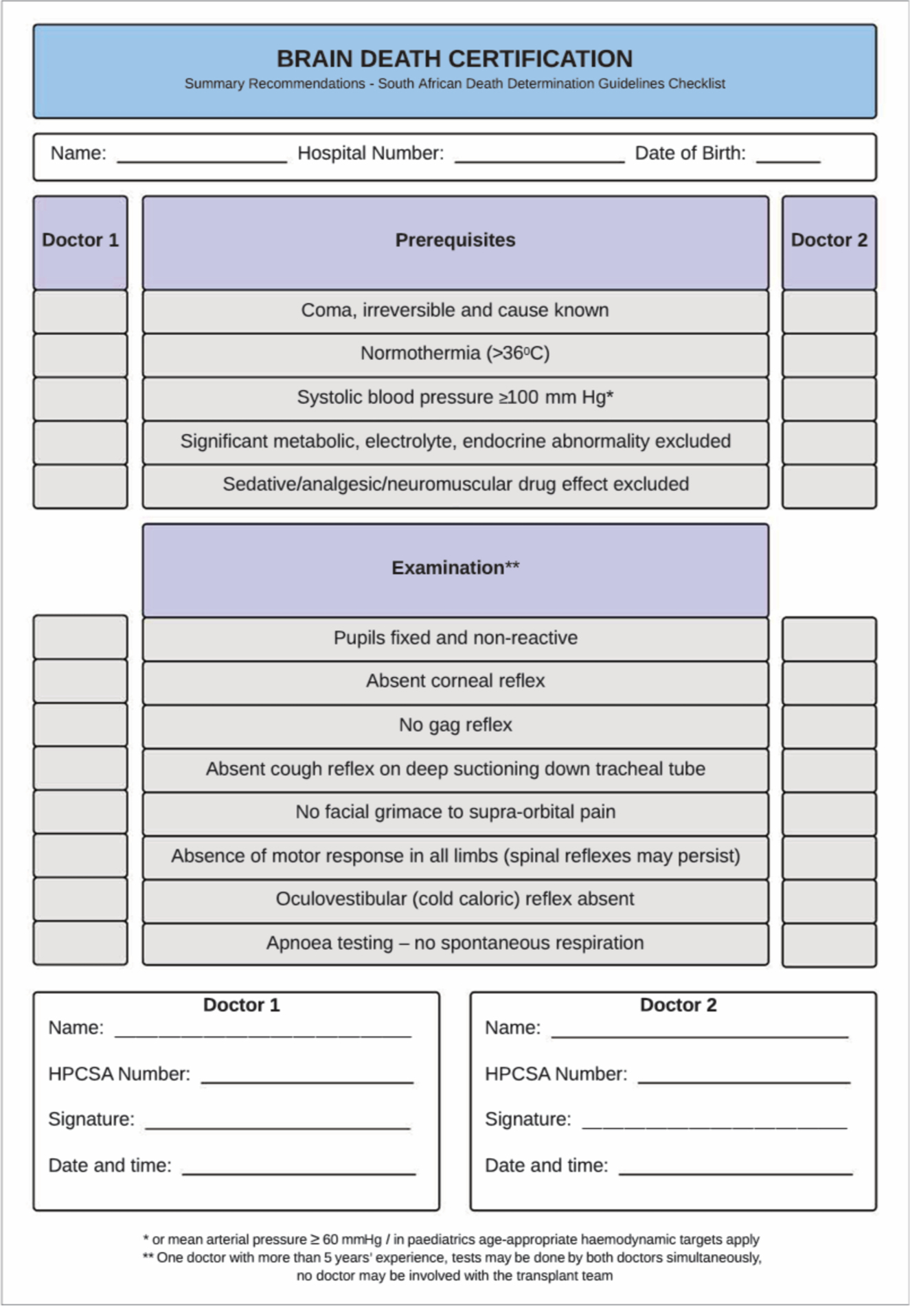
Fig 1. Summary recommendations - Brain death certification checklist (https://criticalcare.org.za/resource/death-determination-checklists/).
Click on the image to enlarge.
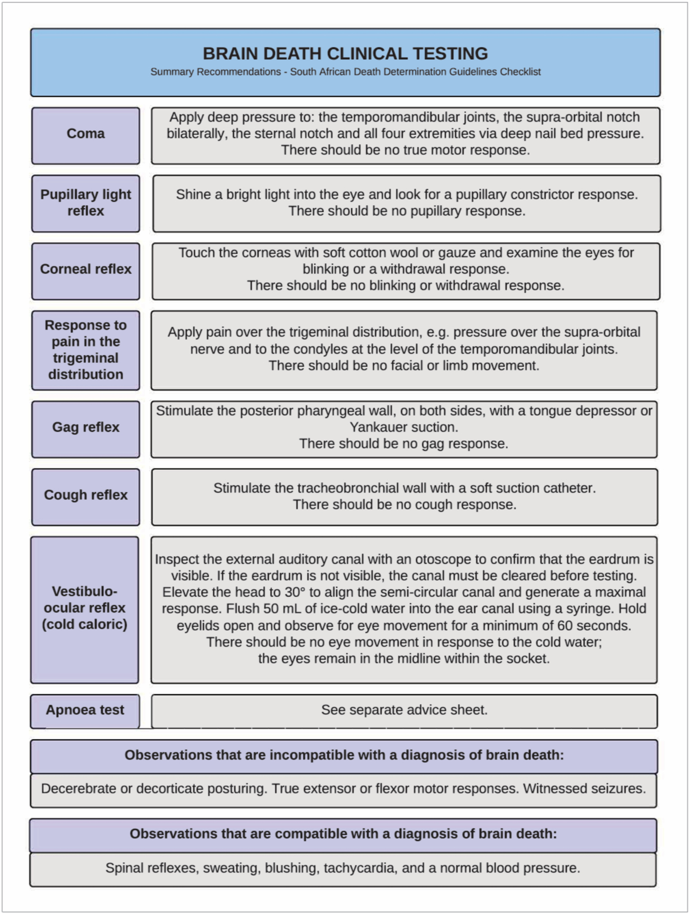
Fig. 2. Summary recommendations – Brain death clinical testing (https://criticalcare.org.za/resource/death-determination-checklists/).
Click on the image to enlarge.
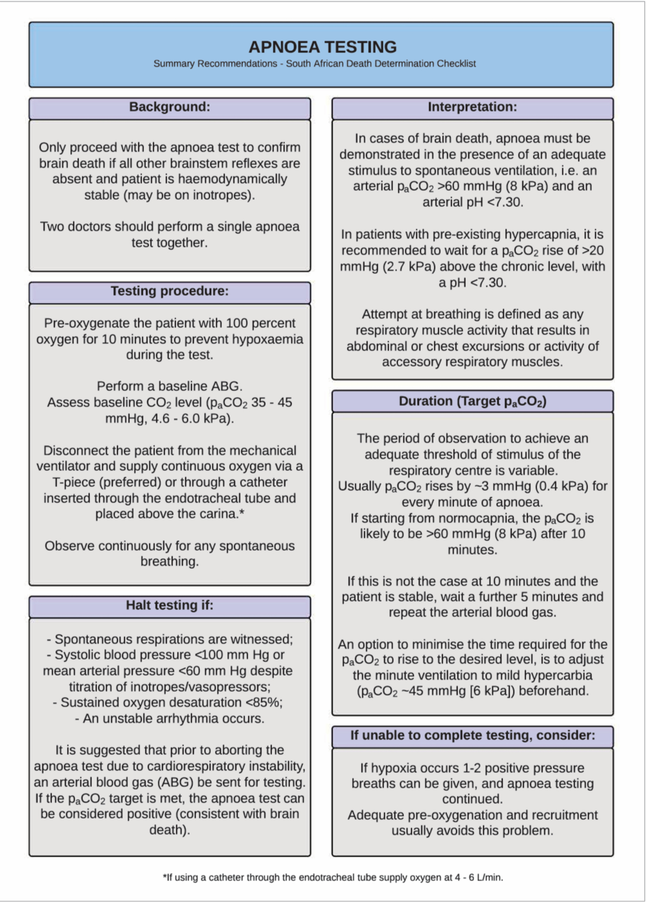
Fig. 3. Summary recommendations – Apnoea testing (https://criticalcare.org.za/resource/death-determination-checklists/).
Click on the image to enlarge.
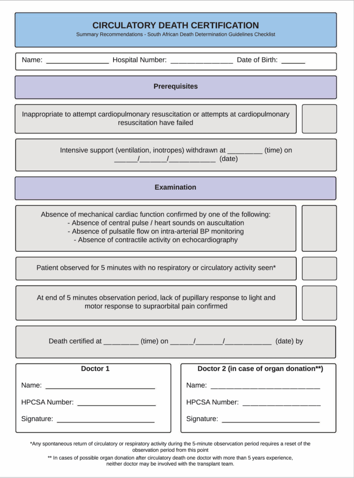
Fig. 4. Summary recommendation – Circulatory death certification checklist (https://criticalcare.org.za/resource/death-determination-checklists/)
Click on the image to enlarge.
Applicability, barriers, facilitators and cost implications of these guidelines
Barriers to effective implementation of death determination guidelines include a lack of uptake and acceptance by both clinicians and the public as well as the challenge of ensuring cultural and religious engagement and support for the medical determination of death. The endorsement and application of these guidelines is to be formally encouraged across all healthcare disciplines.
Undergraduate and postgraduate teaching should consider these guidelines as the standard of care for the determination of death in SA and incorporate them into their training programmes. Hospital policies and standard operating procedures should similarly align themselves with the guidelines to prevent differing practices across institutions, which can cause confusion among clinicians, families and the general public. [9,69] Use of the provided checklists is expressly recommended in the guidelines to facilitate clinicians to apply them in their daily practice. Continuing professional development programmes should incorporate these guidelines in the education of practising clinicians.
Pursuing a diagnosis of brain death may entail additional resources, and the clinician must judge the appropriateness in each clinical context. The beneficial effects of organ and tissue donation to the SA public and healthcare system are large. Exploring the option of organ and tissue donation at the end of life is a standard of care that should be fully assessed and appropriately explored with all patients and their families. Folder reviews and audits of documentation completed at end of life to assess compliance with these guidelines will assist in improving standards in death determination and end-of-life care. [70]
Conclusion
Death can be determined with complete certainty by medical professionals adhering to these guidelines which offer the clinician the latest evidence in best practice for determining death by either neurological or circulatory criteria. Use of the attached checklists is recommended.
Acknowledgements. The Critical Care Society of Southern Africa for facilitating the creation of these guidelines.
Author contributions. DT led the consensus process, developed and supervised the methodology, and chaired the face-to-face round-table meeting; was responsible for initial literature search, drafting of consensus key questions and supervised the Delphi process, including the drafting and grading of recommendations; drafted the manuscript, supervised revisions to the drafts, and approved the final manuscript. IJ, DG, FP, KDV chose the participants, developed and supervised the methodology, participated in the Delphi process and face-to-face roundtable meeting, performed literature searches, participated in manuscript drafting and revisions to the drafts, and approved the final manuscript. SM, RM, MM, BM, DB, BR, NM, GAR, NW, BL, MS, NA participated in the Delphi process and face-to-face round-table meeting, drafted and graded associated recommendations; participated in revisions to the drafts and approved the final manuscript. IC participated in the Delphi process, drafted and graded associated recommendations; participated in revisions to the drafts and approved the final manuscript.
Funding. These guidelines were internally funded by the Critical Care Society of Southern Africa (CCSSA), a registered non-profit organisation dedicated to delivering appropriate, quality care to the critically ill. The authors declare that the views or interests of the CCSSA have not influenced the final recommendations.
Conflicts of interest. All authors declare that there were no potential conflicts of interest.
Critical Care Society of Southern Africa: South African Guidelines on the Determination of Death_SAMJ/SAJCC_March 2021, Vol. 37, No. 1 (Part 2)