The First Antenatal Visit
The guidelines recommend management of common obstetric conditions at primary and referral level, based on local experience and evidence-based practice. They are not intended as strict protocols nor as standard operating procedures, but rather as a guide to safe effective management of common obstetric problems. The guidelines should be most useful to interns, medical officers and registrars.
Screening to detect tuberculosis (TB)
Pregnant women should book for antenatal care as soon as pregnancy is detected, even as early as 5-6 weeks’ gestation.
Make a complete assessment of gestational age & risk factors at the first antenatal visit. It is not necessary to wait until the second visit before such assessments are finalized. After one visit, a pregnant woman can be regarded as ‘booked’.
HISTORY TAKING
This follows the check-list on the antenatal record
- Gestational age – last menstrual period, cycle regularity & previous contraception
- Current pregnancy, especially last menstrual period
- Previous pregnancies, any complications & outcomes
- Medical conditions, previous surgery, & psychiatric problems
- Familial & genetic disorders, also considering ethnic origin, e.g sickle cell
- Allergies
- Use of medications,
- Use of alcohol, tobacco & other substances
- Family & social circumstances
- Plans for contraception after the pregnancy
SCREENING TO DETECT TUBERCULOSIS (TB)
All women infected with human immunodeficiency virus (HIV) should be asked if they have experienced the following symptoms:
- Cough for two weeks
- Fever
- Drenching night sweats
- Weight loss
Women with any one or more of these symptoms should provide sputum for TB testing & be followed up, and/or be further investigated.
PHYSICAL EXAMINATION
- General examination, including weight, heart rate, colour of mucous membranes, mid-upper arm circumference (MUAC), blood pressure (BP), & a check for oedema.
- Systemic examination, including teeth & gums, breasts, thyroid, & heart
- Pregnancy examination, including inspection & palpation of the pregnant uterus, with measurement of the symphysis-fundal height (SFH) in cm, & systematic palpation using Leopold’s method – fundal, lateral, Pawlik & pelvic grips
ESTIMATION OF GESTATIONAL AGE
Indicate clearly on the antenatal card (top left above the symphysis-fundal height graph how the gestational age was estimated. The first estimation of gestational age, with the expected date of delivery, will be used for the remainder of the pregnancy & must not be changed unless important new information becomes available.
Ultrasound
This is extremely useful for measuring gestational age. An ultrasound scan at ≤24 weeks is an accurate indicator of gestation, & overrides other methods of gestational age estimation. An ultrasound estimate of >24 weeks is less reliable but is still useful when there is uncertainty about the date of a woman’s last menstrual period.
Last menstrual period (LMP)
This is valid if the woman is sure of her dates, & where palpation of the uterus & SFH measurement are compatible with the given dates. Use Naegele’s rule: Expected date of delivery (EDD) = first day of last menstrual period plus 7 days, minus 3 months, plus 1 year.
Example: If LMP is on 5 October 2016, EDD will be on 12 July 2017.
Symphysis-fundal height (SFH) measurement
This is used if the dates from the last menstrual period are unknown or wrong, & if there is no ultrasound estimation of gestational age, provided that the pregnancy is otherwise normal. The measured SFH is plotted onto the 50th centile line on the SFH graph, allowing the corresponding gestational age to be read from the graph. Examples are shown below.
Palpation
The SFH measurement is of little value at <20 cm (corresponding to <20 weeks). In early pregnancy, bimanual & abdominal palpation can be used. At term, palpation of the fetal head may be helpful. Gestational age assessment by palpation requires care, skill & experience.
SCREENING INVESTIGATIONS
The following are offered as routine:
- Syphilis serology (RPR)
- Rhesus (D) blood group, using a rapid card test
- Haemoglobin (Hb) level
- HIV serology, following principles of HIV counselling & testing (HCT). A rapid test is done, with confirmation of positive results using a different kit. Post-test counselling follows immediately. A CD4 cell count is done on all women who test HIV positive
- Urine dipstick for protein & glucose
- Ultrasound scan for any woman presenting at ≤24 weeks
- Nuchal translucency & uterine artery Doppler screening (from 11-14 weeks) may be offered to women sent for scan at ≤14 weeks
All of the above tests should ideally be performed at the antenatal clinic, with the results available to the pregnant women before they complete their first visits.
MEDICATIONS
The following are given to all pregnant women:
- Ferrous sulphate 170 mg daily, OR Ferrous fumarate 200 mg daily
- Folic acid 5 mg daily
- Elemental calcium 1 g daily in divided doses
FINAL ASSESSMENT
The final assessment should include:
- A problem list in the appropriate space on the antenatal card
- A note on how the problems should be managed further
- The next follow-up date entered on the symphysis-fundal height graph corresponding the gestational age at that visit
- A delivery plan: the estimated date, & the planned place & mode of delivery
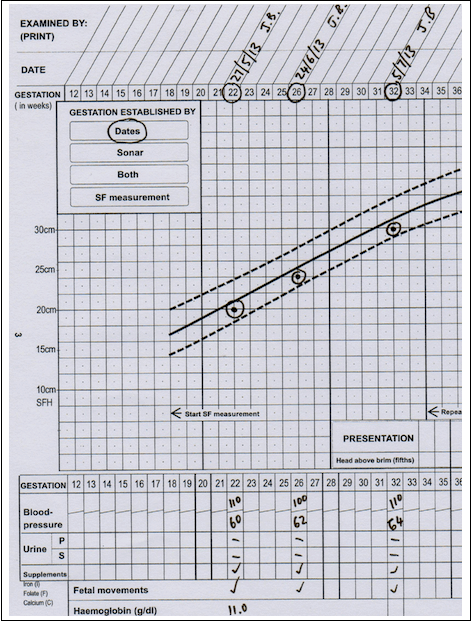
SFH graph of a woman with correct menstrual dates
At booking (27/5/13), she is 22 weeks pregnant by dates. The SFH is 20 cm, in keeping with her dates. Subsequently, SFH growth is normal, between the 10th (lower dotted line) & 50th centile (solid line).
SFH graph of a woman whose menstrual dates are unknown
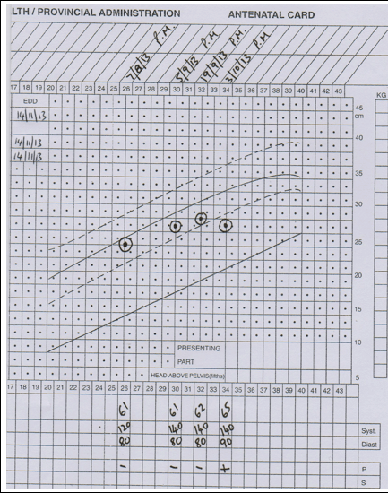
At booking (5/8/13), the SFH of 28 cm is entered on the 50th centile line, giving a gestational age of 29 weeks. At subsequent visits, SFH growth is above the 50th centile line, but is normal as it has not crossed above the 90th centile line (the upper dotted line).

SFH graph of a woman with abnormal SFH growth
At 34 weeks of gestation, the SFH measurement has crossed to below the 10th centile. Possible reasons include
- intrauterine growth restriction;
- olighydramnios; &
- fetal death, each possibly linked to pre-eclampsia (BP 140/90 mmHg with +proteinuria).
These findings necessitate ultrasound scanning, & if the fetus is alive, assessment of amniotic fluid volume, fetal biometry & umbilical artery Doppler studies, & hospital admission.