The Physiology of Acute Pain
Definitions Understanding nociceptive pathways Neurotransmitters Intracellular events Receptors and ligands Nociceptive pathways Receptors Autonomic nervous system Psychological aspects of acute pain The progression of acute to chronic pain Adverse effects of pain Definitions Pain is defined by the International Association for the Study of Pain (IASP) as an “unpleasant sensory and emotional expe
Understanding nociceptive pathways
Psychological aspects of acute pain
The progression of acute to chronic pain
Definitions
Pain is defined by the International Association for the Study of Pain (IASP) as an “unpleasant sensory and emotional experience, associated with actual or potential tissue damage or described in terms of such damage” (Mersky). It is a complex interaction of sensory, emotional and behavioural factors. Nociception is mostly modulated at the spinal cord level and eventually interpreted by the cortex, resulting in varying degrees of discomfort and pain. Perceived pain, however, does not necessarily correlate with the degree of tissue damage.
Pain is divided into physiological pain and pathophysiological or clinical pain.
Physiological pain is the activation of nociceptors in response to a noxious stimulus, whereas clinical pain includes tissue and/or nerve injury and the inflammatory response. Acute and chronic pain can be represented on a continuum of a process which includes inflammatory, neuropathic, visceral and somatic pain. The nervous system is not a hard-wired system. It allows for peripheral, central, intracellular and synaptic modifications. Acute pain can result in longterm changes and, subsequently, modified responses to sensory input (neuroplasticity).
Acute physiological pain is defined as pain of short and limited duration. It is well localised, transient and well differentiated from touch. The pain relates to an identifiable cause (trauma, surgery or inflammation) and its purpose is to act as a protective mechanism.
Clinical pain, on the other hand, outlasts the stimulus and spreads to nondamaged areas, leading to primary hyperalgesia. Peripheral sensitisation occurs as part of the inflammatory response and results in activation of the Aβ-fibres which leads to the pain to touch (allodynia). Antidromic impulses (travelling in the direction opposite of nociceptive impulses conducted to the higher centres) result in the release of neurotransmitters from nerve endings of a primary afferent in response to noxious stimulation. In turn, these additional neurotransmitters have modulating effects on further impulse transmission.
Table I: Pain physiology definitions
| Allodynia | Pain perceived from a normally nonnoxious stimulus. |
| Central sensitisation | Changes in the central nervous system (CNS) that lead to increased pain transmission and perception (central plasticity). |
| Gate control theory (Melzack and Wall) |
Input along large, low threshold (Aβ-) fibres inhibits response of wide dynamic range (WDR) cells to nociceptive input. (E.g. rubbing your leg after bumping it reduces the pain sensation.) |
| Hyperalgesia | Increased response to stimulus normally perceived as painful. |
| Neuroplasticity | Change in neural pathways and synapses due to modulating factors or processes. |
| Peripheral sensitisation | Decreased threshold of nociceptors and increased output, caused by mediators released during tissue injury |
| Primary hyperalgesia | Increased pain perception from the injury site due to the release of inflammatory mediators peripherally. |
| Secondary hyperalgesia | Increased pain perception due to CNS mechanisms sensitising central neurons. |
| Wind-up phenomenon | A form of central plasticity. Repeated stimulation of C-fibres results in an increased number of discharges per stimulus, an expansion of receptive field size and an increase in spontaneous discharge rate. |
Understanding nociceptive pathways
The experience of pain involves a series of neurophysiological processes that reflect the following four components:
- Transduction: A noxious stimulus is converted to electrical impulses in sensory nerve endings.
- Transmission: The conduction/propagation of impulses via peripheral afferent fibres to the CNS. Voltage-gated sodium channels play an important role in this component.
- Peripheral sensory afferent fibres travel from the site of transduction to the spinal cord dorsal horn on the ipsilateral side.
- Second-order neurons cross the midline to ascend in the contralateral spinothalamic tract.
- Third-order neurons project from the thalamus to the sensory cortex.
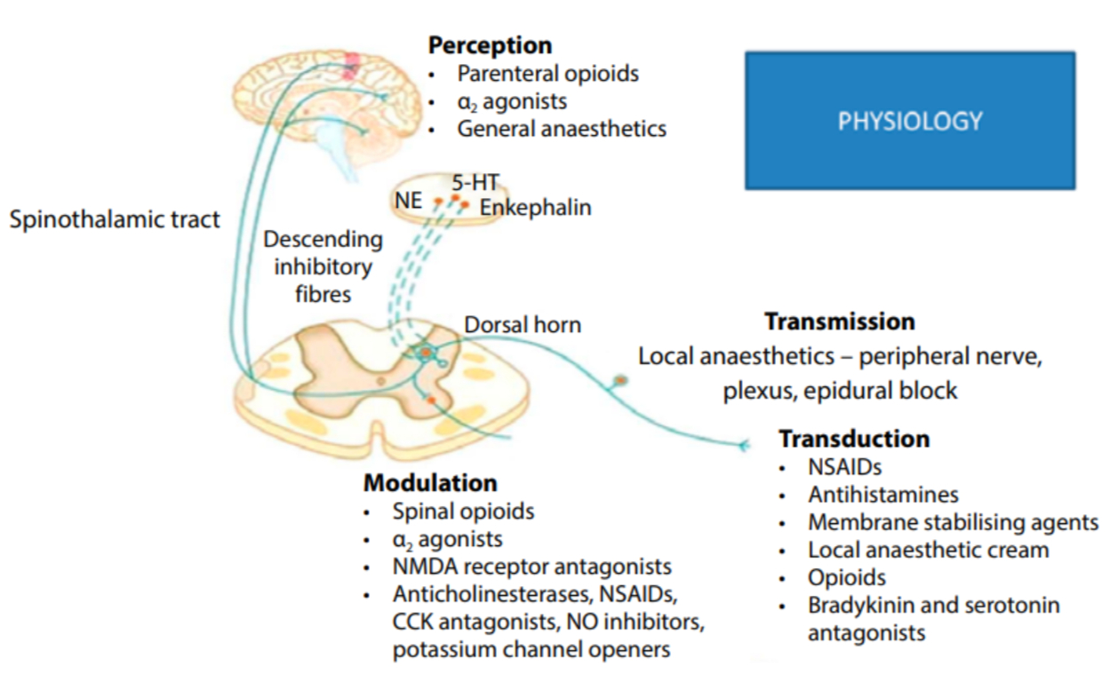
Figure 1: Elements of pain processing and antagonists acting at these sites 5-HT – 5-hydroxytryptamine (serotonin), CCK – cholecystokinin, NE – norepinephrine, NMDA – N-methyl-D-aspartate, NO – nitric oxide, NSAID – nonsteroidal anti-inflammatory drug
Click on the image to enlarge
3. Modulation: This is the alteration of pain transmission. Inhibition or excitation of ascending and descending pathways in the peripheral nervous system (PNS) and CNS result in inhibitory or facilitatory effects. Modulation involves the periaqueductal grey matter (PAG) and rostral ventromedial medulla (RVM) from descending projections from cortical sites to the spinal cord. Modulation involves the gate control theory, wind-up phenomenon and glial activation. Glial cells become activated after an insult (e.g. nerve injury or chronic opioid therapy), which subsequently release several substances. This causes a change in membrane potentials that cause normally inhibitory inputs to become excitatory.
4. Perception: This occurs in the thalamus and the sensory cortex, and produces a subjective pain sensation.
Primary afferent fibres
Peripheral nociceptors are free nerve endings of afferent nerves which respond to pressure, temperature and chemical stimuli. The cell bodies of these afferent nerves are located in the dorsal root ganglia, except for the fibres innervating the head, whose cell bodies are located at the trigeminal ganglion.
Sensory fibres are classified as Aβ-fibres that are large-diameter, myelinated fibres and conduct the fastest. They are activated by nonnociceptive stimuli and not involved in noxious transmission.
There are two main categories of nociceptive afferent fibres.
- Aδ-fibres are thin, myelinated and transmit mechano-thermal stimuli. They terminate in laminae I, III, IV, V of the dorsal horn of the spinal cord, and act as fast-conducting fibres involved with immediate pain.
- C-fibres make up 80–90% of nociceptive afferents. They are thin, unmyelinated and polymodal (i.e. they respond to thermal, mechanical and/or chemical stimuli). They terminate in the dorsal horn laminae I and II, and are slow conducting fibres, involved in dull, burning or throbbing, poorly localised pain. An important subgroup is nociceptors insensitive to mechanical stimuli (mechanically insensitive afferents/nociceptors [MIAs]) and is thought to play an important role in central sensitisation.
The Aδ- and C-fibres are high-threshold fibres. An ‘inflammatory soup’ of chemicals sensitise high-threshold nociceptors after surgery and trauma. Sensitised nociceptors react to a painful stimulus at a lower threshold (peripheral sensitisation). Silent nociceptors can become active in the presence of inflammation and also play a part in peripheral sensitisation.
Dorsal horn
Primary afferents (first-order neurons) interact extensively with other afferents, as well as with interneurons and the endings of descending tract fibres.
Second-order neurons comprise both high-threshold neurons (nociceptive specific) and WDR neurons. Once sensitised, the WDR neurons respond to and discharge upon tactile, nonnoxious stimuli (allodynia).
Central sensitisation can result from the activation of N-methyl-D-aspartate (NMDA) receptors on second-order neurons. Increased activity in the dorsal horn following repetitive stimulation from primary nociceptors causes increased excitability of second-order neurons. This leads to secondary hyperalgesia, wind-up, temporal summation and long-term potentiation. A low-threshold stimulus then results in both an increased magnitude and a prolonged duration of depolarisation at the second-order neurons.
Collateral branches of the small Aδ- and C-fibres may travel in the lateral part of the entry zone for several segments before synapsing in the dorsal horn (Lissauer’s tract).
Neurotransmitters
At the periphery
Peripheral sensitisation occurs due to substances released by the damaged tissues, blood vessels and sympathetic terminals. This is termed the ‘inflammatory soup’ and contains, among others, hydrogen and potassium ions, bradykinin (BK), histamine, noradrenalin (NA), 5-hydroxytryptamine (5-HT), prostaglandins (PGs), leukotrienes, nerve growth factor, glutamate and substance P.
Dorsal horn
Excitatory
Glutamate, substance P and neurokinin 1 (NK-1) activate the low-threshold α-amino-3-hydroxy5-methyl-4-isoxazolepropionic acid (AMPA) and NK-1 receptors, which in turn, sensitise and activate the high-threshold NMDA receptors on the second-order neurons.
Inhibitory
Noradrenalin, dopamine, serotonin, histamine, oxytocin, vasopressin, acetylcholine, γ-aminobutyric acid (GABA), glycine and opioids predominantly occur in the descending pathways.
Table II: Neurotransmitters
| Neurotransmitter | Receptor | Effect on nociception |
| Substance P | Neurokinin-1 | Excitatory |
| Calcitonin gene-related peptide | Excitatory | |
| Glutamate | NMDA, AMPA, kainite, quisqualate | Excitatory |
| Aspartate | NMDA, AMPA, kainite, quisqualate | Excitatory |
| Adenosine triphosphate (ATP) | P1 ,P2 | Excitatory |
| Somatostatin | Inhibitory | |
| Acetylcholine | Muscarinic | Inhibitory |
| Enkephalins | μ, δ, κ | Inhibitory |
| β-endorphine | μ, δ, κ | Inhibitory |
| Norepinephrine | α2 | Inhibitory |
| Adenosine | A1 | Inhibitory |
| Serotonin | 5-HT1 (5-HT3) | Inhibitory |
| γ-aminobutyric acid (GABA) | A, B | Inhibitory |
| Glycine | Inhibitory |
NMDA – N-methyl-D-aspartate, AMPA – 2-(aminomethyl)phenylacetic acid, 5-HT – 5-hydroxytryptamine
Intracellular events
NMDA activation in the CNS (by means of removal of the magnesium plugs) leads to
i) calcium (Ca2+) influx to the cell,
ii) the production of nitric oxide and secondary messengers, and
iii) prostaglandin production.
Receptors and ligands
Ligands transduce the specific stimulus into an action potential which is sodium (Na+) channel dependent. Local anaesthetics (LAs) act at this level, but as Na+ channels are present in all nerve fibres, blocking of the autonomic motor and sensory fibres can occur to some degree.
Pain modulation can be achieved by i) decreasing excitation (opioids, Na+ channel blockers and ketamine) and/or ii) increasing inhibition (NA, glycine, GABA at the level of the spinal cord).
The most common receptors and ligands are outlined in Table III.
Table III: Receptors, stimuli and ligands
| Receptor | Receptor subtypes | Ligands and stimuli |
| Transient receptor potential receptors (TRPs) | TRPV1 TRPV2 TRPA |
Heat, H+ and capsaicin Heat Cold |
| Acid-sensing | ASIC DRASIC |
Protons |
| Purine | P2X3 | ATP |
| Serotonin | 5-HT3 | 5-HT |
| NMDA receptor | NRI Glutamate | Glutamate |
| AMPA | iGlutR1 | Glutamate |
| Kainate | iGlutR5 | Glutamate |
| Prostanoids | EP1-4 IP |
PGE2 PGI2 |
| Histamine | HI | HA |
| Serotonin | 5-HT1A, 5-HT2A and 5-HT4 | 5-HT |
| Bradykinin | BK1 and BK2 | BK |
| Cannabinoids | CB1-2 | Anandamide |
| Opioids | Mu, delta and kappa | Enkephalin, dynorphin and beta-endorphin |
| Tachykinin | NK-1 | Substance P and neurokinin A |
5-HT – 5-hydroxytryptamine, AMPA – α-amino-3-hydroxy-5-methyl-4-isoxazolepropionic acid, ASIC – acid-sensing ion channel, ATP – adenosine triphosphate, DRASIC – dorsal root acid-sensing ion channel, NK-1 – neurokinin 1, NMDA – N-methyl-D-aspartate, PGE2 – prostaglandin 2, PG12 – prostacyclin, TRP – transient receptor potential
Nociceptive pathways
Ascending pathways
The spinothalamic tract originates in laminae I, II and V of the opposite dorsal horn, and after crossing the midline, it ascends to the thalamus and the somatosensory cortex, providing information on the type and the site of the painful stimulus.
The spinomesencephalic tract mainly originates in lamina I and mediates the affective and emotional components of the nociceptive stimulus. Autonomic and sensory coordination is provided by this pathway.
The cingulate cortex, insula, PAG, reticular formation and prefrontal cortex receive multiple inputs from the ascending pathways, and facilitate the coordination of autonomic and emotional responses.
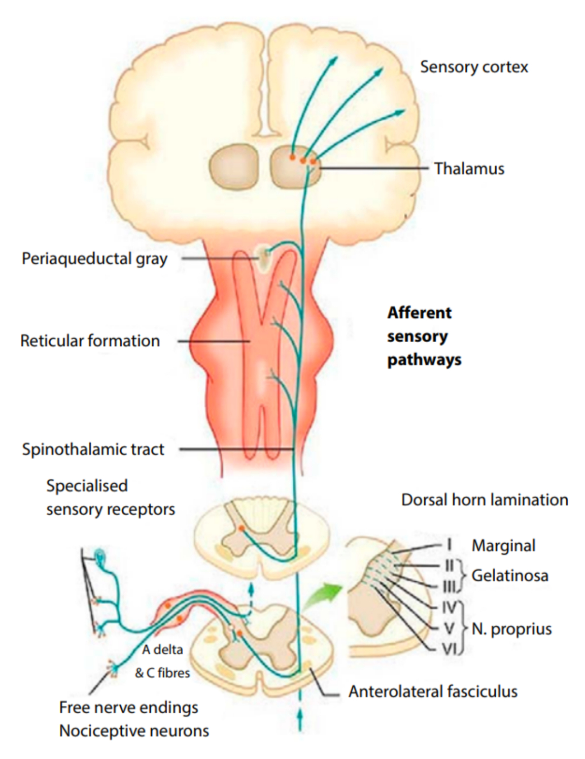
Figure 2: Ascending pathways
Click on the image to enlarge
Descending inhibition
These pathways modulate nociception by action on the primary nociceptive afferents and interneurons at the level of the dorsal horn. They mainly inhibit transmission towards the cortex and other higher centres. These tracts originate in the cortex, PAG and brainstem nuclei. Their fibres terminate in the dorsal horn, facilitating inhibition and modulating nociceptive input. Inhibitory neurotransmitters include opioids, 5-HT, NA and GABA. Tricyclic antidepressants (TCAs), opioids and alpha-2 agonists exert their effects on modulating nociception via the descending pathways.
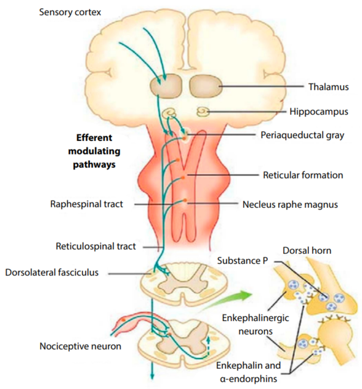
Figure 3: Descending pathways
Click on the image to enlarge
Neuropathic pain
By definition, neuropathic pain is pain that originates in the nervous system. There is no clear distinction between neuropathic and nociceptive pain as clinically defined entities, and they often co-exist. Trauma and surgery cause nociceptive as well as neuropathic pain due to the cutting of nerve endings.
Receptors
Activation of the nociceptors produces depolarisation and eventually triggers an action potential and release of ligands from the nerve endings.
Opiate receptors
They respond to endogenous and exogenous opiates. Note that opiate receptors are also located peripherally along the nerve fibre from the dorsal horn. This explains the effect of opioids when administered intra-articularly or into the subcutaneous (SC) tissue. The opiate receptors are mainly located presynaptically (75%). Activation of the opioid receptors reduces the release of neurotransmitters from the first-order neurons while inflammation and nerve injury result in the presynaptic loss of opioid receptors.
GABA and the glycine receptors
GABA and the glycine receptors in the CNS have an inhibitory function. When GABA binds to its receptor, it increases chloride ion flow through chloride channels resulting in hyperpolarisation of the neuron membrane. GABA-A receptors occur mainly postsynaptically and respond to endogenous GABA ligand and to benzodiazepines. GABA-B receptors occur presynaptically and respond to endogenous GABA and to baclofen. Barbiturates, anaesthetic drugs and corticosteroids are also thought to activate GABA-B receptors.
Adrenoreceptors
Activation of the alpha adrenoreceptors at the dorsal horn has an analgesic effect (endogenous NA and exogenous clonidine). The effect is synergistic with the opioid agonists.
N-methyl-D-aspartic acid receptor
Glutamate and substance P are released from the nociceptive first-order afferents, which activate the low-threshold AMPA and NK-1 receptors which, in turn, activate the NMDA receptors. The removal of the Mg2+ plug is followed by an influx of Ca2+ into the cells and subsequent depolarisation.
Ketamine is an indirect NMDA antagonist with the potential to provide analgesia and modulate the development of chronic pain. The NMDA receptor is involved in the development of tolerance to opioids.
Transient receptor potential receptors
Transient receptor potential (TRP) V1 (TRPV1), previously called VR1, is a nonselective ion channel, activated by capsaicin (a vanilloid compound), heat above 43°C, lipoxygenase, leukotrienes, interleukins (ILs) and N-arachidonoyl dopamine. Stimulation of these receptors, once opened, allows for the influx of charged portions of large molecules (i.e. charged fraction of local anaesthetic drugs, thereby increasing intracellular availability of local anaesthetic drugs to bind to the Na+ channel).
Other TRP ion channels have been described and found to be important in nociceptor activation. TRPV2-4, as well as TRPM8 and TRPA1, are all activated by temperature in the noxious and nonnoxious range, and together encode the entire temperature spectrum.
Autonomic nervous system
The autonomic nervous system (ANS) is closely linked to the nociceptive pathways. The sympathetic system is an efferent system and biofeedback is maintained at the:
- dorsal horn level (extensive synapses between the afferent and sympathetic fibres take place at the dorsal horn level),
- dorsal respiratory group (DRG) level (sympathetic fibres form a ‘basket’ around the DRG, influencing afferent transmission), and
- peripheral level (somatic and visceral nociception causes vasodilatation, tissue damage and the subsequent release of neurotransmitters; circulating catecholamine and NA released from the sympathetic fibres perpetuate the noxious stimulus).
Psychological aspects of acute pain
Pain is an individual biopsychosocial phenomenon and is largely influenced by culture, previous pain experience and the ability to cope. It is a personal and subjective experience. Preoperative anxiety has been shown to contribute to increased postoperative pain, and preoperative depression is a predictor of postoperative pain.
The progression of acute to chronic pain
Chronic pain can develop following an acute pain episode. Postoperative pain, postzoster pain and low back pain are often associated with chronic pain. One and a half per cent of surgical procedures result in chronic pain development.
Risk factors for the development of chronic pain include the following:
- Intense and prolonged preoperative and/or postoperative pain
- Repeated surgery
- Perioperative chemotherapy and/or radiotherapy
- Postoperative complications (e.g. infection)
Some surgical procedures result in an increased incidence of chronic pain (Table IV).
Table IV: Surgical procedures with an increased incidence of chronic pain
| Procedure | Incidence (%) |
| Dental surgery | 5–13 |
| Vasectomy | 0–37 |
| Cholecystectomy | 3–56 |
| Mastectomy | 11–57 |
| Inguinal hernia repair | 0–63 |
| Thoracotomy | 5–67 |
| Amputations | 30–85 |
Central sensitisation and wind-up phenomena are the pathophysiological mechanisms postulated to be involved in chronic pain development.
Adverse effects of pain
Acute pain provokes physiological modification in multiple organ systems. The stress response involves neurohumoral changes with multiple implications. The aim of adequate pain management is to both provide pain relief as a humane measure and minimise the multisystem deleterious effects caused by the stress response.
A catabolic state, sympathetic stimulation and immuno-suppression are hallmarks of the stress response. The psychological effects can create a vicious cycle, maintaining the negative effects. The endocrine system changes result in a catabolic state, increased adrenocorticotropic hormone (ACTH), cortisol, antidiuretic hormone (ADH), catecholamines, angiotensin II, interleukin (IL)-1 and IL-6, and tumour necrosis factor (Table V).
Sympathetic stimulation results in an increased heart rate and blood pressure, increasing the risk of myocardial ischaemia. Pain limits coughing and decreases functional residual capacity, which in turn, increases the risk of atelectasis and pulmonary infection. Decreased mobility results in an increased risk of deep vein thrombosis (DVT). Anxiety, helplessness, loss of control, an inability to interact and sleep deprivation all contribute to psychological disturbances, which can also increase the risk of persistent pain developing.
Table V: Adverse effects of pain
| Endocrine | Increased catabolism Increased ACTH, ADH, GH, catecholamines, angiotensin II, IL-1 and IL-6 and TNF Decreased anabolism Decreased insulin and testosterone |
| Metabolic | Carbohydrates Hyperglycaemia, glucose intolerance and insulin resistance Protein Increased acute phase protein catabolism Lipids Increased lipolysis |
| Water and electrolytes | Water retention Potassium loss |
ACTH – adrenocorticotropic hormone, ADH – antidiuretic hormone, GH – growth hormone, IL – interleukin, TNF – tumour necrosis factor
Bibliography
- Benzon H, Raja SN, Fishman SE, Lui SS, Cohen SP. Essentials of pain medicine (e-book). New York: Elsevier Health Sciences; 2011. p. 3-10.
- Butterworth JF, Mackey DC, Wasnick JD. Morgan and Mikhail’s clinical anaesthesiology, 5th ed. New York: McGraw-Hill Education; 2013. p. 1023-37.
- Feizerfan A. Transition from acute to chronic pain. BJA Educ. 2015;15(2):98-102. https://doi.org/10.1093/bjaceaccp/mku044.
- Mechanism of acute and chronic pain: Can acute pain relief prevent chronic pain? http://gmch.gov.in/sites/default/files/documents/ Mechanism%20of%20acute%20and%20chronic%20pain%202.pdf. e-study slide 4, 5, 8.
South African Society of Anaesthesiologists (SASA) Guidelines for the Management of Acute Pain in Specific Scenarios 2022