Upper Gastrointestinal Bleeding
Surgical Emergencies - Upper Gastrointestinal Bleeding
Assessment
Resuscitation and initial management
Endoscopy
Timing of endoscopy
Endotherapy
Failure to Control Bleeding
Rockall Score
Cape Metro upper GI bleeding algorithm
Endoscopic control for upper GI bleeding
Indications for endoscopic control (Forrest Classification)
Indications for repeat endoscopy (within 24-48 hours)
Acute upper gastrointestinal bleeding is a common medical emergency that has a 10% hospital mortality rate. Despite changes in management, mortality has not significantly improved over the past 50 years. Elderly patients and people with chronic medical diseases withstand acute upper gastrointestinal bleeding less well than younger, fitter patients, and have a higher risk of death. Almost all people who develop acute upper gastrointestinal bleeding present to hospital and the guideline therefore focuses on hospital care.
When seen in the Emergency Unit it is important to confirm if the patient is haemodynamically stable and has any evidence of GI bleeding. This entails reliable historical or witnessed evidence of bleeding as adjudged by red blood vomitus coffee ground vomitus and melaena stool. Those with a normal blood pressure and no evidence melaena do not require admission
The most common causes of life threatening haemorrhage are peptic ulcer and oesophago-gastric varices. it is important as part of the initial assessment to determine if the patient has stigmata of chronic liver disease and signs of portal hypertension he should be assumed to be bleeding from oesophageal varices otherwise he should be classified as non variceal This is important as the initial resuscitative and drug management is different.
The presences of CLD is one of the overall factors to be assessed in the Blatchford risk stratification model which predicts risk of re-bleeding and death and is used to the triage with regard to the intensity of monitoring and the urgency of performing endoscopy and the potential need for surgery. All patients with acute upper gastrointestinal bleeding should have a formal risk score assessment at presentation, and a repeat after endoscopy :
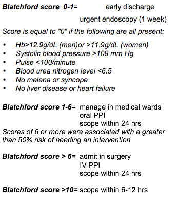
Modified Glasgow-Blatchford Score | |
Admission risk marker | Score |
Blood Urea | |
| ≥6·5 <8·0 | 2 |
| ≥8·0 <10·0 | 3 |
| ≥10·0 <25·0 | 4 |
| ≥25 | 5 |
Haemoglobin (g/L) for men | |
| ≥12.0 <13.0 | 1 |
| ≥10.0 <12.0 | 3 |
| <10.0 | 6 |
Haemoglobin (g/L) for women | |
| ≥10.0 <12.0 | 1 |
| <10.0 | 6 |
| Systolic blood pressure (mm Hg) | |
| 100–109 | 1 |
| 90–99 | 2 |
| <90 | 3 |
| Pulse | |
| Pulse ≥100 (per min) | 1 |
Resuscitation and initial management
Transfuse patients with massive bleeding with blood, platelets and clotting factors in line with local protocols for managing massive bleeding.
Treat patients who are taking warfarin and whose upper gastrointestinal bleeding has stopped in line with local warfarin protocols. NSAID should be discontinued if not taken for cardiovascular risk reduction. However stopping them does not restore platelet function for several days and hence does not aid in achieving acute bleeding control. Most important is appropriate adequate resuscitation tailored to the patients haemodynamic status and their co-morbidities.
In those who are shocked on admission use at least two big intravenous lines for resuscitation. Pass a Foley's catheter to monitor urine output, nasogastric tube to prevent aspiration. Do FBC and INR , urea and electrolytes, liver function tests if cirrhosis is suspected, cross-match blood, chest X-ray and ECG especially if the patient is going to theatre. Monitoring of haemodynamic (pulse, BP, urine output response to resuscitation is key to determining the timing of upper GI endoscopy.
A blood product resuscitation should be instituted in those with suspected variceal haemorrhage, and consider an octreotide (pressor) infusion.
All high risk patients should receive IV acid suppression drugs. This should be continued for 48 hours in those with endoscopic risk factors
Endoscopy is the primary diagnostic investigation in patients with acute upper gastrointestinal bleeding. Endoscopy aids diagnosis, yields information that helps to predict outcome and most importantly allows treatments to be delivered that can stop bleeding reduce the need for transfusion and reduce the risk of re-bleeding and the length of hospital stay. It allows risk assessment for both variceal and non variceal haemorrhage and who is eligible for endotheraphy.
See Ulcer risk stratification Forrest Classification with the Blatchford score
- Patients who remain unstable should be evaluated in theatre with airway protection.
- The vast majority of patients will stabilise with resuscitation
- Those with a high Blatchford score > 8 should have endoscopy within 6 -12 hours. An experienced endoscopist with prior skill in endotherapy should be present for these two categories of patient
- The others should have endoscopy within 24 hours of admission; they should not wait the whole weekend
- The bleeding source should be treated endoscopically
- Dual therapy with Injection 1/10000 adrenaline + heater probe or Endoscopic clip
- Varices should be treated by banding.
Hesitation or delay to surgical intervention is the commonest cause for mortality of a major upper GI bleed.
Decision making can be difficult particularly in elderly patients, or those who are at high surgical risk due to co-morbidities. Discuss with the consultant on call early, if at all in doubt.
Failure of to control bleeding may lead to a variety of strategies, such as temporary use of a Minnesota tube in a patient with varices, or laparotomy for surgical control of the bleeding.
Surgery should be performed if there ongoing haemorrhage or a re-bleed after an adequately controlled endoscopic intervention. Bleeding duodenal ulcers are controlled by under-running the bleeding vessel with suture ligation. Gastric ulcers may require resection of the ulcer or gastrectomy, unless there is a clearly visualised bleeding vessel in the ulcer base, which can be suture ligated.
Interventional embolisation is only available at tertiary centres and timely transfer may allow these approaches to be successful in selected patients.
Discussion with senior colleagues is mandatory- surgery at the referring hospital is the best treatment in most cases.
Rockall Score (to be completed after urgent endoscopy)
Variable | Score 0 | Score 1 | Score 2 | Score 3 |
| Age | <60 | 60- 79 | >80 | |
| Shock | No shock | Pulse >100 BP >100 Systolic | SBP <100 | |
| Co-morbidity | Nil major | CHF, IHD, major morbidity | Renal failure, liver failure, metastatic cancer | |
| Diagnosis | Mallory-Weiss | All other diagnoses | GI malignancy | |
| Evidence of bleeding | None | Blood, adherent clot, spurting vessel |
Interpretation: score is calculated by simple addition.
A score less than 3 carries good prognosis but total score more than 8 carries high mortality.
Stigmata of ulcer haemorrhage | Risk of recurrent bleeding without endoscopic therapy |
| Arterial spurt (active bleeding) | 70-100% |
| Non bleeding visible vessel | 30-50% |
| Non bleeding adherent clot | 8-35% |
| Oozing ulcer | 10-27% |
| Flat spot | <8% |
| Clean base ulcer | <3% |
Cape Metro upper GI bleeding algorithm
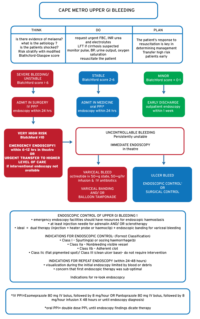
Endoscopic control for upper GI bleeding
- emergency endoscopy facilities should have resources for endoscopic haemostasis
- at least injection needle for adrenaline AND/ OR sclerotherapy
- ideal = dual-therapy (injection + heater probe or haemoclip) + endoscopic banding for variceal bleeding
Indications for endoscopic control (Forrest Classification)
- Class I –Spurting(a) or oozing haemorrhage(b)
- Class IIa –Non-bleeding visible vessel
- Class IIb –Adherent clot
- Class IIc (flat pigmented spot)/ Class III (clean ulcer-base)- do not require intervention
Indications for repeat endoscopy (within 24-48 hours)
- Visualisation during the initial endoscopy limited by blood or debris
- concern that first endoscopic therapy was sub-optimal