Wrist injuries — Approach / Imaging
Wrist injuries — An Emergency Centre approach
Physical evaluation
Test all three major nerves
Imaging
Adequacy
Alignment and angles
Bony shape
Special views
Wrist injuries are common. Most clinicians can diagnose a distal radius fracture with ease but some of the injuries can be complex and involve bones, muscle and ligaments.
If there is doubt in the mind of the primary physician after the exam and radiographic
findings a follow up at a fracture clinic should be made and the wrist placed in a neutral functional position. Moreover, both recognized and occult injuries can lead to significant long-term sequelae. Due to our reliance on our hands for careers and day-to-day activities of all kinds, complete recovery is essential.
Most wrist injuries (90%) are the result of a fall on an outstretched hand. While the characteristics of wrist injuries vary with age, no age group is spared.
- Young children almost never have carpal fractures but may sustain distal radial fractures (especially torus [bulging of the cortex] and greenstick fractures).
- A fall on an outstretched hand in a toddler or young child may also result in a radial head or supercondylar humerus fracture.
- Adolescents and young adults are more likely to injure the carpal bones, especially the scaphoid. When they do sustain a distal radius fracture, it is often complex and associated with other injuries.
- Increasing patient age, scaphoid fractures become less common, while distal radius fractures become more so.
Fall on the outstretched hand, giving us the onomatopoeic acronym “FOOSH”
Physical evaluation
The wrist includes all of the bones and articulations from the distal radius and ulna to the carpometacarpal joint.
Evaluation of any orthopaedic injury starts with confirmation of neurovascular integrity.
Neurological integrity of the hand is best confirmed with two-point discrimination. Most people can detect two points at 0.5 cm apart on the finger pads. Splitting a tongue blade with a twisting motion provides two sharp points that can be placed in the longitudinal axis of the fingertip border (so as not to cross between nerve fields).
Motor testing becomes difficult with pain, but gross function should be confirmed.
Test all three major nerves
- ulnar nerve motor function, ask the patient to abduct his or her fingers (spread them apart against resistance). This will challenge the first dorsal interossei, which are supplied by the ulnar nerve. The sensory branch supplies the volar aspects of the fifth finger and the radial and the ulnar half of the fourth finger.
- median nerve, have the patient lay his or her hand flat with the palm up, and then lift the thumb straight up from the palm. Press against the thumb to determine appropriate resistance (the median nerve innervates the abductor pollicis brevis). The sensory function of the median nerve is best tested at the distal pulp of the index finger.
- radial nerve supplies the motor function to wrist and finger extension via the extensor muscles of the forearm but does not innervate the intrinsic muscles of the hand; therefore, a motor deficit of the radial nerve is unlikely in an isolated wrist injury. To test the motor branch of the radial nerve, have the patient extend the thumb or wrist against gravity. The sensory innervation of the radial nerve is best examined in the first dorsal web space (between the thumb and index finger).
Note should be made of any lacerations to treat as open fractures and to highlight the possibility of foreign bodies in the injury site.
Imaging
The literature about which injuries to image suggest a plain films of the wrist should be obtained if there is a suspicion of a fracture.
2 Basic views are provided in the emergency centre PA and Lateral with the 3rd oblique view done at some of the trauma centres.
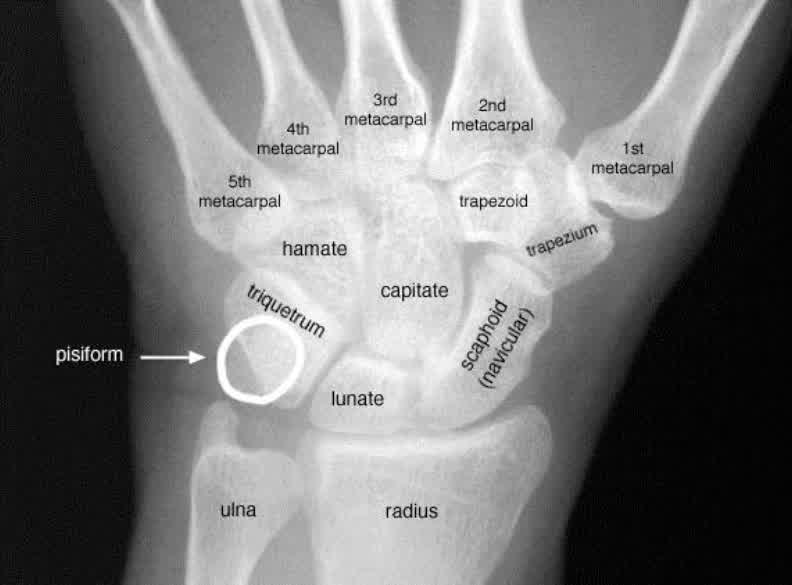
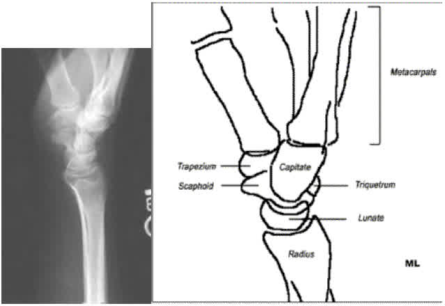
Adequacy
- Carpal-metacarpal junction to distal 5 cm of radius is included.
- Hand is neutral in both the PA and lateral views. The axis of the middle metacarpal lines up with the middle of the radius.
- Rotation (PA view): There is a gap between the two bones at the radioulnar joint. The ulnar styloid projects laterally.
- Rotation (lateral view): The radius and ulna are superimposed or within 3 mm dorsally. The pisiform should overlie the head to the scaphoid.
Alignment and angles
- The three smooth articulating cortical lines of the carpals are visible.
- There is no more than 2-3 mm between individual carpal bones.
- The radius articulates with at least half the lunate.
- Radial inclination is 16-28° on PA view.
- Radial volar (or palmar) tilt is 0-22° on lateral view.
- Scapholunate angle is < 65° on lateral view.
- The radial length is 11-12 mm.
Bony shape
- The scaphoid shows a cortical ring when dislocated on the PA view.
- The lunate should be quadrangular on PA view; triangular shape implies rotation or displacement.
- The pisiform is the last carpal to ossify up to age 12 years.
Special views
- Scaphoid: Views the long axis of the scaphoid.
- Carpal tunnel: Views the pisiform and hamate.
- Semi-pronated: Views the radial elements (trapezium and scaphoid tuberosity).
- Semi-supinated: Views the ulnar elements (pisiform and hamate).