Wrist Injuries - Specific fractures
Specific wrist fractures
Scaphoid fracture
Triquetral Fractures
Other
Scapholunate Injury
Lunate And Perilunate Dislocations
Radius fractures
Other Fractures Of The Distal Radius
Volar Angulation
Radial Styloid Fracture
Radioulnar Joint Injuries
Scaphoid fracture
Scaphoid fractures comprise 60% - 80% of all carpal fractures. Because the scaphoid forms a bridge between the carpal rows, it must withstand dramatic forces across its waist during forced extension of the wrist. Although common, scaphoid fractures can be difficult to visualise on film.
PEARLS
- When patients have clinical signs of scaphoid fracture snuffbox tenderness, pain with axial compression of the thumb, pain on palpation of the tubercle treat them as if they have a fracture. Even in the presence of a negative X-ray.
- Immobilise for 7 – 10 days and bring back for orthopedic review.
- Failure to immobilise these injuries in the emergency centre leads to a high incidence of malunion and avascular necrosis.
- Long-arm casts is suggested in the emergency setting prior to follow up.
Triquetral Fractures
Triquetral fractures, the second most common type of carpal fracture, occur with both direct trauma and with FOOSH injuries. Dorsal chip fractures are due to hyperextension when the ulnar styloid is jammed into the dorsum of the triquetrum. This produces tenderness just distal to the ulnar styloid.
These fractures require splinting and generally do well. Transverse fractures are less frequent and more ominous. Fracture through the body of the triquetrum is associated with perilunate, scaphoid, and ligamentous injury. Patients with triquetral fractures require a long arm splint and early referral.
Other
- Fractures of the Hamate and Lunate should be immobilised and followed up at a fracture clinic within 7-10 days if undisplaced, if displaced should be referred to a centre where reduction can be done.
- Ligament injury of the carpal bones can be present without fractures being present and if swelling persists with disruption of carpal spaces should be immobilised and sent for review.
- Capitate and Trapezium fractures are normally due to direct trauma to the affected bone but are rare.
Scapholunate Injury
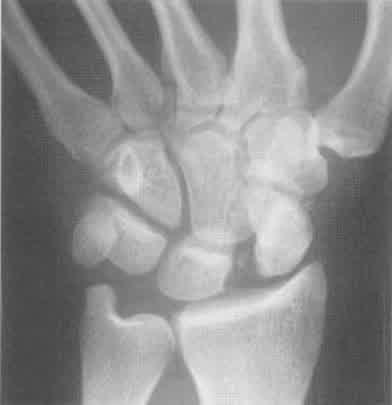
In essence, the scaphoid and distal carpal row are progressively peeled away from the lunate. The first ligamentous complex to rupture is the scapholunate, causing what is variably described as scapholunate instability, scapholunate dislocation, or rotary subluxation.
Physical findings may include tenderness at the scapholunate joint, just ulnar to the snuffbox, or instability of the scaphoid elicited by the scaphoid shift. (Watson’s test can be performed)
The expected radiographic findings of scapholunate injury on the PA view include a widened scapholunate gap. Widening of the scapholunate space as the capitate is forced proximally between the scaphoid and lunate can be increased by taking the PA film with the fist clenched.
Lunate And Perilunate Dislocations
If the scaphoid suffers fracture or ligament damage, it can no longer stabilise the wrist. At this point, further damage to the radiocapitate ligament, part of the volar extrinsic group, allows the capitate to slip out of its position in the lunate cup, leading to perilunate dislocation.
Unless there has been spontaneous reduction of the dislocation this should be obvious on the lateral wrist XR.
PEARLS
- Perilunate : On the lateral view, the capitate will no longer sit in the same line with the lunate, mid-shaft radius, and metacarpals; it will be outside the cup of the lunate.
- Lunate : Further disruption of the ligamentous support leads to lunate dislocation, when the lunate is torn from its normal position on the radius. This is most notable on the lateral view, where the lunate is “spilling over” toward the palm, rather than cupping the capitate.
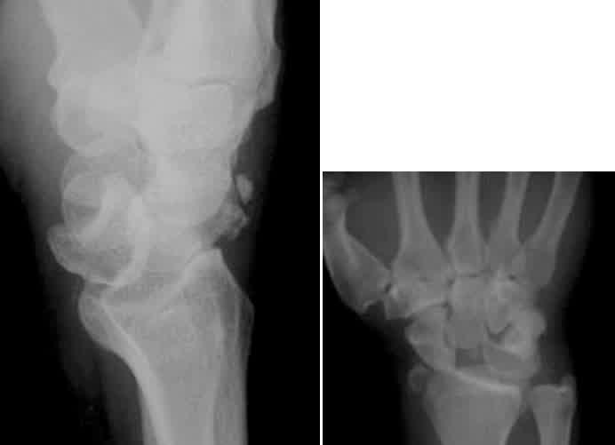
In the lateral and PA view the lunate is now disoriented in relation to the radius on the lateral view and the capitate is no longer coaxial. The lunate is triangular rather than normal quadrangular appearance.
These injuries should be immobilised and analgesia provided and sent to an appropriate centre with orthopaedic operating facility available immediately.
Radius fractures
Distal radius fractures are the most common upper extremity fracture and represent a large portion of the overall fractures seen in the ED. In the elderly due to decrease in bone density (osteopenia) the amount of force needed is significantly less than an young adult and associated ligamentous and carpal injuries should be actively investigated.
PEARLS
- Median nerve function should be routinely examined during a Colles’ fracture.
- Reduction in the emergency centre should be done with either local anaesthetic techniques example haematoma block or conscious sedation to reduce the distal radius fracture.
The 2 keys to successful reduction of the typical Colles’ fracture are as follows:
- Place the hand and wrist in the position of injury and pronate the forearm, which corrects the supination twist of the distal fractured segment. This can be performed with the aid of the Weinberg finger traction apparatus or with an assistant to fix the arm at the elbow. By recreating the mechanism of injury and the position of the bony fragments at injury, the periosteal ligaments are relaxed, which allows for easier reduction of the fracture.
- Extend the wrist to 90°, with the elbow fixed and the forearm supinated, and pull the distal segment back, up, and out at approximately 120°. Use both thumbs to push the distal fragment into alignment as the arm is pronated.
- Arm is then placed in a back-slab keeping the reduction adequately immobilised
- Post reduction films should always be obtained and the reduction is adequate if
- Lateral view the articular surface of distal radius at neutral or 10 degrees volar.
- PA view radial height 1cm above the ulna or ulna must not protrude above the radial articular surface.
Other Fractures Of The Distal Radius
There are other named fractures of the distal radius, each associated with a specific mechanism. Rather than naming the fracture, it is more important to identify and describe the lesion in terms of location, displacement, angulation, and comminution, as these parameters determine treatment and prognosis.
Volar Angulation
The Smith’s fracture (or reverse Colles’ fracture) occurs with direct blow or fall on the dorsum of the wrist. It is often the result of a bicyclist or motorcyclist being thrown over the handlebars. By maintaining a fierce grip on the handlebars, the patient sustains a distal radius fracture with volar, rather than dorsal, angulation.
Treatment criteria are similar to dorsally angulated fractures. (aka Colles’)
Radial Styloid Fracture
The chauffeur fracture (or Hutchinson fracture) is a break in the radial styloid. It is generally due to direct impact on the radial side of the wrist. If markedly displaced might need open reduction so immobilise and refer to fracture clinic.
Radioulnar Joint Injuries
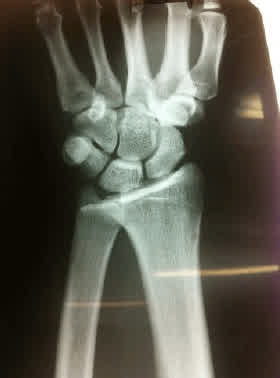
Injuries to this joint are commonly missed in the acute setting. Dislocation of the ulna commonly occurs with distal radius fractures and can be an important component of some fracture-dislocations of the forearm.
Occasionally, ulnar dislocation at the radioulnar joint occurs without bony injury. The injury can result from falls or axial distraction, as in sudden distraction or rotation (such as sudden lifting or grasping a fixed object like the handrails at stairs.
Clinical clues
- Swelling of the wrist particularly on the volar aspect
- Ulna styloid prominence was absent
- Close to full range of palmar flexion and dorsiflexion at the wrist
- The forearm is fixed in supination.
Remember that the ulna has a fixed relationship to the radius in true PA and lateral films.
- The dislocated ulna will project either dorsally or volarly to the radius. (lateral)
- Radioulnar gap will overlap (true PA)
- Fracture of the ulnar styloid base as well as radial shortening very suggestive of radio-ulna joint dislocation.
Treatment
In most cases of isolated volar ulna dislocation, the injury is reduced under general anaesthetic by forceful manipulation of the joint, using manual pressure on the ulna in a medial and dorsal direction, and hyper-pronation of the forearm.
Occasionally, it may be required to reduce the dislocation with an open procedure, particularly when the diagnosis has been delayed or in cases where the ulna is locked under the radius. The arm must then be immobilised for 6 weeks in full pronation in an above-elbow cast with physiotherapy required to regain best range of movement in the wrist joint.
Take home PEARL
- FOOSH (fall on outstretched hand) is a common presentation to the emergency centre
- Placing the wrist in a functional position if fracture is suspected is paramount
- Careful scrutinising of the plain films can unmask subtle injuries.
References:
1. Schwartz DT, Reisdorff EJ, eds. Emergency Radiology. New York: McGraw-Hill Professional Publishing; 2000. (Textbook)
2. Larsen CF, Lauritsen J. Epidemiology of acute wrist trauma. Int J Epidemiol 1993 Oct;22(5):911-916. (Prospective)
3. Kristinsdottir EK, Nordell E, Jarnlo GB, et al. Observation of vestibular asymmetry in a majority of patients over 50 years with fall-related wrist fractures. Acta Otolaryngol 2001 Jun;121(4):481-485. (Observational; 66 patients)
4. Chin HW, Visotsky J. Ligamentous wrist injuries. Emerg Med Clin North Am 1993 Aug;11(3):717-737. (Review)
5. Barnaby W. Fractures and dislocations of the wrist. Emerg Med Clin North Am 1992 Feb;10(1):133-149. (Review)
6. Altman RS, Harris GD, Knuth CJ. Initial management of hand injuries in the emergency patient. Am J Emerg Med 1987 Sep;5(5):400-403. (Review)