Acute Exacerbation of COPD / COAD
Acute Exacerbation of COPD
Introduction
Identification
Severity assessment
Management based on severity
Relevant history, examination
Investigations
Referral criteria
Discharge criteria
Preventative measures
Treatment Algorithm
Introduction/ Description
- COPD is a disease state resulting from an abnormal inflammatory response of the lungs to irritant particles and gases, with resultant progressive airflow limitation that is partially reversible. An exacerbation is defined as an increase in symptoms of COPD above the usual day to-day variation experienced by the patient, and necessitating a change in treatment. The most common cause of exacerbations is tracheobronchial infections.
Identification/recognition/diagnosis of problem
The diagnosis of COPD should be considered in any patient with chronic progressive dyspnoea and/or chronic cough (with or without sputum production) with a smoking history of more than 10 pack-years and/or other risk factors for COPD.
| Features suggesting a diagnosis of COPD | Features that suggest the presence of asthma |
|
|
Differential Diagnosis
- Acute exacerbations must be distinguished from other diseases and complications of COPD (including pneumonia, pneumothorax, congestive heart failure, arrhythmias and pulmonary embolism). When exacerbations are associated with features of infection (pyrexia and purulent sputum), the presence of pneumonia and other forms of lower respiratory infection should be excluded
Severity assessment (Triage)
Key elements from the history include: severity of COPD based on the FEV1 or MRC dyspnea scale, duration of symptom worsening or the development of new symptoms, number of previous episodes (exacerbations/ hospitalisations/respiratory failure/ need for NIV or IPPV), comorbidities and present treatment regimen
Medical research Council (MRC) dyspnoea scale
- SOB only on strenuous exertion
- SOB on hill, or with minimal exertion
- Walks slower than most people on level, or stops at own pace after 15mins
- SOB after about 100m, or few minutes on level
- Too SOB to leave house, SOB with activities of daily living
Signs of severity include: increased dyspnoea, chest tightness, occasional wheezing, and increased cough and sputum, use of accessory respiratory muscles, paradoxical chest wall movements, signs of right heart failure, development of peripheral oedema, hemodynamic instability, reduced alertness, worsening or new onset of central cyanosis
Management based on severity
Immediate action:
Non-drug treatment
Assess exacerbation severity, protect airway, supply oxygen, ensure adequate ventilation and IV access. Call for help if features of a severe / life-threatening exacerbation are present. Perform a focused initial assessment.
Oxygen should be prescribed and titrated based on the oximetry (sats) to avoid worsening of hypercapnoeic (type 2) respiratory failure using oxygen delivery devices as below to effect. Venturi masks are the delivery device of choice although others may have to proxy in its absence. Aim to start with device that can deliver FiO2 of 24-28% (e.g. nasal cannulae 2L/min, simple face or nebuliser mask 2-3L/min) and then titrate up or down to achieve desired saturations
- Saturation aim: 94 – 98% if PaO2 < 8 kPa, pCO2 ≤ 6kPa
- Saturation aim: 88 – 92% if PaO2 < 8 kPa, pCO2 > 6kPa
Measure ABGs 30 minutes later, with any deterioration and to monitor recovery
Mask with reservoir | Nasal cannulae | Venturi* | Simple/ neb mask | ||||
L/min | FiO2 | L/min | FiO2 | Set % | FiO2 | L/min | FiO2 |
| 6 | 60 | 1 | 24 | 24 | 2 | 25 | |
| 7 | 70 | 2 | 28 | 28 | 28 | 3 | 27 |
| 8 | 80 | 3 | 32 | 35 | 35 | 4 | 30 |
| 9 | 85 | 4 | 36 | 40 | 40 | 5 | 35 |
| 10+ | 90 | 5 | 60 | 50 | 6 | 40 | |
* Venturi(fixed oxygen delivery device) is best option for O2 titration
Drug treatment
- First line: inhaled bronchodilators and systemic corticosteroids
- Second line: consider adding intravenous Aminophylline if poor response to above and features of acute severe or life-threatening asthma attack remain
- Antibiotic are commonly indicted in COPD exacerbations: increased sputum volume, sputum purulence, increased dyspnoea and patients who require ventilation.
Magnesium sulphate is not recommended for COPD exacerbation. Intravenous salbutamol is unlikely to add much benefit if first and second line treatment have been started. It should only be used in consultation with the referral centre
Drug tables/ dose charts
Inhaled B2 agonists
- Nebulised 5 mg salbutamol or 1 mg fenoterol every 20 minutes until a satisfactory response is achieved
- No nebulizer available: 10 – 20 puffs (100 mcg/puff) salbutamol MDI via spacer every 20 minutes PRN, taking several deep breaths from spacer after every 2 puffs
Inhaled ipratropium bromide
- Nebulised ipratropium bromide 0.5 mg QID, in addition to salbutamol nebs, until a satisfactory response
- Or if no nebulizer available: up to 20 puffs (20 mcg/puff) ipratropium bromide MDI via spacer QID, taking several deep breaths from spacer after every 2 puffs
Systemic corticosteroids
- Oral prednisone 40mg (0.5 – 1 mg/kg, max 60mg) PO stat, then daily
- Or if severe breathlessness/ unable to swallow: hydrocortisone 100 – 200 mg IVI stat, then 6 hourly
Antibiotics
- Outpatient:
- Amoxicillin 500mg po TDS for 5 days
- Comorbidities, >3 exacerbation per year, antibiotics in the past 3 months: Co-amoxiclav 1g po BD for 5 days
- Inpatient:
- Ceftriaxone 1g IV OD (or Co-amoxiclav 1g po BD/ 1.2g IV TDS)
Methylxanthines
- Loading dose: aminophylline 5 mg/kg IVI over 30 minutes (2.5 mg/kg if patient already taking theophylline)
- Maintenance: aminophylline infusion at 0.5 mg/kg/hr
Intravenous B2 agonists
- Loading dose: salbutamol 15 mcg/kg (max 0.25 mg) in 200 mL 0.9% saline IVI over 10 minutes
- Maintenance: salbutamol infusion at 3 – 20 mcg/min
Relevant history, Examination
A productive cough (usually is worse in the mornings) or an acute chest illness is common. Breathlessness is the most significant symptom, but it usually does not occur until the sixth decade of life. Wheezing may occur in some patients, particularly during exertion and exacerbations. Absence of the following 3 signs nearly rules out COPD or airflow obstruction of another cause:
- Self-reported smoking history of more than 55 pack-years
- Wheezing on auscultation
- Self-reported wheezing
The sensitivity of a physical examination in detecting mild to moderate COPD is relatively poor however, physical signs are quite specific and sensitive for severe disease. Patients with severe disease experience tachypnoea and respiratory distress with simple activities. The respiratory rate increases in proportion to disease severity. Use of accessory respiratory muscles and paradoxical in-drawing of lower intercostal spaces is evident (known as the Hoover sign). In advanced disease, cyanosis, elevated jugular venous pulse (JVP), and peripheral oedema can be observed. Chest examination reveals the following:
- Hyperinflation (barrel chest)
- Wheezing – Frequently heard on forced and unforced expiration
- Diffusely decreased breath sounds
- Hyper-resonance on percussion
- Prolonged expiration
Investigations
- ABG: clinical judgement more valuable, but is useful to detect degree of CO2 retention. Can also be used to monitor K+ levels
- ECG: Usually sinus tachycardia but look for dysrhythmias, ischemia, features of right heart strain
- CXR: pneumonia/pneumothorax although bearing in mind that not seeing features of an infective process does not rule out and infective cause. It should also be borne in mind that some COPD patients present often and that the cumulative x-ray risk could potentially be harmful.
- Bedside ultrasound is useful to rule out a Pneumothorax
Referral criteria
- Monitor patients frequently and closely during treatment. Step up treatment as necessary according to response. Transfer to higher level of care if worsening or failing to respond and who is mechanically ventilated. The decision to admit to hospital should be based on clinical status and lung function, response to treatment, presence of high-risk, co-morbid features and the ability to manage at home.
The criteria for intensive care admission are:
- Satisfactory functional status before the exacerbation (patient coped with activities of daily living); if not known, the patient should be given the benefit of the doubt
- Possible need for mechanical ventilation i.e. PaO2 <6.7 kPa (50 mmHg) on room air, arterial blood pH <7.3, and confusion
- The presence of a reversible factor. Examples are infections, bronchospasm, oxygen-induced carbon dioxide narcosis, sedative administration, heart failure or other associated illnesses.
The criteria for ventilator support are:
- Exhaustion, confusion, coma
- Respiratory acidosis ( pH < 7,3 and declining)
- Inability to clear secretions
- Respiratory or Cardiac arrest
The criteria for non-invasive ventilation are:
- Patient able to protect own airway
- Patient able to clear own airway secretions
- pH < 7,3
- pCO2 > 6,7 kPa (>50mmHg)
- Contra-indications include: haemodynamic instability, decreased level of consciousness, vomiting and excessive secretions
Discharge criteria/checklist
- To qualify for discharge, a patient should have stable clinical symptoms and subjectively feel better. The patient should not require salbutamol more often than every four hours. If the patient is stable and can use a metered dose inhaler, there is no added benefit to using nebulised bronchodilators. Patient education may improve the response to future exacerbations.
- COPD is a chronic disease that is largely incurable. Given the progressive nature of the disease, as well as the morbidity and mortality associated with it, clinicians should always include end-of-life care discussions with patients. These discussions should focus on palliative efforts to improve quality of life, as well as assistance with advanced directives and care planning, and referrals for home care when needed.
Preventative measures
- The following measures have been shown to reduce the frequency and/or severity of COPD exacerbations: smoking cessation, prevention of respiratory infections (influenza and pneumococcal vaccination), bronchodilator treatment, inhaled corticosteroids. Patients should be advised as such on each admission.
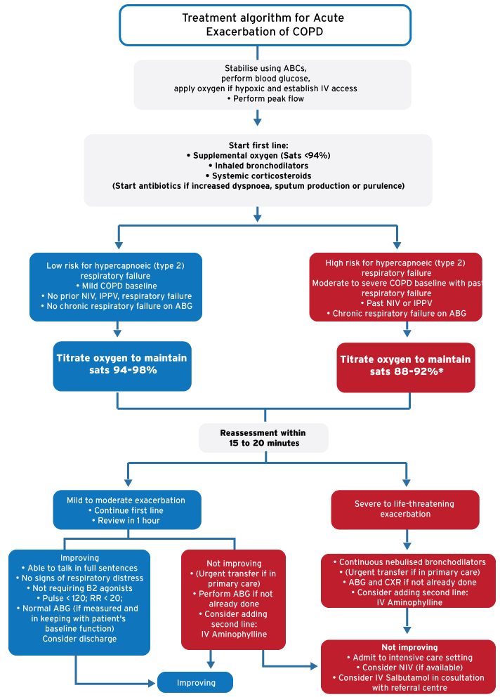
Treatment algorithm for Acute Exacerbation of COPD
*Mask with reservoir | *Nasal cannulae | *Venturi | *Simple/ neb mask | ||||
L/min | FiO2 | L/min | FiO2 | Set % | FiO2 | L/min | FiO2 |
| 6 | 60 | 1 | -- | 24 | 24 | 2 | 25 |
| 7 | 70 | 2 | 28 | 28 | 28 | 3 | 27 |
| 8 | 80 | 3 | 32 | 35 | 35 | 4 | 30 |
| 9 | 85 | 4 | 36 | 40 | 40 | 5 | 35 |
| 10+ | 90 | 5 | -- | 50 | 50 | 6 | 40 |
References
Abdool-Gaffar MS, et al. Guideline for the management of chronic obstructive pulmonary disease-2011 update. S Afr Med J. 2011;101(1 Pt 2):63-73